

Color Atlas of Neurology (Thieme 2004)
.pdfSpinal Circulation
22
Spinal Circulation
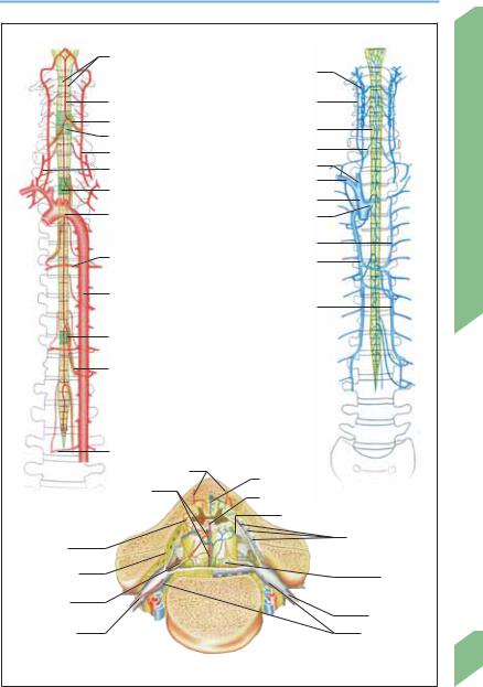
Arteries
Most of the blood supply of the spinal cord is supplied by the segmental spinal arteries, while relatively little comes from the vertebral arteries via the anterior and posterior spinal arteries. The segmental and spinal arteries are linked by numerous anastomoses.
Segmental arteries. The vertebral, ascending cervical, and deep cervical arteries give off cervical segmental branches; the thoracic and abdominal aorta give off thoracolumbar segmental branches via the posterior intercostal and lumbar arteries.
The segmental arteries give off radicular branches that enter the intervertebral foramen and supply the anterior and posterior roots and spinal ganglion of the corresponding level. The spinal cord itself is supplied by unpaired medullary arteries that originate from segmental arteries. The anatomy of these medullary arteries is variable; they usually have 5 to 8 larger ventral and dorsal branches that join up with the anterior and posterior spinal arteries. Often there is a single large radicular branch on one side, the great radicular artery (of Adamkiewicz), that supplies the entire lower twothirds of the spinal cord. It usually enters the spinal canal in the lower thoracic region on the left side.
Spinal arteries. The spinal arteries run longitudinally down the spinal cord and arise from the vertebral artery (p. 14). The unpaired anterior spinal artery lies in the anterior median fissure of the spinal cord and supplies blood to the anterior two-thirds of the cord. The artery’s diameter steadily increases below the T2 level. The two posterior spinal arteries supply the dorsal columns and all but the base of the dorsal horns bilaterally. Numerous anastomoses of the spinal arteries produce a vasocorona around the spinal cord. The depth of the spinal cord is supplied by these arteries penetrating it from its outer surface and by branches of the anterior spinal artery penetrating it from the anterior median fissure (sulcocommissural arteries).
Spinal Veins
Blood from within the spinal cord travels through the intramedullary veins, whose anat-
omy is variable, to the anterior and posterior spinal veins, which form a reticulated network in the pia mater around the circumference of the cord and down its length. The anterior spinal vein drains the anterior two-thirds of the gray matter, while the posterior and lateral spinal veins drain the rest of the spinal cord. These vessels empty by way of the radicular veins into the external and internal vertebral venous plexuses, groups of valveless veins that extend from the coccyx to the base of the skull and communicate with the dural venous sinuses via the suboccipital veins. Venous blood from the cervical spine drains by way of the vertebral and deep cervical veins into the superior vena cava; from the thoracic and lumbar spine, by way of the posterior intercostal and lumbar veins into the azygos and hemiazygos veins; from the sacrum, by way of the median and lateral sacral veins into the common iliac vein.
Watershed Zones
Because blood can flow either upward or downward in the anterior and posterior spinal arteries, the tissue at greatest risk of hypoperfusion is that located at a border zone between the distributions of two adjacent supplying arteries (“watershed zone”). Such vulnerable zones are found in the cervical, upper thoracic, and lower thoracic regions (ca. C4, T3–T4, and T8–T9).
Rohkamm, Color Atlas of Neurology © 2004 Thieme
All rights reserved. Usage subject to terms and conditions of license.
Spinal Circulation
|
Posterior spinal a. |
|
|
|
|
Vertebral v. |
|
|
Anterior spinal a. |
Deep cervical v. |
|
|
Radicular a. |
Spinal v. |
|
|
Watershed |
|
|
|
|
|
|
|
Vertebral a. |
Radicular v. |
|
|
Ascending cervical a. |
Inferior jugular v. |
|
|
Watershed |
Subclavian v. |
|
|
Right brachiocephalic v. |
|
|
|
|
|
|
|
Aortic arch |
Left brachiocephalic v. |
Circulation |
|
|
||
|
|
Accessory hemiazygos v. |
|
|
Thoracic |
Azygos v. |
|
|
intercostal a. |
||
|
|
||
|
|
|
|
|
Aorta |
|
Spinal |
|
|
Hemiazygos v. |
|
|
|
|
|
|
Watershed |
|
|
|
Great radicular a. |
|
|
|
(a. of Adamkiewicz) |
|
|
|
Lumbar a. |
|
|
|
Posterior spinal a. |
Posterior spinal v. |
|
|
Anterior |
|
|
|
Sulcocommissural a. |
Spinal |
|
Spinal arteries |
spinal a. and v. |
|
veins |
|
|
Anterior radicular v. |
|
Vasocorona |
|
|
Posterior external |
|
|
vertebral venous |
|
|
|
|
plexus |
Epidural space |
|
|
Pia mater |
|
|
|
|
Ventral root |
|
|
|
|
|
|
Spinal nerve |
Spinal branch |
|
|
Spinal ganglia |
|
Vessels of spinal cord (left: arteries; right: veins) |
23 |
|
|
|
||
Rohkamm, Color Atlas of Neurology © 2004 Thieme
All rights reserved. Usage subject to terms and conditions of license.

Anatomical and Functional Organization
24
Anatomical and Functional Organization
Cortical Structures
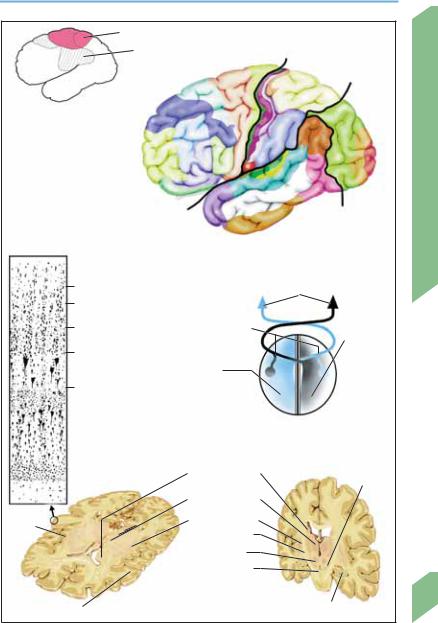
Different areas of the cerebral cortex (neocortex) may be distinguished from one another by their histological features and neuroanatomical connections. Brodmann’s numbering scheme for cortical areas has been used for many years and will be introduced in this section.
Projection areas. By following the course of axons entering and leaving a given cortical area, one may determine the other structures to which it is connected by afferent and efferent pathways. The primary projection areas are those that receive most of their sensory impulses directly from the thalamic relay nuclei (primary somatosensory cortex; Brodman areas 1, 2, 3), the visual (area 17), or the auditory (areas 41, 42) pathways. The primary motor cortex (area 4) sends motor impulses directly down the pyramidal pathway to somatic motor neurons within brainstem and the spinal cord. The primary projection areas are somatotopically organized and serve the contralateral half of the body. Proceeding outward along the cortical surface from the primary projection areas, one encounters the secondary projection areas
(motor, areas 6, 8, 44; sensory, areas 5, 7a, 40; visual, area 18; auditory, area 42), which subserve higher functions of coordination and information processing, and the tertiary projection areas (motor, areas 9, 10, 11; sensory, areas 7b, 39; visual, areas 19, 20, 21; auditory, area 22), which are responsible for complex functions such as voluntary movement, spatial organization of sensory input, cognition, memory, language, and emotion. The two hemispheres are connected by commissural fibers, which enable bihemispheric coordination of function. The most important commissural tract is the corpus callosum; because many tasks are performed primarily by one of the two hemispheres (cerebral dominance), interruption of the corpus callosum can produce various disconnection syndromes. Total callosal transection causes splitbrain syndrome, in which the patient cannot name an object felt by the left hand when the eyes are closed, or one seen in the left visual hemifield (tactile and optic anomia), and cannot read words projected into the left visual hemifield (left hemialexia), write with the left hand (left hemiagraphia), or make pantomimic move-
ments with the left hand (left hemiapraxia). Anterior callosal lesions cause alien hand syndrome (diagonistic apraxia), in which the patient cannot coordinate the movements of the two hands. Disconnection syndromes are usually not seen in persons with congenital absence (agenesis) of the corpus callosum.
Cytoarchitecture. Most of the cerebral cortex consists of isocortex, which has six distinct cytoarchitectural layers. The Brodmann classification of cortical areas is based on distinguishing histological features of adjacent areas of isocortex.
Functional areas. The functional organization of the cerebral cortex can be studied with various techniques: direct electrical stimulation of the cortex during neurosurgical procedures, measurement of cortical electrical cortical activity (electroencephalography and evoked potentials), and measurement of regional cerebral blood flow and metabolic activity. Highly specialized areas for particular functions are found in many different parts of the brain. A lesion in one such area may produce a severe functional deficit, though partial or total recovery often occurs because adjacent uninjured areas may take over some of the function of the lost brain tissue. (The extent to which actual brain regeneration may aid functional recovery is currently unclear.) The specific anatomic patterns of functional localization in the brain are the key to understanding much of clinical neurology.
Subcortical Structures
The subcortical structures include the basal ganglia, thalamus, subthalamic nucleus, hypothalamus, red nucleus, substantia nigra, cerebellum, and brain stem, and their nerve pathways. These structures perform many different kinds of complex information processing and are anatomically and functionally interconnected with the cerebral cortex. Subcortical lesions may produce symptoms and signs resembling those of cortical lesions; special diagnostic studies may be needed for their precise localization.
Rohkamm, Color Atlas of Neurology © 2004 Thieme
All rights reserved. Usage subject to terms and conditions of license.
Anatomical and Functional Organization
|
Hand movement |
|
|
|
|
|
|
|
|
|
|
Speech |
|
|
|
P |
|
|
|
|
|
|
|
|
|
|
|
|
|
|
|
|
|
|
|
|
|
a |
|
|
|
|
|
|
|
|
|
|
riet |
|
|
|
||
|
|
al |
4 |
|
|
|
al |
lobe |
|
|
|
|
lobe |
|
|
|
|
|
|
|
|
|
|
Front |
6 |
|
7a |
|
|
|
|
|
|
|
8 |
|
25 |
|
|
|
|
|
|
|
|
|
|
7b |
|
Occipit |
||||
|
|
9 |
|
1 |
|
|
|
|
||
Functional areas of cortex |
|
|
|
|
|
|
|
al |
||
|
|
|
|
|
|
|
|
|
||
(as determined by measurement |
|
46 |
3 |
40 |
|
|
|
|
19 |
lobe |
of regional blood flow) |
|
|
|
|
|
|
39 |
|
|
18 |
|
|
|
|
|
|
|
|
|
|
|
|
10 |
44 |
4352 |
|
22 |
|
|
|
|
17 |
|
|
|
|
|
|
|
|
|||
|
|
41 |
|
|
|
|
|
|
||
|
|
45 |
|
|
37 |
|
|
|||
|
|
|
|
|
|
|
|
|||
|
|
11 |
|
21 |
|
|
|
|
|
|
|
|
38 |
|
|
|
|
|
|
|
|
|
|
|
|
|
|
lobe |
|
|
||
|
|
|
|
|
|
|
|
|
||
|
|
|
|
|
|
|
l |
|
|
|
|
|
|
20 |
|
ra |
|
|
|
||
|
|
|
o |
|
|
|
|
|||
|
|
|
|
|
mp |
|
|
|
|
|
|
|
|
|
|
e |
|
|
|
|
|
Layers |
|
|
|
T |
|
|
|
|
|
|
|
|
|
|
|
|
|
|
|
|
|
of isocortex
Brodmann areas (lateral view)
I (Molecular layer)
II (Outer granule cell layer)
III (Middle pyramidal cell layer)
IV (Inner granule cell layer)
V (Large pyramidal cells)
|
|
|
Perception (visual, acoustic, |
||
|
|
|
olfactory, somatosensory) |
||
Commis- |
|
|
|
|
|
|
|
|
|
||
sural tracts |
|
|
Right |
||
|
|
||||
|
|
|
|
(stereo- |
|
Left |
|
gnosis, |
|||
|
spatial |
||||
(speech, |
perception, |
||||
writing, |
|
nonverbal |
|||
calculation, |
|
ideation, |
|||
abstraction, |
|
intuition) |
|||
logical analysis) |
|
|
|
||
VI (Polymorphic cells) |
|
|
|
|
Hemispheric dominance |
|
|
|
|
Caudate |
|
|
Susbstantia |
|
|
|
|
|||
|
|
|
nucleus |
|
||
|
|
|
|
nigra |
||
|
|
|
|
|
|
|
|
|
|
Thalamus |
|
|
|
|
|
|
|
|
||
Lentiform nucleus
Insula
Internal capsule
Red nucleus
Cerebral peduncle
Subcortical structures
Frontal operculum (Sections: left, horizontal; right, coronal) Hippocampus
Anatomical and Functional Organization
25
Rohkamm, Color Atlas of Neurology © 2004 Thieme
All rights reserved. Usage subject to terms and conditions of license.

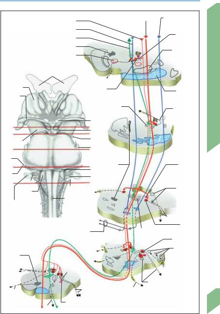
Brain Stem
The brain stem consists of the midbrain (mesencephalon), pons, and medulla. It contains the nuclei of the cranial nerves and ascending and descending tracts running to and from the brain, cerebellum, and spinal cord. It also contains autonomic centers that regulate cardiovascular function, breathing, and eating behavior as well as acoustic and vestibular relay nuclei. The flow of information along afferent and efferent pathways is regulated by reflex systems.
Nerve Pathways
|
All motor (p. 44) and sensory projection systems |
|
|
(p. 104) pass through the brain stem and com- |
|
|
municate with its intrinsic structures at various |
|
|
sites. The central sympathetic pathway (p. 90) |
|
Stem |
originates in the hypothalamus. |
|
Reticular Formation |
||
Brain |
||
The reticular formation (RF) is a network of nu- |
||
|
||
|
clei and interconnecting fibers that is anatomi- |
|
|
cally intertwined with the cranial nerve nuclei |
|
|
and other fiber tracts of the brain stem. Different |
|
|
parts of the reticular formation perform differ- |
|
|
ent functions. The reticular activating system |
|
|
(RAS) provides the anatomical and physiological |
|
|
basis for wakeful consciousness (p. 116). The |
|
|
medullary RF contains the vital centers control- |
|
|
ling the heartbeat, breathing, and circulation as |
|
|
well as reflex centers for swallowing and vomit- |
|
|
ing. The pontine RF contains centers for coordi- |
|
|
nation of acoustic, vestibular, respiratory, and |
|
|
cardiovascular processes. The midbrain RF con- |
|
|
tains centers subserving visuospatial orienta- |
|
|
tion and eating behavior (chewing, sucking, lick- |
|
|
ing). |
|
|
Reflex Systems (pp. 118ff) |
|
|
Pupillary light reflex. The Edinger–Westphal nu- |
|
|
cleus in the midbrain, which is adjacent to the |
|
|
oculomotor nucleus, provides the efferent arm |
|
|
of the reflex loop (p. 90; examination, p. 92.) |
|
|
Vestibulo-ocular reflex (VOR, p. 84). The vestibu- |
|
|
lar nuclei receive their main input from the |
|
|
labyrinthine semicircular canals and collateral |
26input from the cerebellar nuclei; their output is conveyed to the extraocular muscles through
the medial longitudinal fasciculus, and to the
spinal cord through the vestibulospinal tract. Examination: Suppression of visual fixation: the subject extends his arms and stares at his thumbs while spinning on a swivel chair. Nystagmus does not occur in normal subjects. Oculocephalic reflex (doll’s eyes phenomenon): Horizontal or vertical passive rotation of the subject’s head causes the eyes to rotate in the opposite direction; normally suppressible by awake persons, this reflex is seen in patients with impaired consciousness but preserved vestibular function. Caloric testing: The examiner first confirms that the patient’s eardrums are intact, then instills cold water in the external auditory canal with the head elevated at a 30° angle (which inactivates the ipsilateral horizontal semicircular canal). This normally causes nystagmus in the contralateral direction, i.e., slow ipsilateral conjugate deviation of the eyes, followed by a quick jerk to the other side.
Corneal reflex. Afferent arm, CN V/1; efferent arm, CN VII, which innervates the orbicularis oculi muscle. Examination: Touching the cornea from the side while the subject looks forward evokes blinking. The reflex can also be assessed by electromyography (EMG).
Pharyngeal (gag) reflex. Afferent arm, mainly CN IX, X, and V/2; efferent arm, CN IX and X. The gag reflex may be absent in normal persons. Examination: Touching the soft palate or back of the pharynx evokes pharyngeal muscle contraction.
Cough reflex. Afferent arm, CN IX and X; efferent arm, via the solitary tract to the diaphragm and other participating muscle groups. Examination: Tested in intubated patients with endotracheal suction (tracheal reflex).
Masseter (jaw jerk) reflex. Afferent arm, probably CN V/3; efferent arm, CN V. Examination: Tapping the chin evokes jaw closure.
Acoustic reflex (p. 68). Afferent arm, projections of the cochlear nuclei to the RAS. Examination: Sudden, intense acoustic stimuli evoke a fright reaction including lid closure, startle, turning of the head, and increased alertness.
Rohkamm, Color Atlas of Neurology © 2004 Thieme
All rights reserved. Usage subject to terms and conditions of license.
Brain Stem
|
|
|
Corticonuclear tract |
|
Corticospinal tract |
Corticopontine |
|
||
|
|
|
|
|
|
|
tract |
|
|
|
|
|
|
|
|
|
|
|
|
|
|
|
Corticopontine tract |
|
|
|
|
|
|
Medial longitudinal fasciculus |
|
|
|
|
Reticular |
|
|||
|
|
|
|
formation |
|
||||
|
|
|
|
|
|
|
|
|
|
|
|
|
Medial lemniscus |
|
|
|
|
Substantia |
|
|
|
|
|
|
|
|
|
|
|
|
|
|
Red nucleus |
|
|
|
|
nigra |
|
|
|
Lateral ventricle |
|
|
|
|
|
|
|
|
|
|
|
|
III |
|
|
A |
|
Thalamus |
|
|
|
Central |
|
|
Cerebral |
|
|
|
|
|
|
|
|
|
|||
Optic - |
|
|
|
sympathetic tract |
|
|
|
||
|
|
|
|
|
peduncle |
|
|||
|
|
|
|
|
|
|
|
||
tract |
|
|
|
|
Reticular |
|
|
|
|
|
|
|
|
|
|
|
IV |
|
|
|
|
|
|
|
formation |
|
|
|
|
|
|
|
|
|
|
|
|
Stem |
|
|
|
|
|
II |
|
|
|
|
|
A |
|
|
|
III |
|
|
|
|
|
|
|
|
|
|
|
|
Brain |
||
B |
|
|
|
|
|
|
|
||
|
|
|
IV |
|
|
|
|
||
Cerebral |
|
|
|
|
|
|
B |
||
peduncle |
|
|
|
|
|
|
|
||
|
|
|
V |
|
|
|
|
||
C |
|
|
|
|
|
|
|
|
|
|
|
|
|
|
|
|
|
|
|
VII |
|
|
|
|
|
|
|
|
|
D |
|
|
|
|
Medial |
|
|
Central |
|
|
|
|
VI |
lemniscus |
|
|
sympathetic |
|
|
VIII |
|
|
|
|
|
|
|||
|
|
|
X |
|
|
|
tract |
|
|
|
IX |
|
|
|
|
|
|
|
|
E |
|
|
|
|
|
|
|
|
|
|
|
XI |
|
|
|
|
Principal |
|
|
Olive |
|
|
|
|
|
|
|||
|
|
|
|
|
sensory nucleus/ |
|
|||
|
|
|
|
|
|
|
|
||
Choroid plexus |
|
Pyramidal |
|
|
|
|
Spinal tract of |
|
|
|
decussation |
|
|
|
|
CN V |
|
||
(fourth ventricle) |
|
|
|
|
|
||||
|
|
|
|
|
|
|
|||
|
|
|
|
|
|
|
|
C |
|
|
|
|
|
|
|
|
V |
|
|
Brain stem |
|
|
|
|
|
|
|
Motor |
|
(ventral; red lines, planes of section) |
|
|
|
|
|
||||
|
|
Reticular |
|
nucleus V |
|
||||
|
|
|
Middle |
|
|
|
|||
|
|
|
cerebellar peduncle |
formation |
|
Nucleus VI |
|
||
|
|
|
|
|
|
|
|
|
|
|
|
|
|
VII |
|
|
|
Vestibular |
|
Medial |
|
|
|
|
|
|
nuclei |
|
|
|
|
|
|
|
|
|
|
||
|
|
|
VI |
|
|
|
|
|
|
lemniscus |
|
|
Reticular |
|
|
|
|
|
|
X |
|
|
|
|
|
VIII |
D |
|
|
|
|
formation |
|
|
|
|
|||
|
|
|
|
|
|
|
|||
|
|
|
|
E |
|
CN VII and nucleus |
|
||
XII |
|
|
X |
|
VI |
|
|
|
|
|
|
|
|
|
|
|
|
||
|
|
|
|
|
|
|
|
|
|
|
|
|
Central sympathetic tract |
|
|
|
27 |
||
|
|
|
XII |
|
|
|
|
|
|
Rohkamm, Color Atlas of Neurology © 2004 Thieme
All rights reserved. Usage subject to terms and conditions of license.

Cranial Nerves
28
Cranial Nerves
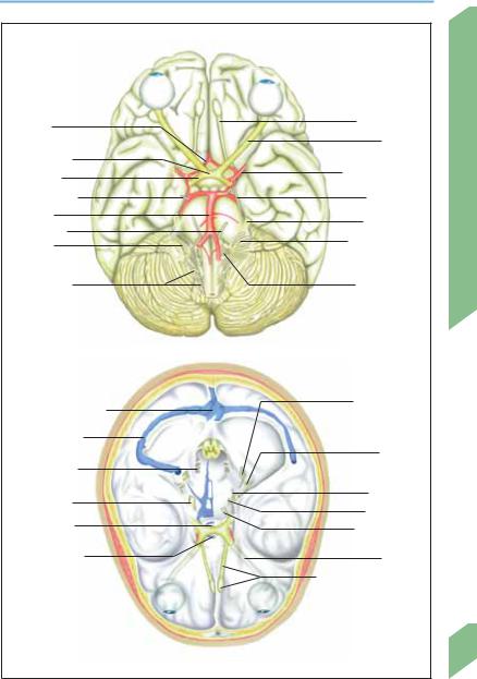
Cranial Nerve Pathways
Cranial Origin/Course (see also pp. 70ff. and 74 ff)
Nerve
IOlfactory nerves cribriform plate olfactory bulb olfactory tract anterior perforated substance lateral olfactory stria ( parahippocampal gyrus) and medial olfactory stria ( limbic system)
IIRetinal ganglion cells optic disk optic nerve orbit optic canal optic chiasm optic tract lateral geniculate body ( optic radiation occipital lobe) and superior colliculi ( pretectal area)
IIIMidbrain interpeduncular fossa between superior cerebellar artery and posterior cerebral artery tentorial edge cavernous sinus medial orbital fissure oculomotor nerve, superior division (levator palpebrae and superior rectus muscles) and inferior division (medial and inferior rectus and inferior oblique muscles) or parasympathetic fibers ciliary ganglion
IV |
Midbrain dorsal brainstem below the inferior colliculi around the cerebral peduncle lateral |
|
wall of the cavernous sinus orbital fissure superior oblique muscle |
VPons ca. 50 root filaments (sensory root = portio major; motor root = portio minor) petrous apex through the dura mater trigeminal ganglion (V/1 orbital fissure; V/2 foramen rotundum, V/3 + portio minor foramen ovale)
VI |
Posterior margin of pons up the clivus through the dura mater petrous apex lateral to |
|
internal carotid artery in the cavernous sinus orbital fissure lateral rectus muscle |
VII |
Pons (cerebellopontine angle) above the olive internal acoustic meatus petrous pyramid |
|
(canal of facial nerve) geniculum of facial nerve ( nervus intermedius/greater petrosal nerve |
|
gustatory fibers) medial wall of the tympanic cavity stylomastoid foramen muscles of fa- |
|
cial expression |
VIII |
Lateral to CN VII vestibular nerve, cochlear nerve |
IX |
Medulla jugular foramen between carotid artery and internal jugular vein root of the |
|
tongue |
X |
Posterolateral sulcus of medulla jugular foramen internal organs |
XI |
Cranial and spinal roots trunk of accessory nerve jugular foramen muscles |
XII |
Medulla hypoglossal canal tongue muscles |
CN = cranial nerve.
See Table 1, p. 356, for the functions of the cranial nerves.
Rohkamm, Color Atlas of Neurology © 2004 Thieme
All rights reserved. Usage subject to terms and conditions of license.
Cranial Nerves
Anterior |
|
Olfactory tract |
|
|
|
|
|
communicating a. |
|
Optic n. |
|
Optic chiasm |
|
|
|
Optic tract |
|
Middle cerebral a. |
|
|
|
|
|
Oculomotor n. |
|
Trochlear n. |
|
Basilar a. |
|
Trigeminal n. |
|
Abducens n. |
|
Nerves |
|
|
Glossopharyn- |
||
Facial n., |
|
||
|
geal n., vagus n. |
||
vestibulo- |
|
||
|
|
||
cochlear n. |
|
|
|
|
|
Cranial |
|
Accessory n., |
|
Hypoglossal n. |
|
spinal root |
|
|
|
|
|
|
|
|
Cranial nerves at the base of the brain |
|
|
Confluence of sinuses |
|
Glossopharyn- |
|
|
geal n., vagus |
|
|
|
|
|
|
|
|
n., accessory n. |
|
Transverse sinus |
|
|
|
|
|
Facial n., |
|
Hypoglossal n. |
|
vestibulo- |
|
|
cochlear n. |
|
|
|
|
|
|
Trigeminal n. |
|
Trochlear n. |
|
|
Abducens n. |
|
|
|
|
|
|
Pituitary stalk |
|
Oculomotor n. |
|
|
|
|
|
Cavernous sinus |
|
Optic n. |
|
|
|
Olfactory bulb and tract |
|
|
Cranial nerves at the base of the skull |
|
29 |
|
|
|
Rohkamm, Color Atlas of Neurology © 2004 Thieme
All rights reserved. Usage subject to terms and conditions of license.

Spine and Spinal Cord
Spine
|
The spine (vertebral column) bears the weight |
|
|
of the head, neck, trunk and upper extremities. |
|
|
Its flexibility is greatest in the cervical region, |
|
|
intermediate in the lumbar region, and lowest in |
|
|
the thoracic region. Its uppermost vertebrae |
|
|
(atlas and axis) articulate with the head, and its |
|
|
lowermost portion, the sacrum (which consists |
|
|
of 5 vertebrae fused together), articulates with |
|
|
the pelvis. There are 7 cervical, 12 thoracic (in |
|
|
British usage dorsal), and 5 lumbar vertebrae, |
|
Cord |
making a total of 24 above the sacrum. Below |
|
the sacrum, the coccyx is composed of 3 to 6 |
||
|
||
Spinal |
coccygeal vertebrae. |
|
Intervertebral Disks |
||
and |
Each pair of adjacent vertebrae is separated by |
|
|
||
Spine |
an intervertebral disk. From the third decade of |
|
life onward, each disk progressively diminishes |
||
in water content, and therefore also in height. Its |
||
|
tensile, fibrous outer ring (annulus fibrosus) |
|
|
connects it with the vertebrae above and below |
|
|
and is held taut by the pressure in the central |
|
|
nucleus pulposus, which varies as a function of |
|
|
the momentary position of the body. The pres- |
|
|
sure that obtains in the sitting position is double |
|
|
the pressure when the patient stands, but that |
|
|
found in the recumbent position is only one- |
|
|
third as great. The interior of the disk has no |
|
|
nociceptive innervation, in contrast to the peri- |
|
|
osteum of the vertebral bodies, which is inner- |
|
|
vated by the meningeal branch of the segmental |
|
|
spinal nerve, as are the intervertebral joint cap- |
|
|
sules, the posterior longitudinal ligament, the |
|
|
dorsal portion of the annulus fibrosus, the dura |
|
|
mater, and the blood vessels. |
|
|
Spinal Canal |
|
|
The spinal canal is a tube formed by the vertebral |
|
|
foramina of the vertebral bodies stacked one on |
|
|
top of another; it is bounded anteriorly by the |
|
|
vertebral bodies and posteriorly by the vertebral |
|
|
arches (laminae). Its walls are reinforced by the |
|
|
intervertebral disks and the anterior and poste- |
|
|
rior longitudinal ligaments. It contains the spinal |
30cord and its meninges, the surrounding fatty and connective tissues, blood vessels, and spinal
nerve roots. Its normal sagittal diameter ranges
from 12 to 22 mm in the cervical region and from 22 to 25 mm in the lumbar region.
Spinal Cord
Like the brain, the spinal cord is intimately enveloped by the pia mater, which contains numerous nerves and blood vessels; the pia mater merges with the endoneurium of the spinal nerve rootlets and also continues below the spinal cord as the filum terminale internum. The weblike spinal arachnoid membrane contains only a few capillaries and no nerves. The denticulate ligament runs between the pia mater and the dura mater and anchors the spinal cord to the dura mater. In lumbar puncture, cerebrospinal fluid is withdrawn from the space between the arachnoid membrane and pia mater (spinal subarachnoid space), which communicates with the subarachnoid space of the brain. The spinal dura mater originates at the edge of the foramen magnum and descends from it to form a tubular covering around the spinal cord. Its lumen ends at the S1–S2 level, where it continues as the filum terminale externum, which attaches to the sacrum, thus anchoring the dura mater inferiorly. The dura mater forms sleeves around the anterior and posterior spinal nerve roots which continue distally, together with the arachnoid membrane, to form the epineurium and perineurium of the spinal nerves. Unlike the cranial dura mater, the spinal dura mater is not directly apposed to the periosteum of the surrounding bone (i.e., the vertebral canal) but is separated from it by the epidural space, which contains fat, loose connective tissue, and valveless venous plexuses (p. 22).
The root filaments (rootlets) that come together to form the ventral and dorsal spinal nerve roots are arranged in longitudinal rows on the lateral surface of the spinal cord on both sides. The ventral root carries only motor fibers, while the dorsal root carries only sensory fibers. (This socalled “law of Bell and Magendie” is actually not wholly true; the ventral root is now known to carry a small number of sensory fibers as well.) The cell bodies of the pseudounipolar sensory neurons are contained in the dorsal root ganglion, a swelling on the dorsal root just proximal to its junction with the ventral root to form the segmental spinal nerve.
Rohkamm, Color Atlas of Neurology © 2004 Thieme
All rights reserved. Usage subject to terms and conditions of license.

Spine and Spinal Cord
C |
= |
Cervical nerves (C1-C8, blue) |
|
|
|
|
|
|
|
|
|
||
T |
= |
Thoracic nerves (T1-T12, purple) |
|
|
|
|
|
|
|
|
|
||
L |
= |
Lumbar nerves (L1-L5, turquoise) |
|
|
|
|
|
|
|
|
|
||
S |
= |
Sacral nerves (S1-S5, light green) |
|
|
|
|
|
|
|
|
|
||
Co |
= |
Coccygeal nerve (Co1, gray) |
|
|
|
|
|
|
|
|
|
||
|
|
|
|
|
|
|
|
|
|
|
|
|
Atlas |
Posterior branch ( |
|
> skin and muscles of back) |
|
|
|
|
|
|
|
|
C 1 |
||
|
|
|
|
|
|
|
|
|
C 2 |
||||
|
|
|
|
|
|
|
|
|
|||||
|
|
|
|
|
|
|
|
|
|||||
|
|
Epidural space |
|
|
|
1 |
|
|
|
C 3 |
|||
|
|
|
|
|
2 |
|
|
C 4 |
|||||
|
|
|
|
|
|
|
|
3 |
|
|
|||
|
|
|
|
|
|
|
|
|
|
C 5 |
|||
|
|
|
|
Pia mater |
|
|
|
4 |
|
|
|||
|
|
|
|
|
|
|
|
|
C 6 |
||||
|
|
|
|
|
|
|
|
5 |
|
|
|
||
|
|
|
|
Subarachnoid space |
|
|
6 |
|
|
|
C 7 |
||
|
|
|
|
|
|
7 |
|
|
|
C 8 |
|||
|
|
|
|
|
|
|
|
|
|
|
|||
|
|
|
|
|
|
|
|
1 |
|
|
|
||
|
|
|
|
|
|
|
|
|
|
|
T 1 |
||
|
|
|
|
|
|
|
|
2 |
|
|
|
||
|
|
|
|
|
|
|
|
|
|
|
T 2 |
||
|
|
|
|
|
|
|
|
3 |
|
|
|
||
|
|
|
|
|
|
|
|
|
|
|
T 3 |
||
Spinal nerve |
|
4 |
|
|
|
|
T 4 |
||||||
|
|
|
|
|
|
5 |
|
|
|
|
|
T 5 |
|
|
|
|
|
|
Spinal |
6 |
|
|
|
|
|
T 6 |
|
Ventral root |
ganglion |
|
|
|
|
|
T 7 |
||||||
7 |
|
|
|
|
|
||||||||
|
|
|
|
|
Root |
|
|
|
|
|
T 8 |
||
|
|
|
|
|
8 |
|
|
|
|
|
|||
|
|
|
|
|
|
|
|
|
|
T 9 |
|||
|
|
|
|
|
sleeve |
|
|
|
|
|
|||
|
|
|
|
|
9 |
|
|
|
|
|
|||
Denticulate |
|
|
|
|
|
|
T 10 |
||||||
|
10 |
|
|
|
|
|
|||||||
|
|
|
|
|
|
|
|||||||
lig. |
|
|
|
|
|
|
|
|
|
|
T 11 |
||
|
|
|
|
|
|
|
|
|
|
|
|
||
|
|
|
|
|
|
11 |
|
|
|
|
|
||
Rib |
|
|
|
|
Intervertebral |
|
|
|
|
|
|
||
|
|
|
|
12 |
|
|
|
|
|
T 12 |
|||
|
|
|
|
|
foramen |
|
|
|
|
|
|
||
|
|
|
|
|
|
|
|
1 |
|
|
|
L 1 |
|
Arachnoid |
|
|
|
2 |
|
|
|
L 2 |
|||||
membrane |
|
|
|
3 |
|
|
|
|
|||||
Costo- |
|
|
|
|
|
|
|
|
|
L 3 |
|||
|
|
|
Meningeal |
|
|
4 |
|
|
|
||||
vertebral |
|
|
|
|
|
|
|||||||
joint |
|
|
|
|
branch |
|
|
5 |
|
|
|
L 4 |
|
|
|
|
|
|
|
|
|
|
|
|
|||
|
|
|
|
|
Sacrum |
|
|
|
|
|
1 |
L 5 |
|
Epidural |
|
|
|
|
|
|
|
2 |
|
||||
|
|
|
|
|
|
|
|
|
|
S 1 |
|||
veins |
|
|
|
|
Posterior |
|
|
|
|
3 |
|
|
|
|
|
|
|
|
|
|
|
|
|
S 2 |
|||
|
|
|
|
|
|
|
|
|
|
|
|||
|
|
|
|
|
longitudinal lig. |
4 |
|
|
|
||||
|
|
|
|
|
|
|
S 3 |
||||||
Inter- |
|
|
|
|
|
|
|
|
|||||
|
|
|
|
|
|
|
|
|
|
||||
|
|
|
|
|
|
5 |
|
S 4 |
|
||||
transverse lig. |
|
|
|
|
|
||||||||
|
|
|
|
|
Dura mater |
|
|
|
|
|
|
S 5 |
|
Vertebral |
Intervertebral disk, |
|
|
|
|
Co 1 |
|||||||
|
|
|
|
|
|
||||||||
body |
|
|
|
|
annulus fibrosus |
|
|
|
|
|
|
|
Coccyx |
|
|
|
|
Spinal cord, spinal canal |
|
|
|
|
Spinal nerves |
||||
|
|
|
|
(thoracic spine, frontal view) |
|
|
|
|
|
|
|
|
|
|
|
|
|
|
|
|
|
|
|
|
|
|
|
Spine and Spinal Cord
31
Rohkamm, Color Atlas of Neurology © 2004 Thieme
All rights reserved. Usage subject to terms and conditions of license.