

Atlas of breast surgery
.pdf106 |
7.9 |
Pedicle TRAM Flap Reconstruction |
7
Fig. 7.20. Positions of the paddle according to the previous positions
Plastic and Reconstructive Breast Surgery |
Chapter 7 |
107 |
a
b
Median
line
c
Fig. 7.21a–c. a Bipedicle flap. b Umbilical closure. c Abdominal closure
108 |
7.9 |
Pedicle TRAM Flap Reconstruction |
Fig. 7.22. Abdominal closure TRAM. Centralization of the umbilicus in the case of a single pedicle TRAM
7
b
a |
c |
Fig. 7.23a–c. Left pedicle TRAM with suture on the right rectus sheath in order to centralize the umbilicus

Plastic and Reconstructive Breast Surgery |
Chapter 7 |
109 |
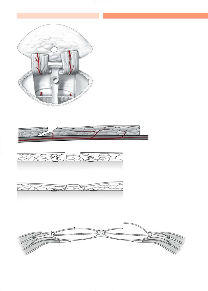
7.10 Nipple Reconstruction
The creation of a nipple–areola complex following breast reconstruction improves the cosmetic outcome, and many patients may request such a procedure. The most popular technique for nipple reconstruction combines a tattoo procedure with a small triangular skin flap that is often referred to as a skate flap, illustrated in Fig. 7.24. The location of the areolar should be drawn preoperatively with the patient
in the standing position to check for bilateral symmetry of the nipple–areola complex. The nipple–are- ola reconstruction is generally performed as a sec- ond-stage procedure under local anesthesia. The tattooing should be done prior to reconstructing the nipple.A skin flap is then developed and folded on itself and sutured with 4–0 absorbable sutures, as shown in the accompanying illustrations. The angle of the skin flap should be oriented appropriately if it is in the vicinity of the previous mastectomy scar (Fig. 7.25).
Fig. 7.24. Nipple–areola reconstruction (color of the areola and the nipple is obtained by tattooing the surface of the circle)
110 |
7.10 |
Nipple Reconstruction |
7
Fig. 7.25. Nipple–areola reconstruction in the vicinity of the previous mastectomy scar
Plastic and Reconstructive Breast Surgery |
Chapter 7 |
111 |
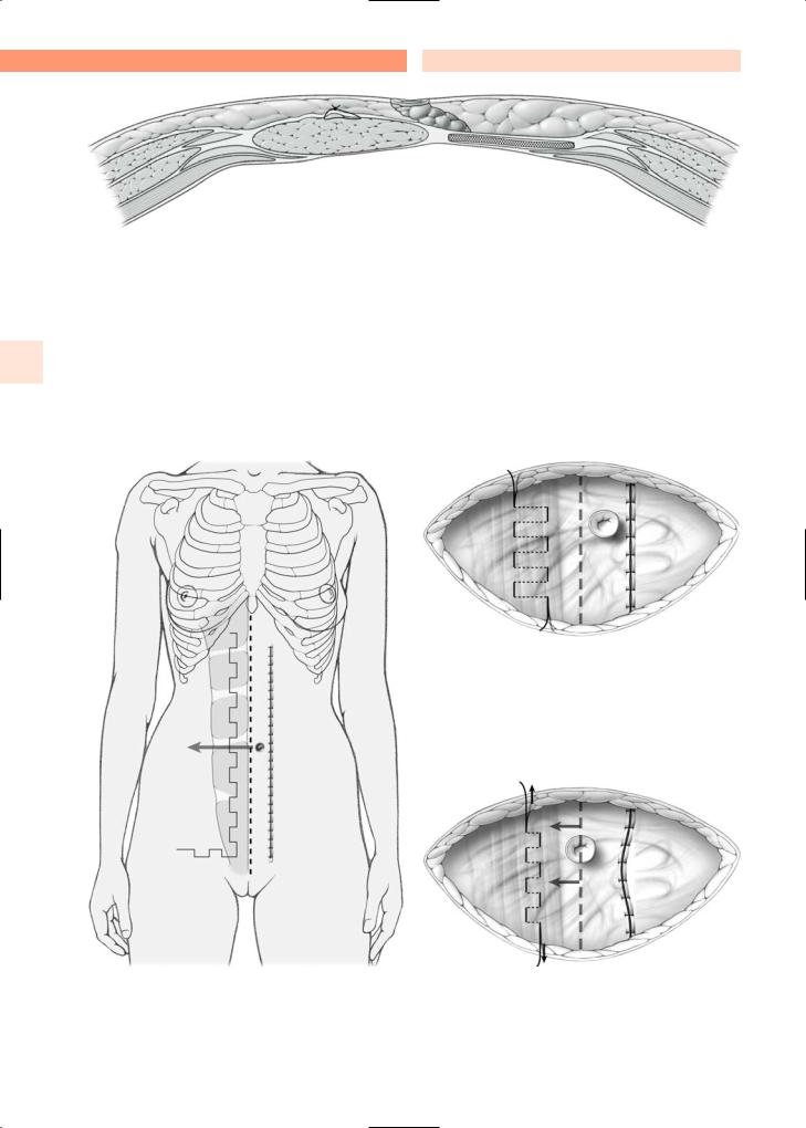
7.11 Thoracoepigastric Cutaneous Flap |
|
in the accompanying illustrations (Fig. 7.26), the flap |
|
can be developed horizontally, with its superior end |
|
for Large Thoracic Wall Defects |
|
|
|
towards the axilla. The fascia of the abdominal mus- |
|
|
|
|
Local recurrences following mastectomy for breast |
cles should remain attached to the flap to preserve its |
|
blood supply. After rotation of the flap, the chest wall |
||
cancer will generally require extensive skin and mus- |
defect should be closed with the patient placed in a |
|
cle resection. The latissimus dorsi or TRAM flaps are |
semi-sitting position on the operating room table to |
|
often used to close such defects. However, when the |
facilitate advancement of the flap. |
|
defect is not too large, a thoracoepigastric cutaneous |
Also, chest wall defects can be closed with mesh, |
|
flap can be used. The blood supply for this flap is de- |
using the suspension technique described in Figs. |
|
rived from the epigastric perforators and, as shown |
7.9a, b and 7.10a, b. |
|
Fig. 7.26. Thoraco-axillary cutaneous flap for large thoracic defects

112 |
7.12 |
Partial (or Total) Reconstruction |
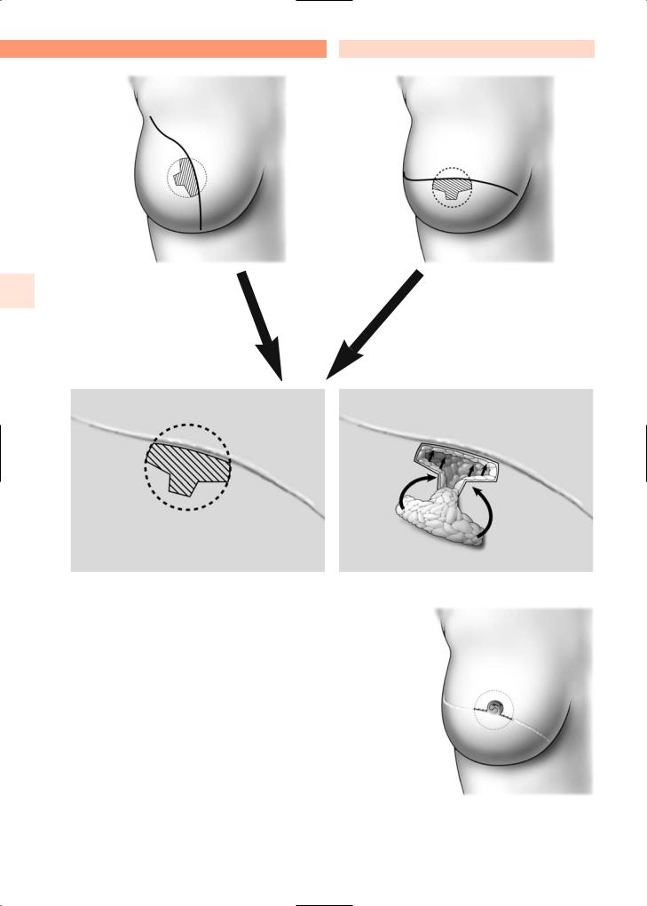
7.12Partial (or Total) Reconstruction with Omental Flaps
Local recurrences following mastectomy for breast cancer can also be managed with omental flaps (Fig. 7.27). Following resection of the area of local recurrence, an omental flap can be used to cover the defect. The omentum is one of the most vascularized tissues of the body and provides excellent material to close huge defects, particularly after radiotherapy.
The omentum should be mobilized with a pedicle consisting of the right gastroepiploic artery and vein. It should be detached from the greater curvature of the stomach. This dissection can also be done by endoscopy, with the omental flap pulled up to the surface of the thoracic wall through a small peritoneal hole in the epigastric area. It can also be used as a free flap because the vessels are wide and can therefore be easily anastomosed. In selected cases, the omental flap can also be used for partial breast reconstruction.
7
Art. gastroepiploica dextra
•
•
Art. gastroepiploica sinistra
Fig. 7.27. Partial or total reconstruction with omental flap

Plastic and Reconstructive Breast Surgery
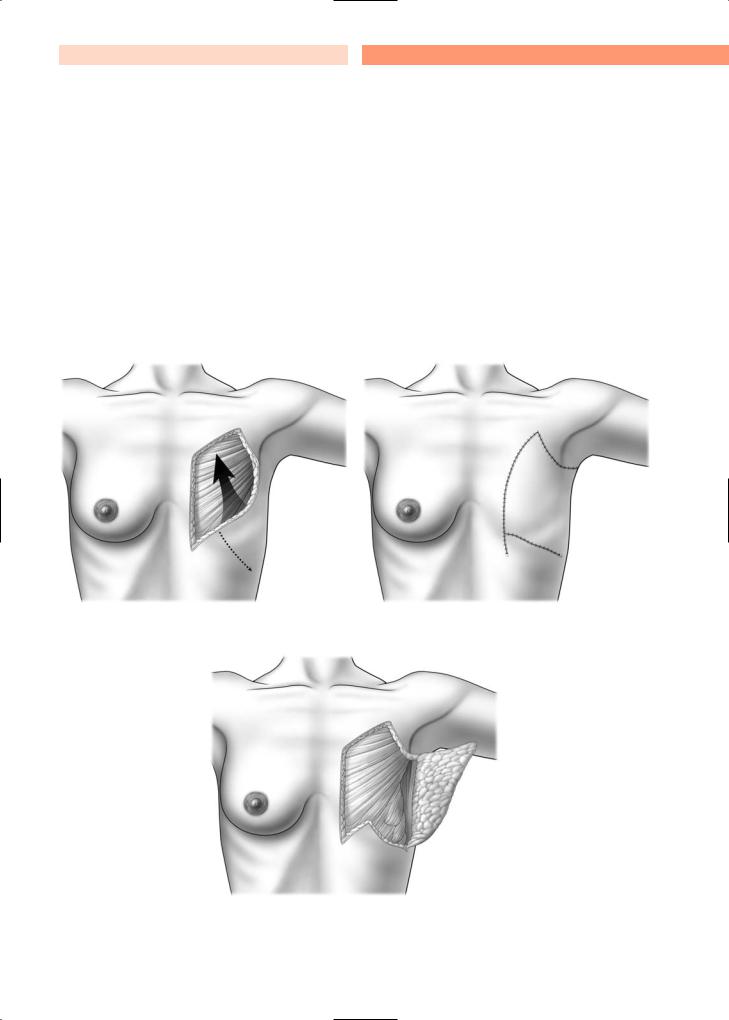
7.13 Reduction Mammoplasty
Heavy, pendulous breasts are often a source of chronic pain and discomfort for many women. Although women may request reduction mammoplasty to relieve pain and discomfort, many also hope that the procedure will improve their appearance.
Prior to undertaking breast reduction mammoplasty, the surgeon should document several measurements that are useful in the procedure. These measurements are indicated in Fig. 7.28.
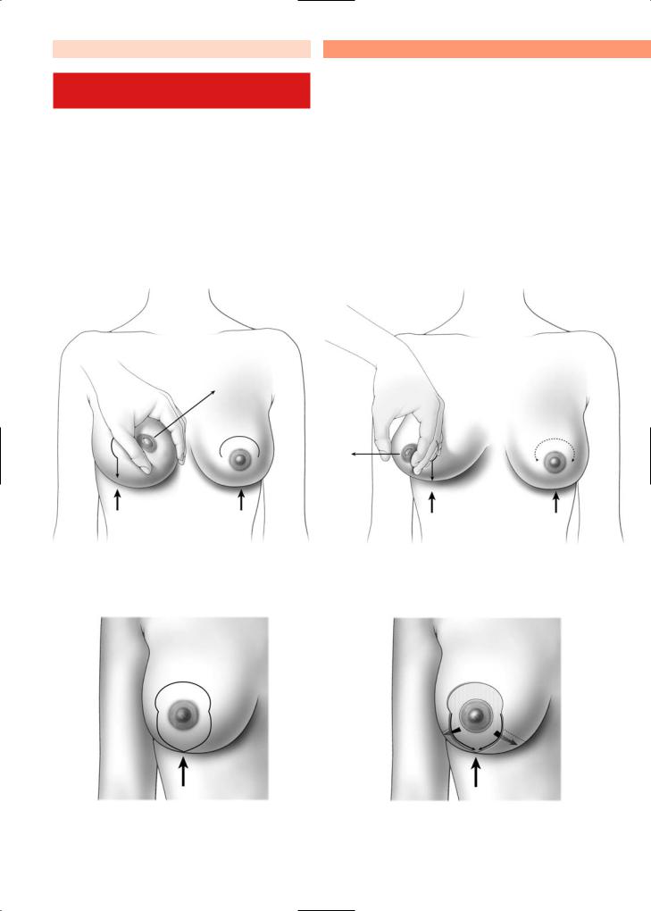
Additionally, the breasts should be appropriately marked with the patient in the standing position, indicating the planned incision pattern, as depicted in the accompanying illustration (Fig. 7.29).
Chapter 7 |
113 |
As shown in the accompanying illustrations, reduction mammoplasty can be performed using a vertical technique, where a superior pedicle is left intact, providing blood supply to the nipple–areola complex (Fig. 7.30a). Alternatively, the procedure can be performed with an inferior pedicle providing the necessary blood supply to the nipple–areola complex (Fig. 7.30b).
The drawings on this page illustrate both the superior pedicle technique and the inferior pedicle technique. Specifically, these drawings depict the contour of the breast tissue specimen that is obtained after using the superior pedicle technique and the contour of the breast tissue specimen that is obtained after using the inferior pedicle technique for reduction mammoplasty (Fig. 7.30c, d).
a
b |
c |
c |
|
|
d |
e |
b |
Fig. 7.28. Measurements required prior to reduction mammoplasty. a=19–21 cm; b=9–11 cm; c=4–5 cm; d=5–8 cm; e=0–2 cm

114 |
7.13 |
Reduction Mammoplasty |
7
a Superior pedicle
a |
b |
b Inferior pedicle
Fig. 7.29. Reduction mammoplasty
a |
b |
Fig. 7.30a–d. Reduction mammoplasty |
c |
d |
Plastic and Reconstructive Breast Surgery
7.14Reduction Mammoplasty: Vertical Technique (Lejour)
Figure 7.31 depicts a surgeon’s preoperative drawing for a reduction mammoplasty, utilizing the vertical scar technique (Lejour). The drawing shows the limit of the supra-areolar de-epithelialization (1, in Fig. 7.32a), the medial limit of infra-areolar de-epithelial- ization (2, in Fig. 7.31a), the lateral limit of infra-areo- lar de-epithelialization (3, in Fig. 7.31b), and the inferior aspect of the resection (4, in Fig. 7.31c).
Chapter 7 |
115 |
The accompanying illustrations briefly summarize the key features of the reduction mammoplasty utilizing the vertical technique (Lejour). A margin of tissue around the nipple–areola complex is de-epi- thelialized (1, in Fig. 7.31a). The surgeon undermines a wide area of tissue anterior to the serratus anterior and pectoralis major muscles. Breast tissue in the inferior quadrant is then resected (Fig. 7.32). Glandular tissue is then re-approximated with absorbable stitches (Fig. 7.33). The skin edges are re-approximat- ed with interrupted 3–0 Monocryl stitches (Fig. 7.33). These stitches are also used to re-approximate the
2 1 3
a |
b |
|
4
c |
d |
Fig. 7.31a–d. Reduction mammoplasty; vertical scar technique (Lejour)