Advanced Veterinary Life Support For the acls Health Care Professional
Written By Henry Feldman, m.D. Lori Feldman, d.V.M.
This document uses dogs as an example (but will work for cats), as most EMS providers are more likely to encounter dogs (such as police or rescue dogs) severely injured in the course of their work. In general animals such as dogs and cats are more like pediatric cases than human adults, in that they go into arrest due to respiratory problems. They also follow the pediatric compensatory shock model of extreme compensation, followed by a sudden drop in systolic blood pressure which is hard to reverse.
There is no AVLS flowchart, as it looks basically like the human ACLS chart. Most techniques that work on humans will work on animals, as they were often developed on dogs. Many medics have learned pediatric/neonatal intubation on cats and ferrets. The good news about this, is that there is little "new" material.
A - Advanced Airway
Intubation of an animal in many ways is easier than a human. The technique is similar with a few exceptions. Remember that animals often have proportionally larger tracheas than humans of the same size; for instance a 100lb human would probably use a 7-8 ET tube, while a 100lb german sheppard might require a 11-12 tube! The only tube currently in use in animals is the Endotracheal tube; other tube types are not recommended or tested.
-
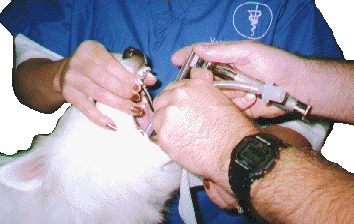
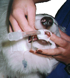
Place the animal sternal
-
Using a gauze 4x4, grab the end of the tip of the tongue, and pull the tongue out of the mouth. This is often sufficient to expose the glottis.
-
Using a long Miller Laryngoscope blade, pull the epiglottis back against the anterior throat.
-
Insert the tube as normal, and inflate the cuff as normal.
-
Check tube placement by 2 separate methods
-
Wrap a foot long (25cm) piece of roller bandage around the tube in the mouth and tie in a bow over the snout. This will hold the tube in place. Remember to check tube placement after any movement.
-
Note: cats are often in laryngospasm naturally, and Lidocaine gel should be used.
-
Note: Make sure that the animal is unresponsive! Remember, those teeth are designed to rip flesh!!!
B - Breathing
After intubation, ventilatory assistance is the same as humans. Bag-Valve resuscitators are the normal instrument of choice, however a demand-valve power-resuscitator should work fine. Some animals will not tolerate a mask of any kind, so use high-flow blow-by on breathing animals in respiratory distress.
remember that animals have a much higher breathing rate than humans. Here are some rules of thumb:
-
Open mouth breathing in a cat usually indicates a problem(they can do this when mad however)
-
"Real" breathing (not panting) rates above 60 BPM in a dog or cat indicate a serious problem
-
Remember that panting is normal for a dog, and can have a rate well in excess of 100bpm
-
Use 20-40BPM as a rate for resuscitation
-
Accessory muscle (abdominal) breathing indicates a severe problem
-
Lack of breath sounds can indicate a pneumothorax
Pneumothorax
A closed chest pneumothorax is often tolerated better by animals than by humans. However, severe intrathoracic pressures can cause other problems. Here is the technique for reducing a tension pneumothorax:
-
Lay the animal lateral with the pneumothorax up
-
Find the 8th intercostal space
-
Using an 18 gauge needle, insert the needle on the caudal (inferior) margin of the 8th intercostal space at the dorsal third of the chest wall
-
Using a stop-cock or other venting technique, reduce the pleural tension
C - Circulation/Cardiac
Dogs and cats do not normally have myocardial infarctions, as they don't live long enough, even with an all meat diet, to build up the necessary plaques; Dogs also have collateral circulation of the coronary arteries, allowing them to "go around" an occlusion that would cause an MI in a human. Almost all cardiac arythmias are congenital, or can be caused by acute trauma/poisons.
EKG/Monitor
EKG monitoring is the same as in humans, except that you can't use pads. Small alligator clips are used in place of the adhesive pads, and are used in conjunction with conductive gel (defib gel works fine). Proper placement is with the animal in right lateral recumbancy, with the clips on the elbows and knees:
-
Right Arm - (Neg)
-
Left Arm - (+)
-
Right Leg - (Common)
Make sure that you use plenty of conductive gel. If you choose to use quick-look paddles, make sure that you use plenty gel, to get through the hair. Arythmias are the same patterns as in humans.
IV Access
Veterinary medicine has a much more aggressive approach to IV access, and typically in hospital, any case which is the least critical, will often get an IV. With the exception of diabetic emergencies, the preferred fluid is Lactated Ringers Solution (LRS). In animals that cannot successfully be started IV, sub- cutaneous fluids can be used, although this is a much slower route. The normal type of catheter is the Intracath, with the catheter over a stylette.
IV Access Technique - Front Leg
-
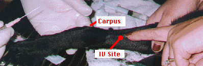
Shave a 1-2 inch (2-5cm) band approximately 2-4 inches (4-8cm) proximal to the capus circumferentially around the leg
-
Clean the site with a scrub solution then alcohol (betadine, nolvasan...)
-
Have one person firmly grasp the arm slightly distal to the elbow, straightening the arm, with the thumb on the medial side of the arm
-
The person inserting the catheter should grasp the distal arm and pull it straight with moderate tension, making sure not to cut off circulation
-
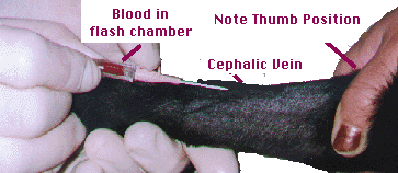
Roll the thumb lateral, twisting the flesh of the arm 60 degrees. This will roll the cephalic vein to an easy to find position as well as act as the tourniquet.
-
With the catheter's bevel up, deliberately plunge the catheter in. Note, that the veins of a dog or cat are much tougher than a humans, especially if the animal is not neutered/spayed (you'll need to roll more). Veins are much more superficial than a human's.
-
Use the appropriate size catheter:
-
Cats typically use a 24 guage
-
Large dogs (over 50lbs/22Kg) use a 20 or 18 guage
-
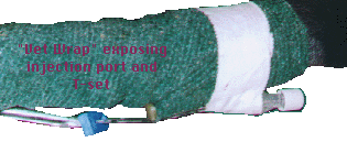
When you see flash in the chamber, feed the catheter into the vein, pull the stylette and insert the T-set or appropriate line set you use in your normal protocol. You should test with a saline/LRS flush to make sure that you have good flow.

-
Circumferentially tape the catheter in, making sure that the tape also goes proximal to the catheter to stick to the leg. Don't worry, unlike humans this doesn't cut off venous supply.

-
Circumferentially roller bandage from several inches below to several inches above, including crossing under the port. Normally a layer of Vet-Wrap or other tough bandage material is wrapped over the roller bandage.