

Advances in small animal wound management
.pdf
|
Published in IVIS with the permission of the editor |
Close window to return to IVIS |
|
|
|
|
|
|
Advances in small animal wound management
Steven Swaim, DVM, MS
Scott-Ritchey Research Center and Department of Clinical Sciences, College of Veterinary Medicine, Auburn University, Alabama, USA
Dr. Swaim graduated from Kansas State University. He was in private small animal practice in Wichita, Kansas, before serving in the U.S. Army Veterinary Corps at a medical research facility. Following 2 years in the Army, he joined a private small animal practice in Denver, Colorado. From 1969 until the present,
Dr. Swaim advanced from Research Associate to Professor emeritus at Auburn University’s College of Veterinary Medicine. He had a joint appointment with the Department of Clinical Sciences and the Scott-Ritchey Research Center. His early work was in neurology and neurosurgery. From 1975 until the present, Dr. Swaim’s clinical and research work has been with small animal wound management.
Mark Bohling, DVM, PhD, Dipl. ACVS
College of Veterinary Medicine, University of
Tennessee, Knoxville, Tennessee, USA
Dr. Bohling graduated from the University of California, Davis and was employed in food animal practice (dairy and poultry) until 1992, when he began in small animal private practice. He did an internship in small animal medicine and surgery in 1999.
In 2001, Mark Bohling began a residency in small animal surgery at Auburn University, where he met Dr. Swaim and became his graduate student. Since 2005, he has been on the faculty at the University of Tennessee in the section of small animal surgery. Dr. Bohling’s clinical and research interests are in reconstructive surgery and wound healing, particularly in the feline species.
 Introduction
As with all fields of medicine and surgery, wound management has seen advances in the science and art of wound care over the last several years. These are in medication, materials, and methods that are used in treating and reconstructing injured tissues. When considered from the standpoint of comparative medicine, some of the advances apply to management of wounds in both animals and humans. It will not be possible to cover all the developments in wound management; thus, the following presents some of the significant aspects in the field.
 Medications
Over time, there has been the instinct to place substances in wounds with the intent of enhancing healing. Recently, there has been a revival in the use and understanding of the mechanisms of action of some old standbys in vulnerary topicals, these being the application of sugar and honey to wounds (1). Other medications have been developed to enhance the healing process in animals and people. These include a tripeptidecopper compound; acemannan, a mannose sugar derivative; a D-glucose polysaccharide; plateletderived products and chitosan, a medication derived from the exoskeleton of shellfish (1) (Table 1).
Vol 18 No 1 / / 2008 / / Veterinary Focus / / 17

Sugar and honey
Sugar has a high osmolality and affects healing by reducing edema, attracting macrophages, accelerating necrotic sloughing, providing cellular energy, and enhancing healthy granulation tissue. Honey has similar action in wound therapy, with an antimicrobial activity coming from hydrogen peroxide. The hydrophilic properties of these 2 topicals warrant close monitoring of hydration, electrolyte, and protein levels when they are used on large wounds (1).
Tripeptide-copper complex
A tripeptide-copper complex has been found to stimulate neovascularization, epithelialization, collagen deposition and wound contraction. Controlled research has shown enhanced healing of open wounds in dogs and open ischemic wounds in rats (1,2).
Table 1.
Selected wound healing stimulants (1)
|
Ingredient |
Brand name |
Action |
||
|
Sugar |
|
Osmolality, reduce edema, |
||
|
|
|
attract macrophages, |
||
|
|
|
necrotic slough, |
||
|
|
|
cell energy, enhanced |
||
|
|
|
granulation tissue |
||
|
Honey |
|
Reduce edema, attract |
|
|
|
|
|
macrophages, necrotic |
||
|
|
|
slough, cell energy, |
||
|
|
|
enhanced granulation |
||
|
|
|
tissue |
||
|
Tripeptide- |
Iamin |
Neovascularization, |
|
|
|
copper |
|
epithelialization, collagen |
||
|
complex |
|
deposition, contraction |
||
|
|
|
|
|
|
|
Acemannan |
CarraVet, |
Macrophage stimulation, |
||
|
|
Carrasorb |
fibroblast proliferation, |
||
|
|
|
neovascularization, |
||
|
|
|
epithelialization, |
||
|
|
|
collagen deposition |
||
|
|
|
|
|
|
|
Maltodextrin – |
Intracell |
Attract |
||
|
D-glucose |
|
polymorphonuclear cells, |
||
|
polysaccharide |
|
lymphocytes and |
||
|
|
|
macrophages, cell energy, |
||
|
|
|
hydrophilic, |
||
|
|
|
necrotic slough |
||
|
|
|
|
|
|
|
Platelet |
|
Epithelialization, |
||
|
products |
|
neovascularization, |
||
|
|
|
contraction |
||
|
|
|
|
|
|
|
Chitan |
Ultrasan |
Enhanced inflammatory |
||
|
|
|
cell function, increased |
||
|
|
|
growth factors, increased |
||
|
|
|
fibroblasts, enhanced |
||
|
|
|
granulation tissue |
||
|
|
|
|
|
|
Acemannan
The mannose sugar derivative acemannan has been found to act as a growth factor to stimulate macrophages to produce interleukin 1 (IL-I) and tumor necrosis factor (TNF-α) (1). The result is enhanced fibroblast proliferation, increased neovascularization, increased epidermal growth, and enhanced collagen deposition (3). A controlled study showed acemannan-treated paw pad wounds were significantly smaller at 7 days than triple antibiotic-treated or untreated wounds (1).
Maltodextrin – D-glucose polysaccharide
Maltodextrin is a D-glucose polysaccharide with ascorbic acid. It is available as a hydrophilic powder and gel that serve as a chemoattractant for polymorphonuclear cells, lymphocytes, and macrophages which increase the level of growth factors needed in healing (1,3). It may also provide cell energy to promote healing (3). The medication has been reported to soften necrotic tissue, penetrate wound irregularities, be nontoxic, have no systemic absorption, and be effective in infected and non-infected wounds (1).
Research by one author (SFS) has shown that if anything has an effect on healing, it has its greatest effect in the first 7 days of use. To take advantage of this, the authors have used a 7-day alternating regimen of three of the above medications (acemannan, D-glucose polysaccharide, and tripeptide-copper complex), in treating challenging wounds, e.g. large deep wounds, large wounds with exposed bone, and chronic wounds. The therapy starts using a freeze dried form of acemannan, followed by the D-glucose polysaccharide therapy and then the tripeptidecopper complex. If therapy extends beyond 21 days, the latter 2 are used in the alternating 7-day pattern. When used in a chronic wound, it is imperative that the cause of the non-healing be found and addressed before use of the wound healing stimulants. Empirically, the regimen has tended to keep wound healing prog ressing rapidly. The question ar ises as to w h a t e f fe c t wo u l d b e p r o d u c e d by 1 ) simultaneous use of these medications, and 2) combinations of other medications (alternating or simultaneous use). Thus, potential research projects arise.
18 / / Veterinary Focus / / Vol 18 No 1 / / 2008

ADVANCES IN SMALL ANIMAL WOUND MANAGEMENT
Platelet products
Platelet-derived products have potential in wound care due to the large number of growth factors available in high concentrations from activated platelets. The topical application of platelet-derived growth factors in non-healing wounds in people has enhanced epithelialization, contraction, and neovascularization. Application of a homologous platelet-rich plasma gel to experimental equine wounds has produced similar results (1). It is possible that platelet-derived medications will be as effective in dogs and cats.
Chitosan
Chitosan is a polysaccharide with glucosamine as an active ingredient. It is derived from chitan extracted from the exoskeleton of shellfish. When applied to wounds, it enhances inflammatory cell function, provides various growth factors and stimulates fibroblasts. The result is promotion of granulation tissue and accelerated healing as seen in a group of experimental dogs (1,4).
 Materials
This section will be devoted to some of the advances that have been made in wound bandaging/dressing materials (Table 2). In general, the philosophy of “conceal and it will heal” is no longer applicable. Dressing materials are now available that interact with wound tissues to enhance wound healing. Three bandaging/dressing materials/techniques will be covered – moisture retentive dressings, collagen dressings, and extracellular matrix dressings.
Moisture retentive dressings
When moisture is retained over a wound by a bandage, healing is enhanced. The process is related to the moisture vapor transmission rate (MVTR). It has been found that when the MVTR is low, i.e. moisture retained, there is a strong correlation with positive wound healing outcome when all other variables are held constant, and infection rates tend to be lower with a lower MVTR
(5). Thus, moisture retentive dressings (MRD) would be indicated for open wound management.
Some of the reasons MRD’s have a positive effect on wound healing include cell proliferation and function in the inflammatory and repair stages which are enhanced by the warm moist environment.
A physiologic ratio of proteases, protease inhibitors, growth factors and cytokines is present at each stage of healing. White blood cells with their enzymes stay in the wound rather than being
Table 2.
Selected bandages and dressings (1,2)
Bandage/ |
Exudate |
Action |
|
Dressing |
level for use |
||
|
|||
Polyurethane |
Moderate to |
Absorbs; can donate |
|
foams (MRD)* |
high |
medication if |
|
|
|
premoistened; non- |
|
|
|
adherent; can be used |
|
|
|
early and late in healing; |
|
|
|
stimulates autolytic |
|
|
|
debridement, granulation |
|
|
|
tissue and epithelium |
|
|
|
|
|
Polyurethane |
None to |
No absorption; non- |
|
films (MRD)* |
minimal |
adherent; use in later |
|
|
|
healing; stimulates |
|
|
|
epithelium |
|
|
|
|
|
Hydrocolloids |
Low to |
Limited absorption; |
|
(MRD)* |
moderate |
stimulates autolytic |
|
|
|
debridement, |
|
|
|
angiogenesis, collagen |
|
|
|
synthesis, epithelium; |
|
|
|
adherence to periwound |
|
|
|
|
|
Hydrogels |
None to |
Can donate (dry |
|
(MRD)* |
moderate |
wounds) or absorb (low |
|
|
|
to moderate draining |
|
|
|
wound) fluids; |
|
|
|
stimulates autolytic |
|
|
|
debridement, granulation |
|
|
|
tissue and epithelium |
|
|
|
|
|
Hydrolyzed |
Moderate to |
Hydrophilic; stimulates |
|
bovine collagen |
high |
epithelium; could help |
|
|
|
bathe wound from inside |
|
|
|
with systemic antibiotics |
|
|
|
|
|
Extracellular |
Low |
Chemotactic for repair |
|
matrix dressings |
|
cells; antibacterial; |
|
|
|
promotes granulation |
|
|
|
tissue; replaced by site |
|
|
|
specific tissue |
|
|
|
|
|
Antimicrobial |
Moderate |
Antibacterial; organisms |
|
dressings |
|
do not develop a |
|
Polyhexamethyl- |
|
resistance |
|
ene biguanide |
|
|
|
|
|
|
|
Ionic silver |
|
Broad spectrum of |
|
|
|
antimicrobial activity, |
|
|
|
to include some fungi |
|
|
|
|
|
*Moisture retentive dressing |
|
||
Vol 18 No 1 / / 2008 / / Veterinary Focus / / 19

absorbed away, and they can perform their autolytic debridement function. Occlusion of the wound also provides a barrier against exogenous bacteria, prevents tissue desiccation, and allows for improved concentration of systemically administered antibiotics in the wound. The low oxygen tension under an occlusive dressing lowers pH, deters bacterial growth, and favors collagen synthesis and neovascularization. Moisture retentive dressings do not adhere to the wound surface, and thus are not painful at removal. In addition, their waterproof nature prevents entry of urine and other fluids (5).
A potential disadvantage to MRD’s is that excess moisture retention can result in damage to periwound skin in the form of maceration (skin softening) and excoriation (skin damage from excessive proteolytic enzymes in wound fluid) (5).
Some MRD’s include polyurethane foams and films, hydrocolloids, and hydrogels. Each has properties conducive to enhancing healing.
Polyurethane foams are highly absorptive and designed for wounds with moderate to high exudate levels, thus, helping prevent periwound maceration and excoriation (6). They absorb excess fluid while maintaining a moist environment. Their absorptive property can be used to absorb liquid medications to be delivered to the wound (5). These dressings have the advantage that they can be used in both the early (inflammatory) and later (repair) stages of healing. They do not adhere to the wound and they promote healthy granulation tissue formation.
Polyurethane films are thin, flexible semi-occlusive, and non-absorptive. Thus, they are indicated for wounds with no or minimal exudate, i.e. wounds in the repair stage of healing to promote epithelialization. Care must be taken that fluid does not accumulate to cause peri-wound skin damage. A disadvantage of this type of dressing is that hair regrowth on animals interferes with the adherence of the perimeter of the dressing (5,6).
A common form of hydrocolloid dressing is a sheet made from a combination of absorbent and elastomeric components. These interact with wound fluids to form a tenacious gel to give a moist wound environment. They are indicated
for wounds with low to moderate exudate. Hydrocolloids stimulate autolytic debridement in the inflammatory healing stage and enhance angiogenesis, collagen synthesis and epithelialization in the repair stage (5,6). On the downside of hydrocolloids, the appearance and odor of the yellow purulent gel over the wound at dressing removal may cause concern about infection. However, when this is removed, healthy tissue underlies it (1,5). Although hydrocolloids would primarily be indicated for wounds in the repair stage, in a canine study there is some evidence that the portion of the dressing that is adhered to the periwound skin may delay wound contraction. Thus, their use in the later repair stage should be with caution (5).
Hydrogels are water or glycerin-based gels that can absorb wound fluid as well as donate moisture to rehydrate tissues. In the latter case, they are beneficial in treating eschar and dry sloughing tissue. In necrotic wounds, they promote autolytic debridement, granulation tissue and epithelialization. The primary use of hydrogels is on low to moderately draining wounds, for example, wounds in the late healing stages (1,5). Canine studies revealed that hydrogels enhanced limb wound contraction, but delayed trunk wound contraction (5).
Collagen dressings
Collagen is a normal component of the repair stage of the healing process. However, placing exogenous collagen in sheet, powder, and gel forms is being used in the treatment of wounds. A canine wound healing study evaluated the effects of hydrolyzed bovine collagen powder dressing on open wound healing in dogs (7). The collagen treated wounds had significantly more epithelialization at 7 days than did control wounds. This was attributed to the hydrophilic nature of the collagen drawing fluid up through the wound to keep it clean and provide a moist environment. With this in mind, such therapy could be helpful in the early treatment of contaminated or infected wounds to draw antibiotic-laden fluid up through the wound when the animal is on systemic antibiotics.
Extracellular matrix dressings
Extracellular matrix (ECM) dressings are acellular biodegradable sterile dressings derived from either
20 / / Veterinary Focus / / Vol 18 No 1 / / 2008

ADVANCES IN SMALL ANIMAL WOUND MANAGEMENT
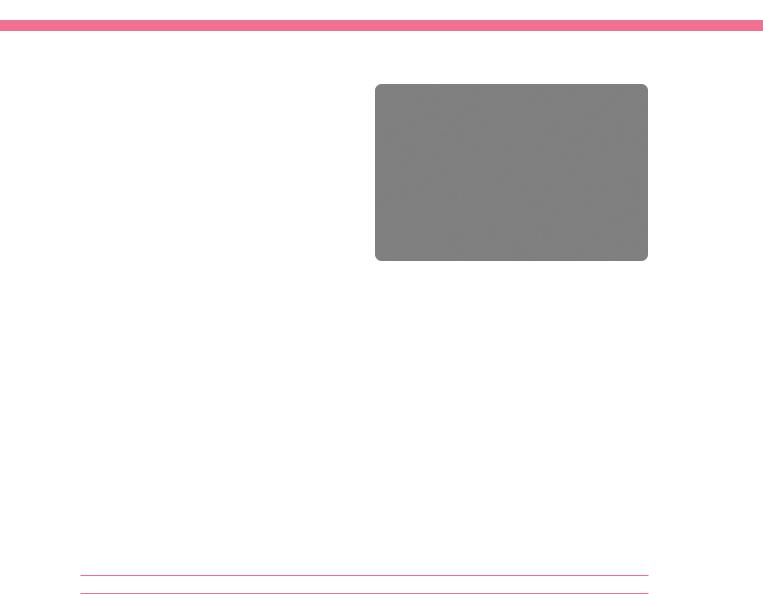
Vet BioSISt™. |
|
©CookBiotech, Inc. Photoscourtesy of Smiths Medical PM, Inc. - SurgiVet®,exclusive worldwide distributor of |
swine intestinal submucosa. |
|
Figure 1. Scanning electron micrograph of an extracellular |
|
matrix bioscaffold wound dressing product derived from |
|
porcine small intestine submucosa (Figure 1) or |
|
porcine urinary bladder submucosa matrix (1,5). |
|
These dressings provide structural proteins, growth |
|
factors, cytokines, and their inhibitors in physiologic |
|
proportions in a three-dimensional ultrastructure. |
|
The scaffold acts as an inductive structure for tissue |
|
replacement (8). As the ECM scaffold is broken |
|
down by mononuclear cells, the degradation |
|
products are chemotactic for repair cells; they |
|
stimulate angiogenesis; and they have antibacterial |
|
properties (5). The end result is the development |
|
of site-specific tissue (8) (i.e. tissues are like |
|
that in which they were placed), with most of |
|
the endothelial cells and fibroblasts coming from |
|
the animal’s bone marrow, i.e. stem cells (9). |
|
Use of ECM’s require some special techniques. The |
|
wound bed should be thoroughly debrided. It |
|
should be free of topical medications, cleansing |
|
agents and exudate. The ECM may be fenestrated |
|
to allow for any drainage. A non-adherent dressing |
|
or absorptive MRD is placed over the ECM followed |
|
by application of the secondary and tertiary |
|
bandage layers. When the bandage is removed |
|
in 3-4 days, the ECM with its degrading center |
|
over the wound is left in place, and another piece |
|
is applied over it followed by outer bandage |
|
replacement. After 2-3 applications in this manner, |
|
the ECM is discontinued (the site-specific tissue |
|
has been established) and wound management |
|
continues with appropriate bandaging (1,5). |
|
Antimicrobial dressings |
|
Dressings that contain antimicrobial agents are |
|
being used and evaluated in veterinary medicine. |
|
Two such dressings are a polyhexamethylene |
|
biguanide (PHMB) dressing and silver ion dressings. |
|
Polyhexamethylene biguanide is a chlorhexidine |
Figure 2. Several sophisticated combination wound dressings are available for open wound management. Pictured is a composite dressing that consists of a polyurethane foam pad with a silverimpregnated calcium alginate coating. Combination dressings such as this allow multiple wound needs to be met with a single product.
related agent that destabilizes bacterial cytoplasmic membranes. Organisms cannot develop a resistance to the chemical. An in vitro study found that PHMB-impregnated dressings decreased or eliminated proliferation of bacterial pathogens that were isolated from dogs and cats in a small animal veterinary teaching hospital, both within and beneath the impregnated dressing (5). The authors have had good success in treating infected wounds with PHMB primary and secondary bandages.
Ionic silver is being used in the treatment of infected wounds (10). It has a very broad spectrum of antimicrobial activity to include some fungal organisms. Silver releasing dressings are available in gauze, gauze rolls, low-adherent, hydrocolloid, hydrogel and alginate forms (Figure 2) (5).
 Methods
As with medication and materials, there have been developments in methods that are being used to treat and reconstruct animal wounds. The following is information on four of these methods – use of omentum to enhance healing, techniques to transpose skin, techniques to stretch skin, and vacuum assisted closure. Space limitation does not allow going into detail on the technical aspect of these procedures; thus, a summary of each will be presented.
Omental flaps
Omental f laps can be used to contribute to circulation and drainage, cover soft tissue defects, enhance healing, control adhesions, and combat infections. They stimulate the formation of
Vol 18 No 1 / / 2008 / / Veterinary Focus / / 21

Figure 3. Thoracodorsal axial pattern flap for closure of a large antebrachial wound. One week postop, uneventful healing.
granulation tissue to allow earlier wound closure with skin grafts and flaps. These flaps are especially useful for chronic non-healing wounds over the thorax, abdomen, inguinal and axillary areas (11). After exposing the omentum and creating the omental flap, it is transposed subcutaneously to the wound site. The appropriate wound closure technique (direct closure, graft or flap) is used in combination with this flap for closure.
Transposition of skin
Surgical closure of wounds involves transposing skin in some way. This involves moving local skin, or use of skin grafts or flaps. Skin flaps have the advantage of having a blood supply via a pedicle all during the time they heal; whereas, grafts have to develop a blood supply after being placed on the wound. Two techniques that have been beneficial in moving large amounts of skin as flaps have been the use of axial pattern flaps and microvascular surgery, which provide a vascular supply to the transposed skin.
Axial pattern flaps are flaps of skin that have a prominent direct cutaneous artery and vein running the length of the flap to help assure a blood supply to a large piece of skin as it heals in a wound (Figure 3). There are numerous named axial pattern flaps and the landmarks and indications for these flaps have been well described in the literature (12).
Microvascular reconstructive surgery requires the harvest of autogenous tissue(s) with a consistent vascular pedicle from a donor site. It is transferred to a recipient bed and circulation is re-established by microvascular anastomosis of the donor artery and vein to an artery and vein in the recipient area. Thus, a microvascular free flap is created.
Such flaps are especially useful for wounds on the distal limb and paw. These flaps have 3 primary disadvantages. They are time consuming, they require expertise in microvascular surgery, and they require special instrumentation (13).
Expansion of skin
Dogs and cats have the advantage of having an abundance of skin on the upper portions of the body. This is beneficial in closing large wounds on the trunk. However, there are occasions when there is a large amount of skin missing on the trunk and when there are large wounds on the limbs where there is a sparsity of skin. Techniques have been developed whereby skin can be stretched to close these wounds. Expansion bands can be used to stretch skin around a wound so it can be used to close a wound. Self-adherent skin pads with VelcroTM are affixed to skin around the wound. Elastic connecting cables with the other part of the VelcroTM material are attached to the pads on one side of the wound and are stretched before attachment to pads on the other side of the wound. The cables are adjusted every 6-8 hours over 24 to 96 hours until enough skin is recruited for wound closure. These are generally used for wounds on the neck and trunk (11,12).
Another technique for stretching skin on the neck and trunk is the use of “walking” sutures. Absorbable sutures are placed under the skin on either side of the wound in a manner that gradually advances/“walks” the skin over the wound (11,12,14). The technique stretches skin and closes the wound at the same time.
Two techniques for stretching skin on the distal limb are presutures and the adjustable horizontal mattress suture. With presutures, Lambert type sutures are placed in skin around the wound such that they cross the wound. They are tied under tension and left for 12-24 hours. After skin stretching, the expanded skin is used to close the wound (12,13,14). The adjustable horizontal mattress suture (Figure 4) is a continuous mono-filament intradermal suture that runs the length of the wound. At each end an adjusting apparatus composed of a sewing button and a split shot fishing weight are placed. At 24 hour intervals tension is placed on the suture ends to advance the wound edges closer together. After tension is applied, it is held by the fishing weights against the buttons (11,13,14).
22 / / Veterinary Focus / / Vol 18 No 1 / / 2008
ADVANCES IN SMALL ANIMAL WOUND MANAGEMENT
Vacuum-assisted closure
Vacuum-assisted closure (VAC) has received considerable attention in human wound management, and it is also being used in veterinary medicine (15). It is used on acute traumatic wounds, chronic non-healing wounds, pressure ulcers, degloving wounds, skin grafts, skin flaps, open abdomens, complex perineal and gynecologic wounds, enterocutaneous fistulae and skull defects (15). In this therapy a closed system is created over the wound with tubing going to a vacuum apparatus. Continuous or intermittent suction is applied to the wound (16). VAC therapy promotes granulation tissue formation and neovascularization with increased blood flow. It also removes excess fluid and edema, and reduces bacterial counts. Micromechanical forces applied to the wound may also be an important factor for inducing cell proliferation and wound healing (17).
 In the future
The field of wound management and reconstructive surgery will undoubtedly continue to advance in both human and veterinary medicine. As tissue engineering and molecular medicine studies increase, they will find application in wound management. There will continue to be studies of comparative medicine-nature, with discoveries in
©(Scardino MS, Swaim SF, Henderson RA, et al.. Enhancing wound closure on the limbs. Comp Cont Educ Pract Vet 1996; 18: 919-933) Veterinary Learning Systems, Yardley, Pennsylvania.
Figure 4. The adjustable horizontal mattress suture may be used to more rapidly reduce the surface area of open wounds thus speeding their final closure by second intention healing.
animals that will benefit both animals and man. One author (SFS) has been involved with such studies, looking at a recombinant vasodilatory protein found in black fly saliva that has shown promise in causing local vasodilation to enhance wound healing (18). Also, a study has been performed investigating pressure-reducing effects of subdermal silicone gel particles in relieving palmar/plantar pressure (19). Respectively, these studies could be applicable in treating chronic wounds in animals and people (e.g. human diabetic foot ulcers), (18) and preventing painful digital calluses in greyhounds as well a preventing plantar calluses and ulcers in human diabetics (19).
REFERENCES
1.Krahwinkel DJ, Boothe HW. Topical and systemic medications for wounds. In: Swaim SF, Krahwinkel DJ (eds). Wound Management, Elsevier/WB Saunders.
Vet Clin North Am Small Anim Pract 2006; 36: 739-757.
2.Canapp SO, Farese JP, Schultz GS, et al. The effect of topical tripetide-copper complex on healing of ischemic open wounds. Vet Surg 2003; 32: 515-523.
3.Swaim SF, Gilette RL. An update on wound medications and dressings.
Comp Cont Educ Pract Vet 1998; 20: 1133-1144.
4.Veno H, Mori T, Fujinaga T. Topical formulations and wound healing applications of chitosan. Adv Drug Deliv Rev 2001; 52: 105-115.
5.Campbell BG. Dressings, bandages, and splints for wound management in dogs and cats. In: Swaim SF, Krahwinkel DJ (eds). Wound Management, Elsevier/WB Saunders. Vet Clin North Am Small Anim Pract 2006; 36: 759-791.
6.Compton-Johnson S, Wilson J. Infected wound management: advanced technologies, moisture-retentive dressings, and die-hard methods.
Crit Care Nursing 2001; 24: 64-77.
7.Swaim, SF, Gillette RL, Sartin EA, et al. Effects of hydrolyzed collagen dressing on the healing of open wounds in dogs. Am J Vet Res 2000; 61: 1574-1578.
8.Badylak SF. The extracellular matrix as a scaffold for tissue reconstruction.
Semin Cell Dev Biol 2002; 13: 377-383.
9.Badylak SF, Park K, Peppas N, et al. Marrow-derived cells populate scaffolds composed of xenogenic extracellular matrix. Exp Hematol 2001; 29: 1310-1318.
10.Dowsett C. The use of silver based dressings in wound care. Nurs Stand 2004; 19: 50-60.
11.Hedlund CS. Large trunk wounds. In: Swaim SF and Krahwinkel DJ (eds).Wound Management, Elsevier/WB Saunders. Vet Clin North Am Small Anim Pract 2006; 36: 847-872.
12.Pavletic MM. Atlas of Small Animal Reconstructive Surgery, 2nd ed. WB Saunders, Philadelphia 1999, pp 131-171, 173-189, 237-274.
13.Fowler D. Distal limb and paw injuries. In: Swaim SF, Krahwinkel DJ (eds). Wound Management. Elsevier/WB Saunders. Vet Clin North Am Small Anim Pract 2006; 36: 819-845.
14.Swaim SF, Henderson RA. Small Animal Wound Management, 2nd ed. Williams and Wilkins, Baltimore 1997, pp. 143-190.
15.Guille AE, Tseng LW, Orsher RJ. Use of vacuum-assisted closure for management of a large skin wound in a cat. J Am Vet Med Assoc 2007; 230: 1669-1673.
16.Morykwas MJ, Argenta LC, Shelton-Brown EI, et al. Vacuum-assisted closure: a new method of wound control and treatment: animal studies and basic foundation. Ann Plast Surg 1997; 38: 553-562.
17.Saxena V, Hwang C, Huang S, et al. Vacuum-assisted closure: microdeformations of wound and cell proliferation. Plast Reconst Surg 2004; 114: 1086-1096.
18.Cupp MS, Swaim SF, Amalsadvala T, et al. Use of a recombinant vasoactive protein (rSVEP) to enhance healing of surgically created wounds. Wounds 2004; 16: 85-90.
19.Swaim SF, Amalsadvala T, Marghitu DB, et al. Pressure reduction effects of subdermal silicone block gel particle implantation: A preliminary study. Wounds 2004; 16: 299-312.
Vol 18 No 1 / / 2008 / / Veterinary Focus / / 23