

Essentials of Orthopedic Surgery, third edition / 02-Skeletal Trauma
.pdf2
Skeletal Trauma
JOHN N. DELAHAY and SCOTT T. SAUER
Skeletal trauma, for the subject of discussion, can be divided into three major groups of injuries to the musculoskeletal system:
Fractures
Dislocations
Fracture/dislocations
A fracture, by definition, is a disruption in the continuity of cortical and/or cancellous bone. A dislocation is a disruption of the normal articulating anatomy of a joint. Dislocations can either be a complete disruption of the normal anatomy or a partial dislocation, in which case the term subluxation is used. A fracture/dislocation is a fracture occurring in or near a joint that results in a subluxation or dislocation of the joint.
Fractures
Fracture Descriptors
A number of different terms can be used to more accurately describe the configuration and features of any given fracture. These descriptors are as follows:
1.Open versus closed: A closed fracture is one in which the skin is intact over the fracture site and an open fracture is one in which the skin is not intact.
2.Simple versus comminuted: A simple fracture is one in which there are only two major fragments and one fracture line. A comminuted fracture is one in which there are multiple fragments of bone and multiple fracture lines.
3.Complete versus incomplete: “Complete” essentially means that the fracture line goes completely across the bone. Incomplete fractures, most typically seen in children, have a fracture line that only crosses one cortex of the bone involved.
40

2. Skeletal Trauma |
41 |
Fracture Deformities
A fracture can be deformed in any one of three possible planes. Classic deformations are described as follows (Fig. 2-1):
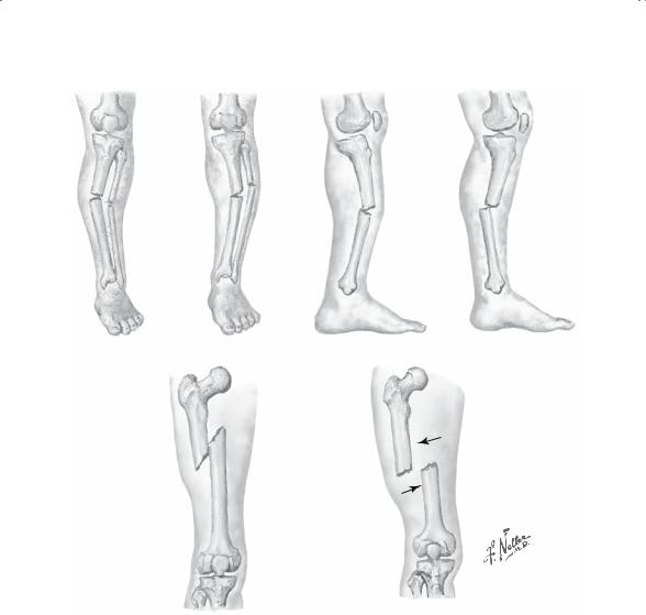
1.Displacement: Translation of the two fragments in relation to each other in one or more planes. Traditionally, displacement refers to the position of the distal fragment in relation to the assumed stationary proximal fragment. Specific types of displacement include overriding, where the two fragments are shortened in relation to one another, and distraction, where essentially the bone ends are pulled apart.
2.Angulation: Occurs when the two fracture fragments are not aligned and an angular deformity is present. Alignment means that the axes of the proximal and distal fragments are parallel to each other and the joint above and below are in the normal (parallel) relationship. Angulation is typically defined by the direction in which the apex of the angle points—medial, lateral, dorsal, volar, etc.
3.Rotation: Present when there is a torsional relationship between the two fracture fragments.
Fracture Patterns
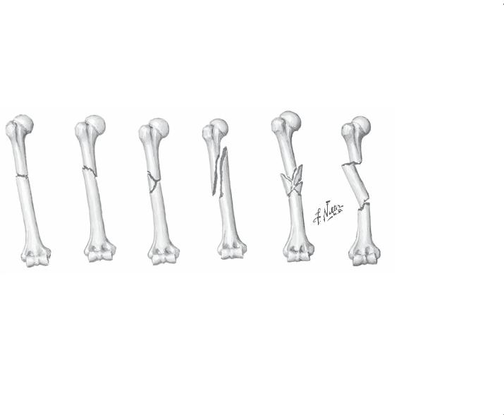
A number of different fracture patterns have been described (Figs. 2-2 to 2-4):
1.Transverse
2.Spiral
3.Oblique
4.Impacted or compressed
5.Avulsion
6.Torus (buckle)
7.Complex (multiple patterns)
Fracture Mode of Loading
The mechanical environment creating a fracture provides a great deal of information as to the mechanism of injury and the extent of that injury. For that reason, biomechanical analyses have been performed to more clearly elucidate the fracture pattern and such specific modes of loading as these:
1.Bending loading produces a transverse fracture
2.Torsional loading produces a spiral fracture
3.Axial loading produces a compression or impacted fracture
4.Tensile loading produces an avulsion fracture
5.Combined loading, such as bending and axial loading, which together produce an oblique fracture.
42 J.N. Delahay and S.T. Sauer
A
Valgus angulation |
Varus angulation |
Anterior angulation |
Posterior angulation |
Shortening |
Translation |
B
FIGURE 2-1. (A) Angulation is described by the direction in which the apex of the fracture is pointing. (B) Displacement (arrows) is defined as the position of the distal fragment in relation to the proximal fragment. (Netter images reprinted with permission from Elsevier. All rights reserved.)
Transverse |
Oblique |
Butterfly |
Spiral |
Comminuted |
Segmental |
fracture |
fracture |
fragment |
fracture |
fracture |
fracture |
FIGURE 2-2. Fracture patterns. (Netter images reprinted with permission from Elsevier. All rights reserved.)
Trauma Skeletal .2
43

44 J.N. Delahay and S.T. Sauer
Type I |
Type II |
|
Type III |
|
|
|
|
|
|
|
|
A,B |
Type IV |
Type V |
C,D |
FIGURE 2-3. Salter classification of epiphyseal plate fractures. Type I: separation of epiphysis. Type II: fracture-separation of epiphysis. Type III: fracture of part of epiphysis. Type IV: (A) fracture of epiphysis and epiphyseal plate; (B) bony union causing premature closure of plate. Type V: (C) crushing of epiphyseal plate;
(D) premature closure of plate on one side with resultant angular deformity. (From Gartland J. Fundamentals of Orthopaedics, 3rd ed. Philadelphia: Saunders, 1987. Reprinted by permission.)
The significance of fracture patterns is that they suggest the amount of force that was applied; hence, an extrapolation can be made that anticipates the amount of soft tissue damage.
Soft Tissues
As already mentioned, a number of soft tissues can be damaged, including the periosteum, blood vessels, nerves, muscles, tendons, and ligaments. The types of injury involving these tissues are delineated in the following subsections.
2. Skeletal Trauma |
45 |
Vascular Injury
Vascular injury is a relatively uncommon event when associated with fractures. When it occurs, it is always an emergent situation. The most common vascular injury is a compartment syndrome. Increased pressure within a fascial compartment can cause muscle necrosis in a relatively short period of time. In the front of the leg, for example, a compartment with the following boundaries exists: the tibia, the syndesmotic membrane, the fibula, and the fascia overlying the tibialis anterior muscle. Because none of these four boundaries can be stretched, the contents of the compartment, that is, the tibialis anterior muscle among others, will necrose from increased pressure caused by an increase in fluid content, occurring after trauma;
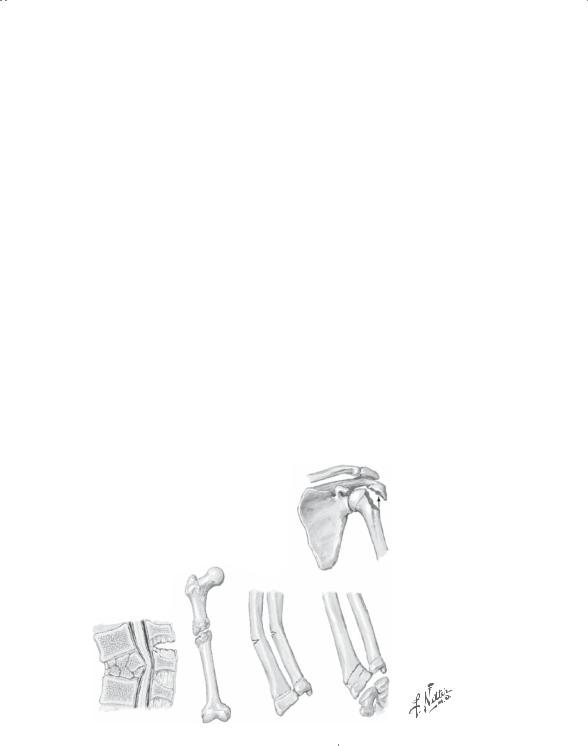
Avulsion (greater tuberosity of humerus avulsed by supraspinatus m.)
Compression fracture |
Greenstick |
Torus (buckle) |
Pathologic fracture |
fracture |
fracture |
(tumor or bone disease) |
|
|
|
|
In children |
FIGURE 2-4. Descriptive terms for typical fracture patterns. (Netter images reprinted with permission from Elsevier. All rights reserved.)

46 J.N. Delahay and S.T. Sauer
this can cause muscle necrosis in a relatively short period of time. The diagnosis of this syndrome is essential. Clinical findings and evaluation methods such as tenderness, pain with passive stretch, and compartmental pressure monitoring assist in diagnosis. Once the diagnosis is confirmed, immediate surgical release of the compartment via fasciotomy is required.
Arterial Injury
Injury to arterial vessels is less common because these vessels are elastic and mobile. The vessels can be damaged when they are either inelastic or fixed by soft tissue structures. The most frequent injury is an intramural hematoma in which the classic signs of arterial injury are usually present. Because of the irreparable damage to the vessel wall, a vein graft or prosthesis is usually required for repair. Injury to the artery is classically associated with several specific fractures involving such sites as the clavicle, the supracondylar region of the elbow (especially in children), the femoral shaft, and the area around the knee.
Nerve Damage
Typically, a nerve is compressed, contused, or stretched as a result of a fracture or other injury. Classic examples include radial nerve injury secondary to fractures of the distal humerus and sciatic nerve injury following posterior fracture dislocations of the hip. The classic grades of neural injury are these:
1.Neuropraxia. Death of the axon does not occur. The condition is generally caused by pressure or contusion and usually improves by itself in a few weeks. The nerve is anatomically intact and physiologically nonfunctional.
2.Axonotmesis. Axonotmesis is an anatomic disruption of the axon in its sheath. Improvement follows regeneration, the axon growing at a slow
rate of 1 mm a day along the existing axonal sheath.
3.Neurotmesis. Neurotmesis is an anatomic disruption of the nerve itself. Surgical repair is required if recovery is to be anticipated.
Muscle and Tendon
It goes without saying that with any fracture or dislocation there is always some associated muscle damage. The extent of this damage and the results will vary depending on the site in question. Myositis ossificans is a specific complication of muscle damage in which heterotopic bone forms within the damaged muscle. The quadriceps and brachialis are specifically predisposed to develop this complication.

2. Skeletal Trauma |
47 |
Ligament
The strength of ligaments is constant throughout life. Certain injuries occurring about the joints can damage the ligaments supporting the joints.
Considering the multiplicity of tissues involved in skeletal trauma, age has been shown to be an important determinant in the results of load application. At any given age, the “weak link,” or the first structure to fail, varies; it could be bone, ligament, or cartilage growth plates. Once the growth plate closes, however, the ligament is the most likely structure to fail. Ligamentous strength, it has already been noted, is constant throughout life. With aging, there is a decrease in cancellous bone volume and an increase in cortical bone porosity. With increasing age, therefore, bone becomes weaker; hence, cartilage, and ligamentous injury are less likely and bone injury more likely. This change means that the same mode of loading can produce a different injury pattern depending on the age of the patient. The same force, such as a tackle in football or a blow by an automobile on the outer side of the knee, is likely to cause a fracture through the distal femoral growth plate in a 12-year-old child, a tear of the medial and anterior cruciate ligaments in a college football player, and a compression fracture of the lateral tibial plateau in a 70-year-old man.
Fractures: Special Types
A number of “special” fractures have been described in the literature. They are defined in the following subsections.
Incomplete Fractures
An incomplete fracture, typical in a child, is one that traverses only a portion of the bone. Two variations have been described:
1.Greenstick fracture: This occurs on the tension side of the bone and involves the diaphysis or cortical bone.
2.Torus or buckle fracture: Known by either name, this occurs on the compression side of bending and involves cancellous bone.
Stress Fractures
Stress fractures are fractures resulting from repetitive loading, each load being below the endurance limit but summated to produce a level of force that indeed causes a fracture. These injuries are typical in the proximal tibia, the second metatarsal, and the femoral neck. They may heal well if the cause of the force ceases soon enough, that is, if the patient stops running for a period of time.

48 J.N. Delahay and S.T. Sauer
Pathologic Fracture
Pathologic fractures occur through abnormal or diseased bone. Among the more common examples are those that occur as a result of tumor or metastatic sites in bone, previously infected bone, or metabolically involved bone such as that resulting from osteoporosis.
Physeal Fractures
In children, a fracture through the cartilaginous growth plate is a common event. The Salter–Harris classification system has allowed such injuries to be more precisely characterized. It is important to remember that physeal fractures heal very rapidly, but they may be complicated by complete or incomplete growth arrest, producing shortening or angular deformity of the limb.
Intraarticular Fractures
Intraarticular fractures enter a joint and disrupt the joint surface and its articular cartilage. Intraarticular fractures can specifically be complicated by joint stiffness and/or the development of premature arthritis.
Pediatric Fractures
Pediatric fractures have a number of special features, which are discussed in Chapter 5.
Fracture Healing
The biology of fracture healing is not particularly complex and parallels that of any nonossified tissue. Essentially, fracture healing occurs in three phases (Fig. 2-5):
1.Vascular phase. This phase begins at the time of the insult and proceeds through the development of a hematoma. This hematoma is then infiltrated by cellular elements, which in turn lay down collagen and cause hematoma organization. A vascularization step follows when the organized hematoma is vascularized by small arterial extensions. The end result of the vascular phase is the development of a soft callus.
2.Metabolic phase. This stage begins about 4 to 6 weeks after the injury. During this period, the soft callus is reworked by a number of specific cellular elements to produce a firm, hard callus satisfactory for meeting the mechanical demands placed upon the fracture in the early phase. There are certain biochemical changes, specifically in pH and oxygen tension, that manipulate the environment during this phase of fracture healing.
3.Mechanical phase. This phase begins once a hard callus is present, which is then manipulated according to the rules of Wolff’s law. Essentially, mechanical stress is required to produce skeletal remodeling during this phase and ultimately to produce a solid, mechanically strong bone.

2. Skeletal Trauma |
49 |
Evaluation of the Patient with Skeletal Trauma
Much more can be said about the evaluation of any surgical patient than the scope of this chapter allows. A number of specific points relative to the orthopedic patient are listed as follows:
1.History of injury. The mechanism of injury and the mode of application are frequently important to determine additional injury.
2.Occupation of the patient. Taking this into account is frequently helpful in planning rehabilitation and recuperative efforts once the fracture has been managed.
3.Activity level before injury. This concern frequently mandates the type of treatment given for a specific injury.
4.Deformity and swelling. These changes must be carefully evaluated physically so that complications can be avoided.
5.Neurovascular status. It is imperative that the neurovascular status of the extremity be carefully evaluated to avoid long-term or permanent sequelae. Similarly, it is critical that the neurovascular integrity of the extremity, or lack thereof, be documented.
6.Integrity of the skin is an absolute. Great care must be taken to ensure that there is no violation of the skin in the area of the fracture site.
Fractures: The Principles of Treatment
All fractures require that two basic goals be accomplished in their treatment: (1) reduction and (2) maintenance of that reduction. Different techniques may be used for achieving these two goals. First, the reduction of a fracture can be accomplished by closed manipulative methods, by surgical open reduction, or through the application of traction. Following reduction, the fracture site must be immobilized so that the fracture will heal in the optimum position. Immobilization can be achieved with external methods such as casts, splints, and external fixators; with internal methods, using various devices such as screws, plates, and intramedullary rods; or by the maintenance of the patient in traction (Fig. 2-6).
Orthopedic Emergencies
Relatively few orthopedic problems mandate immediate intervention. However, those that do exist truly represent emergent situations: these are open fractures, dislocations of major joints, and fractures associated with vascular injury, including compartment syndrome.
Complications of Fractures
A number of complications can occur following fractures and joint dislocations: