

Essentials of Orthopedic Surgery, third edition / 01-Basic Science of Bone and Cartilage Metabolism
.pdf1
Basic Science of Bone and Cartilage Metabolism
JOHN N. DELAHAY
Normal Bone Growth and Development
Bone is a biphasic connective tissue consisting of an inorganic mineral phase and an organic matrix phase. The hardness of bone allows it to provide several specialized mechanical functions: the protection of internal organs, the scaffold that provides points of attachment for other structural elements, and the levers needed to improve the efficiency of muscle action. In addition, bone serves two biologic functions: a site for hematopoietic activity and a reservoir of minerals needed for metabolic interchange.
Embryology
The major components of the musculoskeletal system originate from the mesoderm layer of the trilaminar embryo. This “middle layer” is populated by mesenchymal cells that are totipotent and capable of differentiating into a number of tissues. The sequence of events important in bone growth and development begins with the appearance of the limb bud around the fifth week of life. It is at that time that a tubular condensation of mesenchyme develops centrally in the limb bud. Discrete areas, called interzones, are seen between these condensations (Fig. 1-1) and represent the primitive joints.
During the sixth week, the mesenchyme differentiates into cartilage through the process of chondrification (Fig. 1-2). Interstitial and appositional growth occurs from within and from the surface, respectively. In the seventh week, the cartilage model is penetrated by a vascular spindle, which occurs coincidentally with the necrosis of the central cartilage cells. Once this vascular spindle is established, the central portion of the model is populated by osteoblasts. Matrix is secreted and this in turn is ossified, making immature (woven) bone.
1
2J.N. Delahay
FIGURE 1-1. Histologic study of fetus, approximately 6 weeks gestation, depicting early joint formation. Note the identifiable cartilage and the condensed mesenchymal tissue of the interzone destined to become the joint. (From Bogumill GP. Orthopaedic Pathology: A Synopsis with Clinical Radiographic Correlation. Philadelphia: Saunders, 1984. Reprinted by permission.)
FIGURE 1-2. Histologic study of fetus, approximately 8 weeks gestation. Earliest ossification is depicted here. A sleeve, or collar, of bone is present on the outer surface of the cartilage model. (From Bogumill GP. Orthopaedic Pathology: A Synopsis with Clinical Radiographic Correlation. Philadelphia: Saunders, 1984. Reprinted by permission.)
1. Basic Science of Bone and Cartilage Metabolism |
3 |
Once the central portion of the model is ossified, it is referred to as a primary ossification center (Fig. 1-3). Further ossification of the skeleton occurs via one of two mechanisms: (1) enchondral ossification within a cartilage model (i.e., long bones), and (2) intramembranous ossification within a mesenchymal model (i.e., most flat bones and the clavicle).
From the second through the sixth embryonic months, progressive changes occur in the tubular bones. First, the central (medullary) canal cavitates, leaving a hollow tube of bone with a large mass of cartilage persisting at each end (Fig. 1-4). Within these masses of cartilage, the secondary ossification center, or epiphysis, will form (Fig. 1-5). A cartilage plate, the physis or growth plate (Fig. 1-6), persists between the developing epiphysis and metaphysis. This structure is responsible for growth in length, whereas the covering of the bone, the periosteum, is primarily responsible for growth in girth.
FIGURE 1-3. Primary ossification center of fetus, approximately 14 weeks gestation. The cartilage cells have been removed almost entirely from the center, leaving remnants of acellular cartilage matrix. Bone deposits on the cartilage remnants will form primary trabeculae. Note that the primary sleeve, or collar, of bone has extended along both margins and is located adjacent to the hypertrophied cartilage at each epiphyseal end. (From Bogumill GP. Orthopaedic Pathology: A Synopsis with Clinical Radiographic Correlation. Philadelphia: Saunders, 1984. Reprinted by permission.)
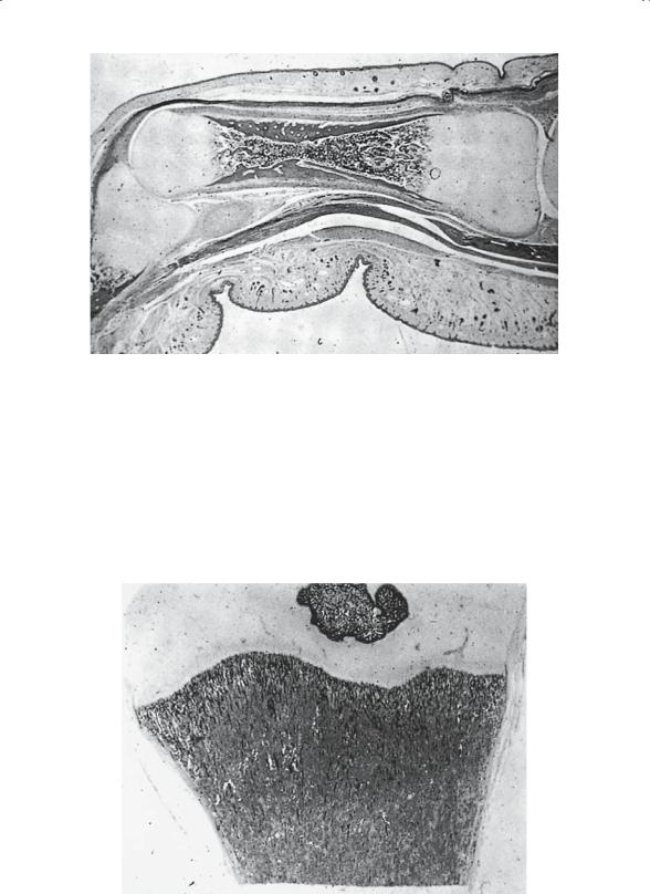
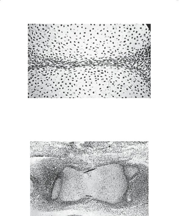
FIGURE 1-4. Primary ossification center, near term. There is complete replacement of cartilage in the diaphyseal portion of the cartilage model. The remaining cartilage is confined to both epiphyseal ends of the model. Note the increasing thickness of the cortical portion of bone, which is a result of conversion of periosteum to bone. A light-staining cambium layer is identifiable. The narrowest portion of the shaft is the site of initial vascular invasion and remains identifiable throughout life in many bones, especially in hands and feet. The eccentric position of this narrowed area indicates the disproportionate contribution to growth in length from each epiphysis. (From Bogumill GP. Orthopaedic Pathology: A Synopsis with Clinical Radiographic Correlation. Philadelphia: Saunders, 1984. Reprinted by permission.)
1. Basic Science of Bone and Cartilage Metabolism |
5 |
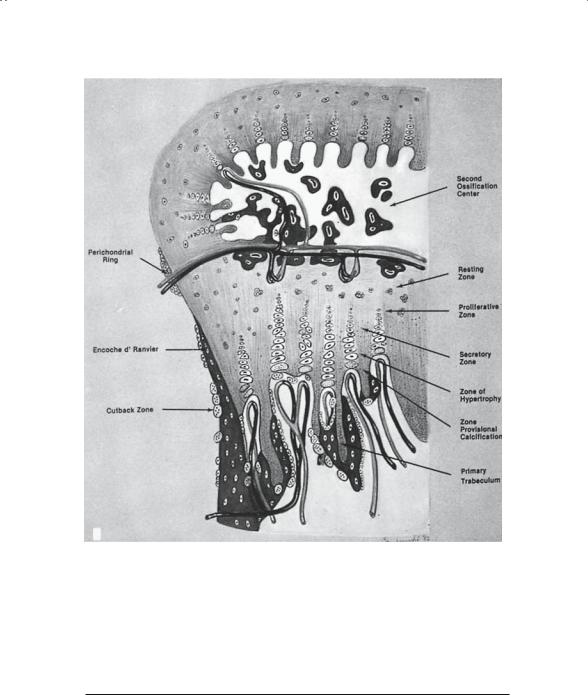
FIGURE 1-6. Schematic diagram of growth plate, consisting of resting zone, proliferative zone, secretory zone, zone of hypertrophy, and zone of calcification. The cross-sectional view helps place events at the growth plate in three-dimensional perspective. (From Bogumill GP. Orthopaedic Pathology: A Synopsis with Clinical Radiographic Correlation. Philadelphia: Saunders, 1984. Reprinted by permission.)
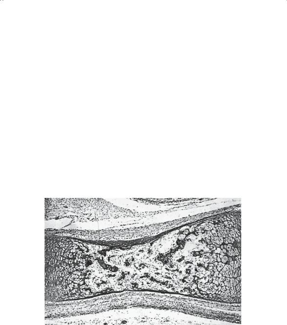
FIGURE 1-5. Early secondary ossification center of mature fetus. The formation of the secondary ossification centers in the lower tibia and upper femur coincide with fetal maturity. The secondary center begins not in the center of the epiphysis but nearer the growth plate. Expansion, therefore, is eccentric. (From Bogumill GP. Orthopaedic Pathology: A Synopsis with Clinical Radiographic Correlation. Philadelphia: Saunders, 1984. Reprinted by permission.)

6J.N. Delahay
Postnatal Development
The physis and the periosteum continue to function postnatally in the growth and development of the infantile skeleton. Numerous local and systemic factors impact on their activity; vascular, hormonal, and genetic effects all play important roles. In essence, the reworking or remodeling of bone that is already present occurs so that the bone can meet the mechanical and biologic demands placed on it.
Bone: The Tissue
Bone, whether it is immature or mature, consists of cells and a biphasic blend of mineral and matrix that coexist in a very exact relationship. The matrix phase consists of collagen and glycosaminoglycans, which are dimeric disaccharides. Both are products of the osteoblast. Calcium hydroxyapatite is the basic mineral crystal in bone. Despite the presence of some less structured amorphous calcium phosphate, the bulk of calcium in the skeletal reservoir is bound in the crystals of hydroxyapatite.
Osteoblasts are bone-forming cells that secrete the matrix components described. As ossification progresses, the osteoblasts become trapped in the matrix they produce and are then referred to as osteocytes. These cells are rather inert but are capable of a small degree of bone resorption. Osteoclasts are those cells whose primary function is the degradation and removal of mineralized bone. It is important to remember that the osteoclasts can remove only mineralized bone, and not unmineralized matrix.
Bone Organization
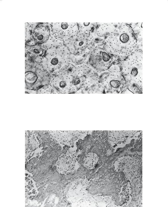
Microscopically, bone is generally described as mature or immature. Mature bone (Fig. 1-7) has an ordered lamellar arrangement of Haversian systems and canalicular communications that give it its classic histologic appearance. Immature bone (Fig. 1-8), in contrast, has a much more random appearance of collagen fibers dispersed in a matrix of irregularly spaced cells. It is produced rapidly by osteoblasts and “remodeled” by the local cell population, until the mature lamellar pattern is achieved. Immature bone is seen in the adult skeleton only under pathologic conditions (i.e., fracture callus, osteogenic sarcoma, myositis, etc.). Macroscopically (Fig. 1-9), the lamellar bone is configured either as dense cortical bone or as delicate spicules called trabeculae. In both areas, the cortex and the trabecular metaphysis, the bone is histologically the same (i.e., mature lamellar bone).
Turnover and Remodeling
Although the tendency is to think of adult bone as an inert tissue, nothing could be further from the truth. Throughout adult life there is a constant ebb and flow of bone formation and bone resorption. These two processes
1. Basic Science of Bone and Cartilage Metabolism |
7 |
FIGURE 1-7. Mature bone: osteonal structure as seen in undecalcified material. Numerous interstitial fragments (osteonal fragments without an associated Haversian canal) are readily observed. (From Bogumill GP. Orthopaedic Pathology: A Synopsis with Clinical Radiographic Correlation. Philadelphia: Saunders, 1984. Reprinted by permission.)
FIGURE 1-8. Immature bone (early callus). Note the large number of osteoblasts and osteocytes. (From Bogumill GP. Orthopaedic Pathology: A Synopsis with Clinical Radiographic Correlation. Philadelphia: Saunders, 1984. Reprinted by permission.)
8J.N. Delahay
METRIC 1
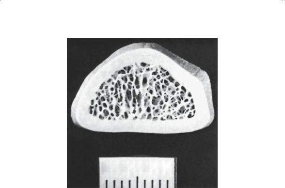
FIGURE 1-9. Cross section of the radius at the distal metaphysis. The majority of bone is cortical bone, in which the annual rate of turnover is only 2%.
are delicately balanced and keep the skeletal mass in a state of equilibrium. A number of authors have popularized the concept of “coupling”; bone formation and bone resorption generally increase or decrease in the same direction. When one process increases, so does the other, and vice versa.
It is important, however, to consider the net effect of the rate changes in these two processes. For example, in osteoporosis, both formation and bone resorption increase, but resorption increases at a much greater rate, so that despite a coupled increase in bone formation the net effect is an overall decrease in bone mass. A number of factors, systemic and local, affect these processes and hence impact bone turnover and remodeling. Perhaps the most well defined factor is mechanical stress, which forms the basis for the classic Wolff’s law. Simply stated, trabecular, and to a lesser degree cortical, bone remodels along lines of mechanical stress. Bone forms where it is needed to meet mechanical demands, and it is resorbed where the need is less. Current research suggests that bone functions as a transducer, converting mechanical energy from the applied load into electrical energy and a voltage gradient. In turn, this voltage gradient that is generated modulates cellular differentiation. Osteoblastic activity is thus seen in regions where the mechanical demands are the greatest. Osteoclas-

1. Basic Science of Bone and Cartilage Metabolism |
9 |
tic activity predominates the pattern when those mechanical demands decrease and less bone is required. This phenomenon has been called the “piezoelectric effect.” Specifically, the deformation of bone apatite crystals by superimposed load generates the voltage gradient, which in turn alters the cell population to respond to that load.
Cartilage: The Tissue
Cartilage, like bone, is a connective tissue. Its histologic organization, however, is far less structured. There are three histologic types of cartilage, each serving a different function:
1.Hyaline cartilage covers the ends of long bones and provides a smooth, frictionless surface for articulation in a diarthrodial (synovial lined) joint.
2.Fibrocartilage is typically found in certain nondiarthrodial joints such as the pubic symphysis. It is also located at the margins of certain diarthrodial joints, forming structures such as the glenoid labrum and acetabular labrum. Following injury to hyaline cartilage, repair of the chondral defect is typically accomplished in the form of fibrocartilage.
3.Elastic cartilage is found in certain areas where resiliency is important. Examples include the tip of the nose and the ear lobe.
The most important of the three, hyaline cartilage, is a relatively aneural, avascular, and relatively hypocellular connective tissue. By weight, it is 70% water and 30% ground substance and cells. The ground substance of hyaline cartilage is composed primarily of type II collagen and GAG proteins (glycosaminoglycans). The collagen endows the cartilage with tensile strength, and the GAGs are critical for resiliency.
The cells are called chondrocytes and are dispersed throughout the chondral layers in four zones: tangential (most superficial), transitional, radial, and calcified. These chondrocytes are found in individual lacunae, where they maintain healthy cartilage by actively synthesizing new ground substance components.
The chondral layer receives the bulk of its nutrition by diffusion from the synovial fluid above and from the vasculature at the subchondral plate below. Normal diarthrodial (synovial lined) joint function depends on the presence of normal hyaline cartilage. In its fully hydrated state, hyaline cartilage provides an almost frictionless bearing, hence minimizing wear on the articular surface.
Abnormal Bone Development and Metabolism
Most skeletal diseases are the result of disruption of normal bone growth and development, breakdown of bone once it has been normally formed, or alteration of the normal mechanisms of bone formation or bone resorp-

10 J.N. Delahay
tion. The etiologies of the pathologic states, as one would expect, are quite varied; but the final manifestations within the musculoskeletal system frequently show striking similarities.
Despite the etiology, damage to the growing skeleton will alter the overall shape of one or more bones, depending on whether the adverse process is localized or generalized. Similarly, disruption of osteoblast function will decrease the amount or the quality of the bone formed. Multiple factors are known to stimulate osteoclast activity, such as parathyroid hormone, the presence of particulate polyethylene, and certain neoplasms, resulting in localized or generalized bone resorption.
As one considers the etiology of skeletal disease, it is helpful to first group the possible differential diagnoses by disease category, which permits one to develop a comprehensive list of possible diagnoses that may explain the findings manifested by the skeleton. The seven disease categories are best remembered using the acronym “VITAMIN”:
V, vascular disease
I, infection
T, tumor
A, arthritis
M, metabolic bone disease
I, injury
N, neurodevelopmental causes
The remainder of this chapter focuses on these diagnostic groups and the way in which they affect the skeleton. Specific emphasis is placed on generalized afflictions of the skeleton. In that light, certain disease categories are more likely to adversely affect the skeleton in a generalized fashion, specifically vascular, metabolic, systemic arthritis, and neurodevelopmental etiologies. The other etiologies—infection, injury, and tumor—are more likely to produce localized changes and, therefore, are considered in individual subsequent chapters.
Last, as a reminder, a differential diagnosis is a listing of plausible specific diagnoses that may explain observed findings such as physical or radiographic. It is not adequate to simply list a disease category because appropriate treatment of a given condition depends on identifying a specific etiology.
Metabolic Bone Disease
General Concepts
Disease processes affecting bone often can be understood as a change in the relationship of bone formation and bone resorption. It is therefore important to understand this relationship. Only by doing so can the net effect on the skeleton be appreciated.