

Structure 87
Chapter 3: Virology of Human Influenza
Lutz Gürtler
Human influenza viruses are members of the orthomyxovirus family, which consists of the genera: influenza A, B, and C virus, and Thogovirus (in ticks). In humans, only influenza A and B viruses are of epidemiological interest.
The main antigenic determinants of influenza A and B viruses are the haemagglutinin (H or HA) and neuraminidase (N or NA) transmembrane glycoproteins. Based on the antigenicity of these glycoproteins, influenza A viruses are further subdivided into sixteen H (H1–H16) and nine N (N1–N9) subtypes. The full nomenclature for influenza virus isolates requires connotation of the influenza virus type (A or B), the host species (omitted if human in origin), the geographical site, serial number, year of isolation, and lastly, the H and N variants in brackets, for example: A/goose/Guangdong/1/96 (H5N1).
Influenza viruses are usually transmitted via air droplets, and subsequently contaminate the mucosa of the respiratory tract. They are able to penetrate the mucin layer of the outer surface of the respiratory tract, entering respiratory epithelial cells, as well as other cell types. Replication is very quick: after only 6 hours the first influenza viruses are shed from infected cells. Part of the viral proteins, such as the fusion peptide and NS2, act as toxins to promote the production of influenza virus. Rapid bacterial growth, most commonly Streptococcus pneumoniae, Staphylococcus aureus, and Haemophilus influenzae, may begin in the very early phase of viral replication (for more details, see the chapter on Pathogenesis).
Structure
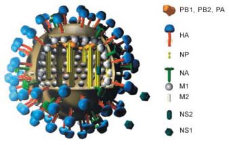
Influenza viruses are enveloped single-stranded RNA viruses with a pleomorphic appearance, and an average diameter of 120 nm. Projections of haemagglutinin and neuraminidase cover the surface of the particle (Figure 1).
The influenza A and B virus genomes consist of 8 separate segments covered by the nucleocapsid protein. Together these build the ribonucleoprotein (RNP), and each segment codes for a functionally important protein:
1.Polymerase B2 protein (PB2)
2.Polymerase B1 protein (PB1)
3.Polymerase A protein (PA)
4.Haemagglutinin (HA or H)
5.Nucleocapsid protein (NP)
6.Neuraminidase (NA or N)
7.Matrix protein (M): M1 constructs the matrix; and in influenza A viruses only, M2 acts as an ion channel pump to lower or maintain the pH of the endosome
8.Non-structural protein (NS); the function of NS2 is hypothetical
The active RNA-RNA polymerase, which is responsible for replication and transcription, is formed from PB2, PB1 and PA. It has an endonuclease activity and is
88 Virology of Human Influenza
linked to the RNP. The NS1 and NS2 proteins have a regulatory function to promote the synthesis of viral components in the infected cell (see below).
The envelope of the virus is a lipid bilayer membrane which originates from the virus-producing cell and which contains prominent projections formed by HA and NA, as well as the M2 protein. The lipid layer covers the matrix formed by the M1 protein.
Influenza C virus harbours only 7 genome segments, and its surface carries only one glycoprotein. As it has a low pathogenicity in humans, it will not be discussed here in detail.
Figure 1. Structure of an influenza A virus. Image copyright by Dr. Markus Eickmann, Institute for Virology, Marburg, Germany. Used with permission. – http://www.biografix.de
Haemagglutinin
Haemagglutinin (HA or H) is a glycoprotein containing either 2 of 3 glycosylation sites, with a molecular weight of approximately 76,000. It spans the lipid membrane so that the major part, which contains at least 5 antigenic domains, is presented at the outer surface. HA serves as a receptor by binding to sialic acid (N-acetyl- neuraminic acid) and induces penetration of the interior of the virus particle by membrane fusion. Haemagglutinin is the main influenza virus antigen; the antigenic sites being A, B (carrying the receptor binding site), C, D, and E. The antigenic sites are presented at the head of the molecule, while the feet are embedded in the lipid layer. The body of the HA molecule contains the stalk region and the fusiogenic domain which is needed for membrane fusion when the virus infects a new cell. At low pH, the fusion peptide is turned to an interior position. The HA forms trimers and several trimers form a fusion pore.
Prominent mutations in the antigenic sites reduce or inhibit the binding of neutralising antibodies, thereby allowing a new subtype to spread within a non-immune
Structure 89
population. This phenomenon is called antigenic drift. The mutations that cause the antigenic drift are the molecular explanation for the seasonal influenza epidemics during winter time in temperate climatic zones. The immune response to the HA antigenic sites is followed by the production of neutralising antibody, which is the basis for resolving infection in an individual, and is sometimes part of the cross immunity found in elderly individuals when a new pandemic virus strain occurs.
Antigenic shift – also termed genome reassortment or just reassortment – arises when the HA is exchanged in a virus, for example H1 replaced by H5 resulting in the formation of a mosaic virus. This may happen when a cell is infected by 2 different influenza viruses and their genome segments are exchanged during replication.
This phenomenon of genome reassortment is frequently seen in water birds, especially ducks. Although the birds are seldomly symptomatic after infection, the virus is shed in their faeces for several months.
Neuraminidase
Like HA, neuraminidase (NA or N) is a glycoprotein, which is also found as projections on the surface of the virus. It forms a tetrameric structure with an average molecular weight of 220,000. The NA molecule presents its main part at the outer surface of the cell, spans the lipid layer, and has a small cytoplasmic tail.
NA acts as an enzyme, cleaving sialic acid from the HA molecule, from other NA molecules and from glycoproteins and glycolipids at the cell surface. It also serves as an important antigenic site, and in addition, seems to be necessary for the penetration of the virus through the mucin layer of the respiratory epithelium.
Antigenic drift can also occur in the NA. The NA carries several important amino acid residues which, if they mutate, can lead to resistance against neuraminidase inhibitors. Mutations that have been observed include:
•R292K
•H274Y, R152K, E119V
The letters represent amino acids (R, arginine; K, lysine; H, histidine; Y, tyrosine; E, glutamic acid; V, valine): the former letter is the original amino acid, and the latter the amino acid after mutation occurred.
When the amino acid arginine (R) is replaced by lysine (K) at position 292 of the neuraminidase glycoprotein, complete resistance may result. The mutation of R to K is linked to a single nucleotide exchange of AGA to AAA in the N gene. Position 292 is so significant because mutation may induce resistance not only against the substance oseltamivir, but also against zanamavir and two other new prodrugs.
M2 protein
When the virus particle is taken up in the endosome, the activity of the M2 ion channel is increased so that ions flood into the particle, inducing a low pH. As a result of this, the HA-M1 linkage is disturbed, the particle opens, the fusion peptide within the HA is translocated, and the HA fuses with the inner layer of the endosome membrane. The ribonucleoproteins are liberated into the cytoplasm of the cell and transported to the nucleus, where the complex is disrupted, and viral RNA synthesis is initiated.