

МРТ - в диагностике болезней суставов
.pdfANATOMY AND MRI OF THE JOINTS
A Multiplanar Atlas

ANATOMY AND MRI OF THE JOINTS
A MULTIPLANAR ATLAS
Editors
William D. Middleton, M.D. Thomas L. Lawson, M.D.
Department of Radiology
Medical College of Wisconsin
Milwaukee, Wisconsin
RAVEN PRESS |
NEW YORK |
|
|

Raven Press, 1185 Avenue of the Americas, New York, New York 10036
© Raven Press, Ltd. All rights reserved. This book is protected by copyright. No part of it may be reproduced, stored in a retrieval system, or transmitted, in any form or by any means, electronical, mechanical, photocopying, or recording, or otherwise, without the prior written permission of the publisher.
Library of Congress Cataloging-in-Publication Data
Anatomy and MRI of the joints.
Includes bibliographies and index.
1. Joints__Anatomy__Atlases. |
2. Joints__Imaging__ Atlases. 3. Magnetic resonance imaging__Atlases. |
||
I. Middleton, William D. II. Lawson, Thomas L. |
|||
[DNLM: 1. Joints__anatomy__atlases. 2. Magnetic Resonance Imaging__atlases. WE 17 A535] |
|||
QM33I.A53 |
611'.72 |
87-42729 |
|
ISBN 0-88167-455-9 |
|
|
|
|
|
|
|
The material contained in this volume was submitted as previously unpublished material, except in the instances in which credit has been given to the source from which some of the illustrative material was derived.
Great care has been taken to maintain the accuracy of the information contained in the volume. However, neither Raven Press nor the editors can be held responsible for errors or for any consequences arising from the use of the information contained herein.
9 8 7 6 5 4 3 2 1

This book is dedicated to my parents, William and Joyce
W.D.M

Preface
As techniques that are used to image the human body improve, our knowledge of anatomy must expand and adapt to enable us to accurately interpret the images. In the 1970s, the development of computed tomography (CT) forced us to become intimately familiar with axial cross-sectional anatomy. The current development and continuing refinement of magnetic resonance imaging (MRI) makes it necessary to understand cross-sectional anatomy in multiple planes. In general, this understanding requires only a reorientation of thinking since we are already well acquainted from our experience with CT with the anatomic structures and their axial relationships.
However, the imaging of the joints is more involved. The lack of respiratory motion and the use of small surface coils allows for images of extremely high contrast and resolution, resulting in a clarity of visualization of many anatomic structures not previously achieved. Therefore, not only are we imaging the joints in orientations that are unfamiliar, but we are seeing structures that we have not been able to see in the past. The resulting displays of anatomy can be as intimidating as they are impressive. The ability to image the complex multiplanar anatomy of the joints with MRI and the resulting need to understand this anatomy served as the motivation for the development of this atlas. Our intent was to produce an atlas to be used as a ready reference for the identification of articular and periarticular structures seen on MR images. To accomplish this we have imaged live volunteers on a 1.5 Tesla system. All images were obtained using commercially available surface coils and standard spin-echo pulse sequences. For all the joints except the vertebral column, T-1-weighted images were obtained since they showed the overall anatomy best. Two excitations were used for all the images. The slice thickness was 3 mm for the smaller joint and 5 mm for the larger joints (the vertebral column and hip). The imaging field of view varied depending on the size of the joint.
To confirm the identity of the structures seen on the MR images, we carefully compared the images with actual anatomic sections obtained from frozen cadavers. The joints of these cadavers were cut into tissue blocks and mounted on the stage of a cryomicrotome unit. The frozen tissue specimens were then sequentially cut and photographs of the surface taken at less than 1-mm intervals. Representative color photographs of these serial sections have been included in the atlas, not only to allow for MR-anatomic correlation but, more importantly, to guard against obsolescence. The MR images are state-of-the-art. Although MR imaging will undoubtedly continue to improve, the cryomicrotomes, however, cannot be improved upon, and will remain a valuable reference regardless of advances in MRI or other imaging
techniques.
William D. Middleton Thomas L. Lawson
vii

Acknowledgments
Many people contributed to the preparation of this atlas and their efforts have been greatly appreciated.
Dr. Victor Haughton was instrumental in obtaining cadavers for anatomic sections as well as providing the cryomicrotome unit used to cut the sections. Without his help, this book would not have been possible.
The cryomicrotomes were produced and photographed by Bruce Nowicki. Their beauty is the result of his skilled work.
The MR images were obtained with the excellent assistance of the technologists in the MRI facility at the Medical College of Wisconsin. They include Beth Beck, Marie Bye, Steve Censky, Lisa Klotz, and Julie Strandt.
The artwork was provided by Robert Fenn at the Medical College of Wisconsin and Leslie MacConnellClubbs at the Mallinckrodt Institute of Radiology.
Preparation of the manuscript was the result of the tireless efforts of Marilyn Bell and Lynn Losse. Finally, the support, assistance, and advice that was received from Dr. Mary Middleton throughout the
preparation of this atlas provided constant encouragement for its ultimate completion.
ix

Contents |
|
1. The Temporomandibular Joint ....................................................................................................... |
1 |
J. Bruce Kneeland, M.D. |
|
2. The Shoulder........................................................................................................................................ |
13 |
William D. Middleton, M.D. |
|
3. The Elbow ............................................................................................................................................ |
49 |
Stephan J. Macrander, M.D. |
|
4. The Wrist ............................................................................................................................................... |
83 |
William D. Middleton, M.D. |
|
5. The Finger............................................................................................................................................ |
121 |
Scott Erickson, M.D. |
|
6. The Vertebral Column.......................................................................................................................... |
139 |
Lowell Sether, Ph.D. |
|
7. The Hip .................................................................................................................................... |
153 |
Thomas L. Lawson, M.D. and William D. Middleton, M.D. |
|
8. The Knee.................................................................................................................................. |
205 |
Gary W. Hinson, M.D. and William D. Middleton, M.D. |
|
9. The Ankle ................................................................................................................................. |
251 |
William D. Middleton, M.D. |
|
Recommended Reading ...................................................................................................... |
293 |
Subject Index ....................................................................................................................... |
297 |
xi

Contributors
Scott Erickson, M.D., Medical College of Wisconsin, Milwaukee County Medical Complex, 8700 West Wisconsin Avenue, Milwaukee, Wisconsin 53226
Gary W. Hinson, M.D., Menorah Medical Center, Kansas City, Missouri 64110
J. Bruce Kneeland, M.D., Medical College of Wisconsin, Milwaukee County Medical Complex, 8700 West Wisconsin Avenue, Milwaukee, Wisconsin 53226
Thomas L. Lawson, M.D., Medical College of Wisconsin, Milwaukee County Medical Complex, 8700 West Wisconsin Avenue, Milwaukee, Wisconsin 53226
Steven Macrander, M.D., Medical College of Wisconsin, Milwaukee County Medical Complex, 8700 West Wisconsin Avenue, Milwaukee, Wisconsin 53226
William D. Middleton, M.D., Mallinckrodt Institute of Radiology, 510 South Kingshighway, St. Louis, Missouri 63110
Lowell Sether, Ph.D., Medical College of Wisconsin, Milwaukee County Medical Complex, 8700 West Wisconsin Avenue, Milwaukee, Wisconsin 53226

ANATOMY AND MRI OF THE JOINTS
A Multiplanar Atlas
CHAPTER 1
THE TEMPOROMANDIBULAR
JOINT
J. Bruce Kneeland, M.D.
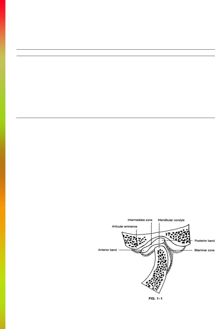
The temporomandibular joint (TMJ) is relatively simple compared to other joints in this atlas. Figure 1-1 illustrates the important articular structures. The osseous portion consists of the articular eminence and mandibular fossa of the temporal bone above and the mandibular condyle below. The articular surfaces are covered with fibrocartilage. Within the joint is an ovoid plate consisting of fibrous tissue called the disc. The disc has a biconcave structure with thick anterior and posterior bands connected by a thin intermediate zone. The joint is separated into two synovial spaces (superior and inferior) by the disc.
Attached to the posterior band is the bilaminar zone which consists of a superior lamella of fibroelastic tissue and a lower lamella of fibrous tissue. These two lamellae are attached to the posterior margin of the mandibular fossa and the posterior margin of the condyle, respectively. Anteriorly the disc is attached to the lateral pterygoid muscle.
In "normal" individuals the posterior margin of the posterior band of the disc is located at the highest point of the condyle (approximately 12 o'clock) with the jaw closed, although it may be anteriorly displaced a few degrees. With jaw opening, the condyle moves anteriorly under the disc.
Although not a part of the TMJ, the external auditory canal is a useful landmark that permits an evaluation of the degree of opening of the jaw. With closed jaw the posterior surface of the condyle abuts the canal. As the jaw opens the condyle moves anteriorly away from the canal.
As previously noted, the disc is attached anteriorly to the lateral pterygoid muscle.
The lateral pterygoid has two parts: a superior head which arises from the sphenoid and temporal bones and inserts on the disc, and an inferior head which arises from the lateral pterygoid plate and inserts on the neck of the mandible. Although not directly attached to the joint, the medial pterygoid muscle is located anterior to the lateral pterygoid. It attaches to the ramus and angle of the mandible.
The TMJ is supplied by the superficial temporal artery, which is one of two terminal branches of the external carotid artery and originates in the parotid gland posterior to the neck of the mandible. Also seen in this region is the maxillary artery, which is the second terminal branch of the external carotid and likewise originates in the parotid gland.
1