

187-2017
.pdfCHAPTER 14 Agents Used in Cardiac Arrhythmias |
233 |
Channels available, percent of maximum
100
|
|
|
|
|
|
|
|
|
|
Control |
|||
|
|
|
|
|
|
|
Drug |
|
|
|
|||
0 |
|
|
|
|
|
|
|
|
|
|
|
|
|
–120 |
–100 |
–80 |
–60 |
||||||||||
|
|||||||||||||
Resting membrane potential (mV)
Recovery time constant (ms)
100,000
10,000
1000
100
10
0
Drug
Control
–120 –100 –80 –60 Resting membrane potential (mV)
FIGURE 14 4 Dependence of sodium channel function on the membrane potential preceding the stimulus. Left: The fraction of sodium channels available for opening in response to a stimulus is determined by the membrane potential immediately preceding the stimulus. The decrease in the fraction available when the resting potential is depolarized in the absence of a drug (control curve) results from the voltagedependent closure of h gates in the channels. The curve labeled Drug illustrates the effect of a typical local anesthetic antiarrhythmic drug. Most sodium channels are inactivated during the plateau of the action potential. Right: The time constant for recovery from inactivation after repolarization also depends on the resting potential. In the absence of drug, recovery occurs in less than 10 ms at normal resting potentials (−85 to −95 mV). Depolarized cells recover more slowly (note logarithmic scale). In the presence of a sodium channel-blocking drug, the time constant of recovery is increased, but the increase is far greater at depolarized potentials than at more negative ones.
maximum upstroke velocity of the action potential, which will in turn reduce action potential conduction velocity.
In cells like those found in the SA and AV nodes, where excitability is determined by the availability of calcium channels, excitability is most sensitive to drugs that block these channels. As a result, calcium channel blockers can decrease pacemaker activity in the SA node as well as conduction velocity in the AV node.
MECHANISMS OF ARRHYTHMIAS
Many factors can precipitate or exacerbate arrhythmias: ischemia, hypoxia, acidosis or alkalosis, electrolyte abnormalities, excessive catecholamine exposure, autonomic influences, drug toxicity (eg, digitalis or antiarrhythmic drugs), overstretching of cardiac fibers, and the presence of scarred or otherwise diseased tissue. However, all arrhythmias result from (1) disturbances in impulse formation and/or (2) disturbances in impulse conduction.
Disturbances of Impulse Formation
Pacemaking activity is regulated by both sympathetic and parasympathetic activity (see above). Therefore, factors that antagonize or enhance these effects can alter normal impulse formation, producing either bradycardia or tachycardia. Genetic mutations have also been found to alter normal pacemaking activity.
Under certain circumstances, abnormal activity can be generated by latent pacemakers, cells that show slow phase 4 depolarization even under normal conditions (eg, Purkinje cells). Such cells are particularly prone to accelerated pacemaker activity, especially under conditions such as hypokalemia. Abnormalities in impulse formation can also be the result of afterdepolarizations (Figure 14–5). These can be either early afterdepolarizations (EADs), which occur during
phase 3 of the action potential, or delayed afterdepolarizations (DADs), which occur during phase 4. EADs are usually triggered by factors that prolong action potential duration. When this prolongation occurs in ventricular cells, there is often a corresponding increase in the QT interval of the electrocardiogram (ECG). Such an
|
|
Prolonged |
Early afterdepolarization |
0 mV |
|
(arises from the plateau) |
|
|
plateau |
|
|
|
|
–70
0.5 sec
0 mV
Delayed afterdepolarization (arises from the resting potential)
–70
FIGURE 14 5 Two forms of abnormal activity, early (top) and delayed afterdepolarizations (bottom). In both cases, abnormal depolarizations arise during or after a normally evoked action potential. They are therefore often referred to as“triggered”automaticity; that is, they require a normal action potential for their initiation.

234 |
SECTION III Cardiovascular-Renal Drugs |
Molecular & Genetic Basis of Cardiac Arrhythmias
It is now possible to define the molecular basis of several congenital and acquired cardiac arrhythmias. The best example is the polymorphic ventricular tachycardia known as torsades de pointes (Figure 14–8), which is associated with prolongation of the QT interval (especially at the onset of the tachycardia), syncope, and sudden death. This represents prolongation of the action potential of at least some ventricular cells (Figure 14–1). The effect can, in theory, be attributed to either increased inward current (gain of function) or decreased outward current (loss of function) during the plateau of the action potential. Action potential prolongation is thought to generate early afterdepolarizations (Figure 14–5) that then trigger torsades de pointes.
Recent molecular genetic studies have identified up to 300 different mutations in at least eight ion channel genes that produce congenital long QT (LQT) syndrome (Table 14–1). Loss-of-function mutations in potassium channel genes (HERG, KCNE2, KCNQ1, KCNE1, and KCNJ2) result in decreased outward plateau current, while gain-of-function mutations in the sodium channel gene (SCN5A) or calcium channel gene (CACNA1c) cause increases in inward plateau current.
The identification of the precise molecular mechanisms underlying various forms of the LQT syndromes now raises the possibility that specific therapies may be developed for individuals with defined molecular abnormalities. Indeed, preliminary
effect can be caused by genetic mutations associated with congenital long QT (LQT) syndrome (see Box: Molecular & Genetic Basis of Cardiac Arrhythmias). A number of drugs (antiarrhythmic as well as non-antiarrhythmic agents) can produce “acquired” or drug-induced LQT syndrome, which is typically due to block of rapidly activating delayed rectifier potassium channels. Many forms of LQT syndrome are exacerbated by other factors that prolong action potential duration, including hypokalemia and slow heart rates. DADs, on the other hand, often occur when there is an excess accumulation of intracellular calcium (see Chapter 13), especially at fast heart rates. They are thought to be responsible for arrhythmias associated with digitalis toxicity, excess catecholamine stimulation, and myocardial ischemia.
Disturbances of Impulse Conduction
The most common form of conduction disturbance affects the AV node, causing various degrees of heart block. The result can be a simple slowing of impulse propagation through the AV node, which is reflected by an increase in the PR interval of the ECG. At the extreme, the result can be complete heart block, where no impulses are conducted from the atria to the ventricles. In this situation, ventricular activity is generated by a latent pacemaker, such as a Purkinje cell. Because the AV node is typically under the tonic influence of the parasympathetic nervous system, which slows conduction, AV block can sometimes be relieved by antimuscarinic agents like atropine.
reports suggest that the sodium channel blocker mexiletine can correct the clinical manifestations of congenital LQT subtype 3, while β-blockers have been used to prevent arrhythmias triggered by sympathetic stimulation in patients with LQT subtype 1.
The molecular basis of several other congenital cardiac arrhythmias associated with sudden death has also recently been identified. At least three forms of short QT syndrome have been identified that are linked to gain-of-function mutations in different potassium channel genes (KCNH2, KCNQ1, and KCNJ2). Catecholaminergic polymorphic ventricular tachycardia, a disease that is characterized by stressor emotion-induced syncope, can be caused by mutations in at least two different genes (hRyR2 and CASQ2) of proteins expressed in the sarcoplasmic reticulum that control intracellular calcium homeostasis. Mutations in two different ion channel genes (HCN4 and SCN5A) have been linked to congenital forms of sick sinus syndrome. Several forms of Brugada syndrome, which is characterized by ventricular fibrillation associated with persistent ST-segment elevation, and progressive cardiac conduction disorder (PCCD), which is characterized by impaired conduction in the His-Purkinje system and right or left bundle block leading to complete AV block, have been linked to loss-of-function mutations in the sodium channel gene (SCN5A). At least one form of familial atrial fibrillation is caused by a gain- of-function mutation in a potassium channel gene (KCNQ1).
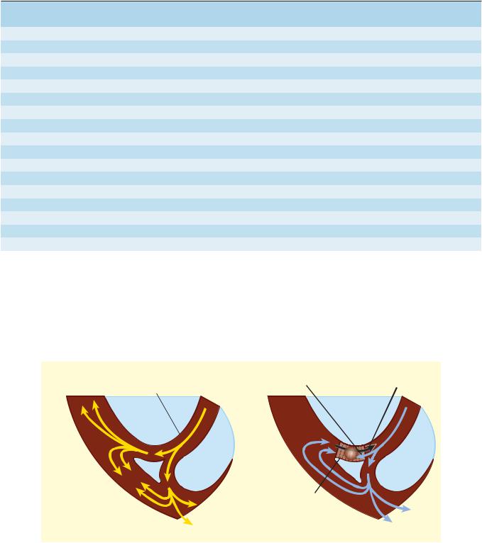
A serious form of conduction abnormality involves reentry (also known as “circus movement”). In this situation, one impulse reenters and excites areas of the heart more than once. The path of the reentering impulse may be confined to very small areas, such as within or near the AV node or where a Purkinje fiber makes contact with the ventricular wall (Figure 14–6), or it may involve large portions of the atria or ventricles. Some forms of reentry are strictly anatomically determined. For example, in Wolff-Parkinson-White syndrome, the reentry circuit consists of atrial tissue, the AV node, ventricular tissue, and an accessory AV connection (bundle of Kent, a bypass tract). Depending on how many round trips through the pathway a reentrant impulse makes before dying out, the arrhythmia may be manifest as one or a few extra beats or as a sustained tachycardia. Circulating impulses can also give off “daughter impulses” that can spread to the rest of the heart. In cases such as atrial or ventricular fibrillation, multiple reentry circuits may meander through the heart in apparently random paths, resulting in the loss of synchronized contraction.
An example of how reentry can occur is illustrated in Figure 14–6. In this scenario, there are three key elements: (1) First is an obstacle (anatomic or physiologic) to homogeneous impulse conduction, thus establishing a circuit around which the reentrant wave front can propagate. (2) The second element is unidirectional block at some point in the circuit. That is, something has occurred such that an impulse reaching the site initially encounters refractory tissue. This can occur under conditions such as ischemia,
CHAPTER 14 Agents Used in Cardiac Arrhythmias |
235 |
TABLE 14 1 Molecular and genetic basis of some cardiac arrhythmias.
|
Chromosome |
|
Ion Channel or |
|
Type |
Involved |
Defective Gene |
Proteins Affected |
Result |
|
|
|
|
|
LQT-1 |
11 |
KCNQ1 |
IKs |
LF |
LQT-2 |
7 |
KCNH2 (HERG) |
IKr |
LF |
LQT-3 |
3 |
SCN5A |
INa |
GF |
LQT-4 |
4 |
Ankyrin-B1 |
|
LF |
LQT-5 |
21 |
KCNE1 (minK) |
IKs |
LF |
LQT-6 |
21 |
KCNE2 (MiRP1) |
IKr |
LF |
LQT-72 |
17 |
KCNJ2 |
IKir |
LF |
LQT-83 |
12 |
C ACNA1c |
ICa |
GF |
SQT-1 |
7 |
KCNH2 |
IKr |
GF |
SQT-2 |
11 |
KCNQ1 |
IKs |
GF |
SQT-3 |
17 |
KCNJ2 |
IKir |
GF |
CPVT-14 |
1 |
hRyR2 |
Ryanodine receptor |
GF |
CPVT-2 |
1 |
CASQ2 |
Calsequestrin |
LF |
Sick sinus syndrome |
15 or 3 |
HCN4 or SCN5A5 |
|
LF |
Brugada syndrome |
3 |
SCN5A |
INa |
LF |
PCCD |
3 |
SCN5A |
INa |
LF |
Familial atrial fibrillation |
11 |
KCNQ1 |
IKs |
GF |
1Ankyrins are intracellular proteins that associate with a variety of transport proteins including Na+ channels, Na+/K+-ATPase, Na+, Ca2+ exchange, and Ca2+ release channels. 2Also known as Andersen syndrome.
3Also known as Timothy syndrome; multiple organ dysfunction, including autism.
4CPVT, catecholaminergic polymorphic ventricular tachycardia; mutations in intracellular ryanodine Ca2+ release channel or the Ca2+ bu er protein, calsequestrin, may result in enhanced sarcoplasmic reticulum Ca2+ leakage or enhanced Ca2+ release during adrenergic stimulation, causing triggered arrhythmogenesis.
5HCN4 encodes a pacemaker current in sinoatrial nodal cells; mutations in sodium channel gene (SCN5A) cause conduction defects.
GF, gain of function; LF, loss of function; LQT, long QT syndrome; PCCD, progressive cardiac conduction disorder; SQT, short QT syndrome.
Forward impulse |
Retrograde |
obstructed and extinguished |
impulse |
Purkinje twig |
|
|
Depressed region |
A. Normal conduction |
B. Unidirectional block |
FIGURE 14 6 Schematic diagram of a reentry circuit that might occur in small bifurcating branches of the Purkinje system where they enter the ventricular wall. A: Normally, electrical excitation branches around the circuit, is transmitted to the ventricular branches, and becomes extinguished at the other end of the circuit due to collision of impulses. B: An area of unidirectional block develops in one of the branches, preventing anterograde impulse transmission at the site of block, but the retrograde impulse may be propagated through the site of block if the impulse finds excitable tissue; that is, the refractory period is shorter than the conduction time. This impulse then reexcites tissue it had previously passed through, and a reentry arrhythmia is established.
236 |
SECTION III Cardiovascular-Renal Drugs |
which cause an increase in extracellular potassium that partially depolarizes the resting membrane potential, slowing sodium channel recovery from inactivation and prolonging the refractory period in the affected area. (3) Finally, conduction time around the circuit must be long enough so that by the time the impulse returns to the site after traveling around the obstacle, the tissue is no longer refractory. In other words, conduction time around the circuit must exceed the effective refractory period duration in the area of unidirectional block. Representative ECGs of important arrhythmias are shown in Figures 14–7 and 14–8.
Unidirectional block can be caused by prolongation of refractory period duration due to depression of sodium channel activity in atrial, ventricular, and Purkinje cells. In the AV node, it may also be a result of depressed calcium channel activity. Drugs that
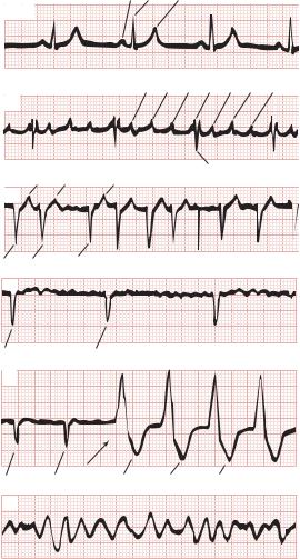
P R T
Panel 1: aVF Normal
sinus rhythm
P' P' P' R P' P' P'
Panel 2: V2 Atrial
flutter
|
T |
T |
|
T |
|
S |
|
|
|
|
|||
|
V1 |
|
|
|
|
|
Panel 3: |
S S |
|
S |
|
|
Before digitalis |
Atrial |
|
|
|
|||
|
|
|
|
|||
fibrillation |
V1 |
|
|
|
|
|
|
S |
|
S |
|
|
After digitalis |
|
|
R |
R |
R |
||
|
|
|
|
|||
Panel 4: |
V1 |
|
|
|
|
|
Ventricular |
|
|
|
|
|
|
tachycardia |
|
|
|
|
|
|
(starting at |
|
|
|
|
|
|
arrow) |
|
|
|
|
|
|
|
QS |
QS |
|
T |
T |
T |
Panel 5: |
V4 |
|
|
|
|
|
Ventricular |
|
|
|
|
|
|
fibrillation |
|
|
|
|
|
|
Electrocardiograms of normal sinus rhythm and some common arrhythmias. Major deflections (P, Q, R, S, and T) are labeled in each electrocardiographic record except in panel 5, in which electrical activity is completely disorganized and none of these deflections is recognizable.
MJ: Principles of Clinical Electrocardiography, 11th ed. McGraw-Hill, 1982. Copyright © The McGraw-Hill Companies, Inc.)
abolish reentry may do so by further reducing excitability by blocking sodium (Figure 14–4) or calcium channels, thus converting an area of unidirectional block to bidirectional block. Drugs that block repolarizing potassium currents may also be effective in converting a region of unidirectional block to bidirectional block by prolonging action potential duration, and thereby increasing the refractory period duration.
■ BASIC PHARMACOLOGY OF THE ANTIARRHYTHMIC AGENTS
Mechanisms of Action
Arrhythmias are caused by abnormal pacemaker activity or abnormal impulse propagation. Thus, the aim of therapy of the arrhythmias is to reduce ectopic pacemaker activity and modify conduction or refractoriness in reentry circuits to disable circus movement. The major pharmacologic mechanisms currently available for accomplishing these goals are (1) sodium channel blockade, (2) blockade of sympathetic autonomic effects in the heart, (3) prolongation of the effective refractory period, and
(4) calcium channel blockade.
Antiarrhythmic drugs decrease the automaticity of ectopic pacemakers more than that of the SA node. They also reduce conduction and excitability and increase the refractory period to a greater extent in depolarized tissue than in normally polarized tissue. This is accomplished chiefly by selectively blocking the sodium or calcium channels of depolarized cells (Figure 14–9). Therapeutically useful channel-blocking drugs bind readily to activated channels (ie, during phase 0) or inactivated channels (ie, during phase 2) but bind poorly or not at all to rested channels. Therefore, these drugs block electrical activity when there is a fast tachycardia (many channel activations and inactivations per unit time) or when there is significant loss of resting potential (many inactivated channels during rest). This type of drug action is often described as use-dependent or state-dependent; that is, channels that are being used frequently, or are in an inactivated state, are more susceptible to block. Channels in normal cells that become blocked by a drug during normal activation-inactivation cycles will rapidly lose the drug from the receptors during the resting portion of the cycle (Figure 14–9). Channels in myocardium that is chronically depolarized (ie, has a resting potential more positive than −75 mV) recover from block very slowly if at all (see also right panel, Figure 14–4).
In cells with abnormal automaticity, most of these drugs reduce the phase 4 slope by blocking either sodium or calcium channels, thereby reducing the ratio of sodium (or calcium) permeability to potassium permeability. As a result, the membrane potential during phase 4 stabilizes closer to the potassium equilibrium potential. In addition, some agents may increase the threshold (make it more positive). Beta-adrenoceptor-blocking drugs indirectly reduce the phase 4 slope by blocking the positive chronotropic action of norepinephrine in the heart.
In reentry arrhythmias, which depend on critically depressed conduction, most antiarrhythmic agents slow conduction further by one or both of two mechanisms: (1) steady-state reduction in
CHAPTER 14 Agents Used in Cardiac Arrhythmias |
237 |
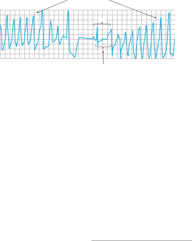
Polymorphic ventricular tachycardia
(torsade de pointes)
NSB
Prolonged QT interval
Electrocardiogram from a patient with the long QT syndrome during two episodes of torsades de pointes. The polymorphic ventricular tachycardia is seen at the start of this tracing and spontaneously halts at the middle of the panel. A single normal sinus beat (NSB) with an extremely prolonged QT interval follows, succeeded immediately by another episode of ventricular tachycardia of the torsades type. The usual symptoms include dizziness or transient loss of consciousness.
McGraw-Hill, 2007. Copyright © The McGraw-Hill Companies, Inc.)
the number of available unblocked channels, which reduces the excitatory currents to a level below that required for propagation (Figure 14–4, left); and (2) prolongation of recovery time of the channels still able to reach the rested and available state, which increases the effective refractory period (Figure 14–4, right). As a result, early extrasystoles are unable to propagate at all; later impulses propagate more slowly and are subject to bidirectional conduction block.
By these mechanisms, antiarrhythmic drugs can suppress ectopic automaticity and abnormal conduction occurring in depolarized cells—rendering them electrically silent—while minimally affecting the electrical activity in normally polarized parts of the heart. However, as dosage is increased, these agents also depress conduction in normal tissue, eventually resulting in drug-induced arrhythmias. Furthermore, a drug concentration that is therapeutic (antiarrhythmic) under the initial circumstances of treatment may become “proarrhythmic” (arrhythmogenic) during fast heart rates (more development of block), acidosis (slower recovery from block for most drugs), hyperkalemia, or ischemia.
■ SPECIFIC ANTIARRHYTHMIC AGENTS
The most widely used scheme for the classification of antiarrhythmic drug actions recognizes four classes:
1. Class 1 action is sodium channel blockade. Subclasses of this action reflect effects on the action potential duration (APD) and the kinetics of sodium channel blockade. Drugs with class 1A action prolong the APD and dissociate from the channel with intermediate kinetics; drugs with class 1B action shorten the APD in some tissues of the heart and dissociate from the channel with rapid kinetics; and drugs with class 1C action
238 |
SECTION III Cardiovascular-Renal Drugs |
Sodium current (microamps / cm2)
Unblocked |
R |
A |
I |
Blocked |
R-D |
A-D |
I-D |
0
–460
–920
–1380
–1840
–2300
0 |
1 |
2 |
3 |
4 |
5 |
Time (ms)
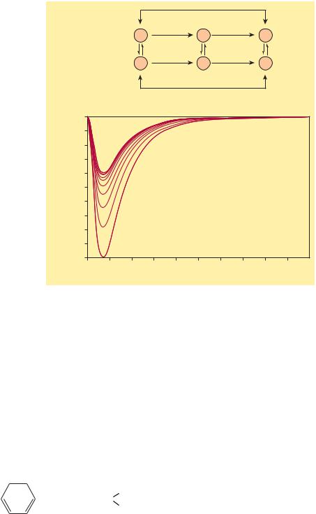
Stateand frequency-dependent block of sodium channels by antiarrhythmic drugs. Top: Diagram of a mechanism for the selective depressant action of antiarrhythmic drugs on sodium channels. The upper portion of the figure shows the population of channels moving through a cycle of activity during an action potential in the absence of drugs: R (rested) → A (activated) → I (inactivated). Recovery takes place via the I → R pathway. Antiarrhythmic drugs (D) that act by blocking sodium channels can bind to their receptors in the channels, as shown by the vertical arrows, to form drug-channel complexes, indicated as R-D, A-D, and I-D. Binding of the drugs to the receptor varies with the state of the channel. Most sodium channel blockers bind to the active and inactivated channel receptor much more strongly than to the rested channel. Furthermore, recovery from the I-D state to the R-D state is much slower than from I to R. As a result, rapid activity (more activations and inactivations) and depolarization of the resting potential (more channels in the I state) will favor blockade of the channels and selectively suppress arrhythmic
cells. Bottom: Progressive reduction of inward sodium current (downward deflections) in the presence of a lidocaine derivative. The largest curve is the initial sodium current elicited by a depolarizing voltage step; subsequent sodium current amplitudes are progressively reduced owing to prior accumulated block and block during each depolarization.
sodium channels in excitable membranes by local anesthetics. Biophys J 1984;46:15. Copyright Elsevier.)
may be somewhat less effective than quinidine (see below) in suppressing abnormal ectopic pacemaker activity but more effective in blocking sodium channels in depolarized cells.
|
|
|
|
|
|
O |
|
H |
|
C2H5 |
||||||
|
|
|
|
|
|
|
|
|
|
|
||||||
|
|
|
|
|
|
|
|
|
|
|||||||
H2N |
|
|
|
|
|
C |
|
N |
|
CH2 |
|
CH2 |
|
N |
||
|
|
|
|
|
|
|||||||||||
|
|
|
|
|
|
|
|
|
|
|
|
|
|
|
|
C2H5 |
|
|
|
|
|
Procainamide |
|
|
|||||||||
Procainamide has direct depressant actions on SA and AV nodes, and these actions are only slightly counterbalanced by drug-induced vagal block.
Extracardiac Effects
Procainamide has ganglion-blocking properties. This action reduces peripheral vascular resistance and can cause hypotension,
particularly with intravenous use. However, in therapeutic concentrations, its peripheral vascular effects are less prominent than those of quinidine. Hypotension is usually associated with excessively rapid procainamide infusion or the presence of severe underlying left ventricular dysfunction.
Toxicity
Procainamide’s cardiotoxic effects include excessive action potential prolongation, QT-interval prolongation, and induction of torsades de pointes arrhythmia and syncope. Excessive slowing of conduction can also occur. New arrhythmias can be precipitated.
A troublesome adverse effect of long-term procainamide therapy is a syndrome resembling lupus erythematosus and usually consisting of arthralgia and arthritis. In some patients, pleuritis, pericarditis, or parenchymal pulmonary disease also occurs. Renal lupus is rarely induced by procainamide. During long-term
CHAPTER 14 Agents Used in Cardiac Arrhythmias |
239 |
TABLE 14 2 Membrane actions of antiarrhythmic drugs.
|
Block of Sodium Channels |
Refractory Period |
Calcium |
Effect on |
|
||
|
|
|
|
|
|
||
|
|
|
|
|
|
||
|
Normal |
Depolarized |
Normal |
Depolarized |
Channel |
Pacemaker |
Sympatholytic |
Drug |
Cells |
Cells |
Cells |
Cells |
Blockade |
Activity |
Action |
|
|
|
|
|
|
|
|
Adenosine |
0 |
0 |
0 |
0 |
+ |
0 |
+ |
Amiodarone |
+ |
+++ |
↑↑ |
↑↑ |
+ |
↓↓ |
+ |
Diltiazem |
0 |
0 |
0 |
0 |
+++ |
↓↓ |
0 |
Disopyramide |
+ |
+++ |
↑ |
↑↑ |
+ |
↓ |
0 |
Dofetilide |
0 |
0 |
↑ |
? |
0 |
0 |
0 |
Dronedarone |
+ |
+ |
na |
na |
+ |
na |
+ |
Esmolol |
0 |
+ |
0 |
na |
0 |
↓↓ |
+++ |
Flecainide |
+ |
+++ |
0 |
↑ |
0 |
↓↓ |
0 |
Ibutilide |
0 |
0 |
↑ |
? |
0 |
0 |
0 |
Lidocaine |
0 |
+++ |
↓ |
↑↑ |
0 |
↓↓ |
0 |
Mexiletine |
0 |
+++ |
0 |
↑↑ |
0 |
↓↓ |
0 |
Procainamide |
+ |
+++ |
↑ |
↑↑↑ |
0 |
↓ |
+ |
Propafenone |
+ |
++ |
↑ |
↑↑ |
+ |
↓↓ |
+ |
Propranolol |
0 |
+ |
↓ |
↑↑ |
0 |
↓↓ |
+++ |
Quinidine |
+ |
++ |
↑ |
↑↑ |
0 |
↓↓ |
+ |
Sotalol |
0 |
0 |
↑↑ |
↑↑↑ |
0 |
↓↓ |
++ |
Verapamil |
0 |
+ |
0 |
↑ |
+++ |
↓↓ |
+ |
Vernakalant1 |
+ |
+ |
+ |
+ |
na |
0 |
na |
1Not available in the USA. na, data not available.
therapy, serologic abnormalities (eg, increased antinuclear antibody titer) occur in nearly all patients, and in the absence of symptoms, these are not an indication to stop drug therapy. Approximately one third of patients receiving long-term procainamide therapy develop these reversible lupus-related symptoms.
Other adverse effects include nausea and diarrhea (in about 10% of cases), rash, fever, hepatitis (<5%), and agranulocytosis (approximately 0.2%).
Pharmacokinetics & Dosage
Procainamide can be administered safely by intravenous and intramuscular routes and is well absorbed orally. A metabolite (N-acetylprocainamide, NAPA) has class 3 activity. Excessive accumulation of NAPA has been implicated in torsades de pointes during procainamide therapy, especially in patients with renal failure. Some individuals rapidly acetylate procainamide and develop high levels of NAPA. However, the lupus syndrome appears to be less common in these patients.
Procainamide is eliminated by hepatic metabolism to NAPA and by renal elimination. Its half-life is only 3–4 hours, which necessitates frequent dosing or use of a slow-release formulation (the usual practice). NAPA is eliminated by the kidneys. Thus, procainamide dosage must be reduced in patients with renal failure. The reduced volume of distribution and renal clearance associated with heart failure also require reduction in dosage.
The half-life of NAPA is considerably longer than that of procainamide, and it therefore accumulates more slowly. Thus, it is important to measure plasma levels of both procainamide and NAPA, especially in patients with circulatory or renal impairment.
If a rapid procainamide effect is needed, an intravenous loading dose of up to 12 mg/kg can be given at a rate of 0.3 mg/kg/ min or less rapidly. This dose is followed by a maintenance dosage of 2–5 mg/min, with careful monitoring of plasma levels. The risk of gastrointestinal (GI) or cardiac toxicity rises at plasma concentrations greater than 8 mcg/mL or NAPA concentrations greater than 20 mcg/mL.
To control ventricular arrhythmias, a total procainamide dosage of 2–5 g/d is usually required. In an occasional patient who accumulates high levels of NAPA, less frequent dosing may be possible. This is also possible in renal disease, where procainamide elimination is slowed.
Therapeutic Use
Procainamide is effective against most atrial and ventricular arrhythmias. However, many clinicians attempt to avoid long-term therapy because of the requirement for frequent dosing and the common occurrence of lupus-related effects. Procainamide is the drug of second or third choice (after lidocaine or amiodarone) in most coronary care units for the treatment of sustained ventricular arrhythmias associated with acute myocardial infarction.
240 |
SECTION III Cardiovascular-Renal Drugs |
TABLE 14 3 Clinical pharmacologic properties of antiarrhythmic drugs.
|
|
Effect on |
|
|
|
Usefulness in Arrhythmias |
||
|
|
AV Nodal |
|
|
|
|
|
|
|
|
|
|
|
Supra- |
|
|
|
|
Effect on SA |
Refractory |
|
QRS |
|
|
|
|
Drug |
Nodal Rate |
Period |
PR Interval |
Duration |
QT Interval |
ventricular |
Ventricular |
Half-Life |
|
|
|
|
|
|
|
|
|
Adenosine |
↓↑ |
↑↑↑ |
↑↑↑ |
0 |
0 |
++++ |
? |
<10 s |
Amiodarone |
↓↓1 |
↑↑ |
Variable |
↑ |
↑↑↑↑ |
+++ |
+++ |
(weeks) |
Diltiazem |
↑↓ |
↑↑ |
↑ |
0 |
0 |
+++ |
− |
4–8 h |
Disopyramide |
↑↓1,2 |
↑↓2 |
↑↓2 |
↑↑ |
↑↑ |
+ |
+++ |
7–8 h |
Dofetilide |
↓(?) |
0 |
0 |
0 |
↑↑ |
++ |
None |
7 h |
Dronedarone |
|
|
|
|
↑ |
+++ |
− |
24 h |
Esmolol |
↓↓ |
↑↑ |
↑↑ |
0 |
0 |
+ |
+ |
10 min |
Flecainide |
None,↓ |
↑ |
↑ |
↑↑↑ |
0 |
+3 |
++++ |
20 h |
Ibutilide |
↓(?) |
0 |
0 |
0 |
↑↑ |
++ |
? |
6 h |
Lidocaine |
None1 |
None |
0 |
0 |
0 |
None4 |
+++ |
1–2 h |
Mexiletine |
None1 |
None |
0 |
0 |
0 |
None |
+++ |
8–20 h |
Procainamide |
↓1 |
↑↓2 |
↑↓2 |
↑↑ |
↑↑ |
+ |
+++ |
3–4 h |
Propafenone |
0, ↓ |
↑ |
↑ |
↑↑↑ |
0 |
+ |
+++ |
5–7 h |
Propranolol |
↓↓ |
↑↑ |
↑↑ |
0 |
0 |
+ |
+ |
5 h |
Quinidine |
↑↓1,2 |
↑↓2 |
↑↓2 |
↑↑ |
↑↑ |
+ |
+++ |
6 h |
Sotalol |
↓↓ |
↑↑ |
↑↑ |
0 |
↑↑↑ |
+++ |
+++ |
7–12 h |
Verapamil |
↓↓ |
↑↑ |
↑↑ |
0 |
0 |
+++ |
− |
7 h |
Vernakalant |
|
↑ |
↑ |
|
|
+++ |
− |
2 h |
|
|
|
|
|
|
|
|
|
1May suppress diseased sinus nodes.
2Anticholinergic e ect and direct depressant action.
3Especially in Wol -Parkinson-White syndrome.
4May be e ective in atrial arrhythmias caused by digitalis.
QUINIDINE SUBGROUP 1A
Cardiac Effects
Quinidine has actions similar to those of procainamide: it slows the upstroke of the action potential, slows conduction, and prolongs the QRS duration of the ECG, by blockade of sodium channels. The drug also prolongs the action potential duration by blockade of several potassium channels. Its toxic cardiac effects include excessive QT-interval prolongation and induction of torsades de pointes arrhythmia. Toxic concentrations of quinidine also produce excessive sodium channel blockade with slowed conduction throughout the heart. It also has modest antimuscarinic actions in the heart.
OCH3
HN
N |
C |
CH CH2 |
|
|
OH
Quinidine
Extracardiac Effects
Adverse GI effects of diarrhea, nausea, and vomiting are observed in one third to one half of patients. A syndrome of headache, dizziness, and tinnitus (cinchonism) is observed at toxic drug concentrations. Idiosyncratic or immunologic reactions, including thrombocytopenia, hepatitis, angioneurotic edema, and fever, are observed rarely.
Pharmacokinetics & Therapeutic Use
Quinidine is readily absorbed from the GI tract and eliminated by hepatic metabolism. It is rarely used because of cardiac and extracardiac adverse effects and the availability of better-tolerated antiarrhythmic drugs.
DISOPYRAMIDE SUBGROUP 1A
Cardiac Effects
The effects of disopyramide are very similar to those of procainamide and quinidine. Its cardiac antimuscarinic effects are even more marked than those of quinidine. Therefore, a drug that slows

AV conduction should be administered with disopyramide when treating atrial flutter or fibrillation.
N
O
CH(CH3)2
H2N C C CH2 CH2 N
CH(CH3)2
Disopyramide
Toxicity
Toxic concentrations of disopyramide can precipitate all of the electrophysiologic disturbances described under quinidine. As a result of its negative inotropic effect, disopyramide may precipitate heart failure de novo or in patients with preexisting depression of left ventricular function. Because of this effect, disopyramide is not used as a first-line antiarrhythmic agent in the USA. It should not be used in patients with heart failure.
Disopyramide’s atropine-like activity accounts for most of its symptomatic adverse effects: urinary retention (most often, but not exclusively, in male patients with prostatic hyperplasia), dry mouth, blurred vision, constipation, and worsening of preexisting glaucoma. These effects may require discontinuation of the drug.
Pharmacokinetics & Dosage
In the USA, disopyramide is only available for oral use. The typical oral dosage of disopyramide is 150 mg three times a day, but up to 1 g/d has been used. In patients with renal impairment, dosage must be reduced. Because of the danger of precipitating heart failure, loading doses are not recommended.
Therapeutic Use
Although disopyramide has been shown to be effective in a variety of supraventricular arrhythmias, in the USA, it is approved only for the treatment of ventricular arrhythmias.
LIDOCAINE SUBGROUP 1B
Lidocaine has a low incidence of toxicity and a high degree of effectiveness in arrhythmias associated with acute myocardial infarction. It is used only by the intravenous route.
CH3 |
|
|
||||||||||
|
|
|
|
O |
|
|
||||||
|
|
H |
|
|
|
|
|
|
|
C2H5 |
||
|
|
|||||||||||
|
|
N |
|
C |
|
CH2 |
|
N |
||||
|
|
|
|
|
||||||||
|
|
|
|
|
|
|
|
|
|
|
|
C2H5 |
CH3 |
|
|
||||||||||
|
|
Lidocaine |
|
|
||||||||
CHAPTER 14 Agents Used in Cardiac Arrhythmias |
241 |
Cardiac Effects
Lidocaine blocks activated and inactivated sodium channels with rapid kinetics (Figure 14–10); the inactivated state block ensures greater effects on cells with long action potentials such as Purkinje and ventricular cells, compared with atrial cells. The rapid kinetics at normal resting potentials result in recovery from block between action potentials and no effect on conduction. In depolarized cells, the increased inactivation and slower unbinding kinetics result in the selective depression of conduction. Little effect is seen on the ECG in normal sinus rhythm.
Toxicity
Lidocaine is one of the least cardiotoxic of the currently used sodium channel blockers. Proarrhythmic effects, including SA node arrest, worsening of impaired conduction, and ventricular arrhythmias, are uncommon with lidocaine use. In large doses, especially in patients with preexisting heart failure, lidocaine may cause hypotension—partly by depressing myocardial contractility.
(mV) |
|
|
|
|
potential |
0 |
|
|
|
|
|
|
|
|
Membrane |
–85 |
–80 |
–75 |
–70 |
|
||||
|
|
|||
|
–100 |
|
|
|
(%) |
100 |
|
|
|
blocked |
|
|
|
|
|
|
|
|
|
Channels |
0 |
|
|
|
|
800 |
|
|
|
|
|
Time (ms) |
|
|
FIGURE 14 10 Computer simulation of the effect of resting membrane potential on the blocking and unblocking of sodium channels by lidocaine as the membrane depolarizes. Upper tracing: Action potentials in a ventricular muscle cell. Lower tracing: Percentage of channels blocked by the drug. An 800-ms time segment is shown. Extra passage of time is indicated by breaks in the traces. Left side: At the normal resting potential of −85 mV, the drug combines with open (activated) and inactivated channels during each action potential, but block is rapidly reversed during diastole because the affinity of the drug for its receptor is so low when the channel recovers to the resting state at −85 mV. Middle: Metabolic injury
is simulated, eg, ischemia due to coronary occlusion, that causes gradual depolarization over time. With subsequent action potentials arising from more depolarized potentials, the fraction of channels blocked increases because more channels remain in the inactivated state at less negative potentials (Figure 14–4, left), and the time constant for unblocking during diastole rapidly increases at less negative resting potentials (Figure 14–4, right). Right: Because of marked drug binding, conduction block and loss of excitability in this tissue result; that is, the “sick”(depolarized) tissue is selectively suppressed.
242 |
SECTION III Cardiovascular-Renal Drugs |
Lidocaine’s most common adverse effects—like those of other local anesthetics—are neurologic: paresthesias, tremor, nausea of central origin, lightheadedness, hearing disturbances, slurred speech, and convulsions. These occur most commonly in elderly or otherwise vulnerable patients or when a bolus of the drug is given too rapidly. The effects are dose-related and usually short-lived; seizures respond to intravenous diazepam. In general, if plasma levels above 9 mcg/mL are avoided, lidocaine is well tolerated.
Pharmacokinetics & Dosage
Because of its extensive first-pass hepatic metabolism, only 3% of orally administered lidocaine appears in the plasma. Thus, lidocaine must be given parenterally. Lidocaine has a half-life of 1–2 hours. In adults, a loading dose of 150–200 mg administered over about 15 minutes (as a single infusion or as a series of slow boluses) should be followed by a maintenance infusion of 2–4 mg/min to achieve a therapeutic plasma level of 2–6 mcg/mL. Determination of lidocaine plasma levels is of great value in adjusting the infusion rate. Occasional patients with myocardial infarction or other acute illness require (and tolerate) higher concentrations. This may be due to increased plasma α1-acid glycoprotein, an acute-phase reactant protein that binds lidocaine, making less free drug available to exert its pharmacologic effects.
In patients with heart failure, lidocaine’s volume of distribution and total body clearance may both be decreased. Therefore, both loading and maintenance doses should be decreased. Since these effects counterbalance each other, the half-life may not be increased as much as predicted from clearance changes alone. In patients with liver disease, plasma clearance is markedly reduced and the volume of distribution is often increased; the elimination half-life in such cases may be increased threefold or more. In liver disease, the maintenance dose should be decreased, but usual loading doses can be given. Elimination half-life determines the time to steady state. Although steady-state concentrations may be achieved in 8–10 hours in normal patients and patients with heart failure, 24–36 hours may be required in those with liver disease. Drugs that decrease liver blood flow (eg, propranolol, cimetidine) reduce lidocaine clearance and so increase the risk of toxicity unless infusion rates are decreased. With infusions lasting more than 24 hours, clearance falls and plasma concentrations rise. Renal disease has no major effect on lidocaine disposition.
Therapeutic Use
Lidocaine is the agent of choice for termination of ventricular tachycardia and prevention of ventricular fibrillation after cardioversion in the setting of acute ischemia. However, routine prophylactic use of lidocaine in this setting may actually increase total mortality, possibly by increasing the incidence of asystole, and is not the standard of care. Most physicians administer IV lidocaine only to patients with arrhythmias.
MEXILETINE SUBGROUP 1B
Mexiletine is an orally active congener of lidocaine. Its electrophysiologic and antiarrhythmic actions are similar to those of lidocaine. (The anticonvulsant phenytoin [see Chapter 24] exerts
similar electrophysiologic effects and has been used as an antiarrhythmic.) Mexiletine is used in the treatment of ventricular arrhythmias. The elimination half-life is 8–20 hours and permits administration two or three times per day. The usual daily dosage of mexiletine is 600–1200 mg/d. Dose-related adverse effects are seen frequently at therapeutic dosage. These are predominantly neurologic, including tremor, blurred vision, and lethargy. Nausea is also a common effect.
CH3
O CH2 CH CH3
NH2
CH3
Mexiletine
Mexiletine has also shown significant efficacy in relieving chronic pain, especially pain due to diabetic neuropathy and nerve injury.The usual dosage is 450–750 mg/d orally. This application is off label.
FLECAINIDE SUBGROUP 1C
Flecainide is a potent blocker of sodium and potassium channels with slow unblocking kinetics. (Note that although it does block certain potassium channels, it does not prolong the action potential or the QT interval.) It is currently used for patients with otherwise normal hearts who have supraventricular arrhythmias. It has no antimuscarinic effects.
O CH2 CF3
O N

 C NH CH2
O CH2 CF3
Flecainide
Flecainide is very effective in suppressing premature ventricular contractions. However, it may cause severe exacerbation of arrhythmia even when normal doses are administered to patients with preexisting ventricular tachyarrhythmias and those with a previous myocardial infarction and ventricular ectopy. This was dramatically demonstrated in the Cardiac Arrhythmia Suppression Trial (CAST), which was terminated prematurely because of a two and one-half-fold increase in mortality rate in the patients receiving flecainide and similar group 1C drugs. Flecainide is well absorbed and has a half-life of approximately 20 hours. Elimination is both by hepatic metabolism and by the kidney. The usual dosage of flecainide is 100–200 mg twice a day.
PROPAFENONE SUBGROUP 1C
Propafenone has some structural similarities to propranolol and possesses weak β-blocking activity. Its spectrum of action is very similar to that of quinidine, but it does not prolong the action potential. Its sodium channel-blocking kinetics are similar