

ЧПНЛТ Атлас
.pdfsurgery illustrated KNOLL
et al.
|
1 |
2 |
3 |
4 |
5 |
6 |
7 |
8 |
|
|
|
|
|
|
|
|
|
|
|
|
|
|
|
|
|
|
|
|
|
a |
b |
a |
b |
a |
b |
c
b
a
c
Surgical Atlas
Percutaneous nephrolithotomy: the Mannheim technique
Thomas Knoll, Maurice S. Michel and Peter Alken
Department of Urology, University Hospital Mannheim, Mannheim, Germany
ILLUSTRATIONS by STEPHAN SPITZER, www.spitzer-illustration.com
INTRODUCTION
Proper establishment of the percutaneous tract is the most important step in percutaneous stone removal. If appropriately done the efficacy is optimal, with low complication rates. Depending on the availability of fluoroscopic facilities, in different countries the tract is established either by radiologists or urologists; this is still under debate in reports in the American literature [1–5]. Recently, Watterson et al. [5] compared renal access acquired by urologist vs radiologist and showed a higher stone-free rate and fewer complications by the former. This confirms our data indicating that the urologist should obtain renal access to achieve the optimum renal access for later stone removal [6].
To establish proper access to the collecting system (CS), as much information as possible should be obtained about the stone and the patient’s anatomy. For this, the combination of fluoroscopy and ultrasonography (US) is optimal. Opacification of the renal CS by i.v. injection with dye or by a retrograde ureteric catheter renders the CS visible and helps to identify its distribution accurately. US will visualize all other structures, e.g. the renal parenchyma, stones of all compositions and all surrounding organs like spleen, intestine, liver, pleura or ribs and, most importantly, simultaneously the needle during puncture. The advantage of this combined visualization is to obtain real-time pseudo-three-
dimensional information while establishing the percutaneous access [6].
PLANNING AND PREPARATION
Percutaneous nephrolithotomy (PNL) is recommended by the Guidelines of the European Association of Urology for the following indications [7]:
•Large stone burden >2 cm or 1.5 cm for lower calyceal stones.
•Staghorn stones.
•Stones that are difficult to disintegrate by ESWL (calcium-oxalate monohydrate, brushite, cystine).
•Stones refractory to ESWL or ureteroscopy.
•Urinary tract obstructions that need simultaneous correction (e.g. PUJ obstruction).
•Malformations with reduced probability of fragment passage after ESWL (e.g. horseshoe or dystopic kidneys, calyceal diverticula.
•Obesity
All patients need preoperative imaging including a plain abdominal film and IVU. If the use of contrast agents is not possible, the CS can by opacified via a retrograde ureter catheter during the intervention. CT-guided access has been described for difficult cases but was never necessary in our experience [8]. Thorough US before intervention, to evaluate if the access to the stone can be established with no problems, is essential for planning of the procedure. Ideally, the urologist who will do the PNL should also do this.
© 2 0 0 7 T H E A U T H O R S |
2 1 3 |
J O U R N A L C O M P I L A T I O N © 2 0 0 7 B J U I N T E R N A T I O N A L | 9 9 , 2 1 3 – 2 3 1 | doi:10.1111/j.1464-410X.2007.06613.x |
K N O L L E T A L .
|
1 |
2 |
3 |
4 |
|
5 |
6 |
7 |
8 |
|
Figure 1 |
|
|
|
|
|
|
||||||||
|
|
|
|
|
|
|
|
|
|
|
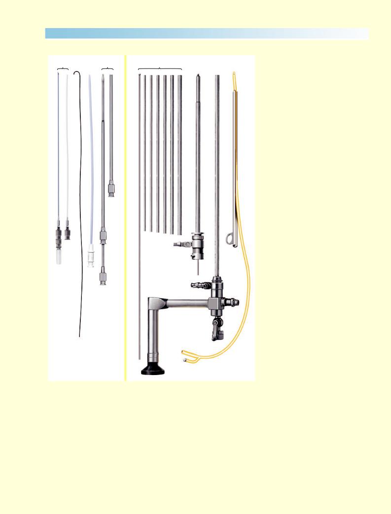
Equipment for PNL includes: |
|
|
|
|
|
|
|
|
|
|
|
|
• |
Fluoroscopy unit (Fig. 2a). |
|
|
|
|
|
|
|
|
|
|
|
• |
Video-endoscopy. |
|
|
|
|
|
|
|
|
|
|
|
• |
US scanner with needle-guide adapter, |
|
|
|
|
|
|
|
|
|
|
|
• |
Balloon occlusion catheter 5 F (i.e. |
|
|
|
|
|
|
|
|
|
|
|
#340–80, Rüsch, Germany). |
|
|
|
|
|
|
|
|
|
|
|
|
• |
Foley catheter 18 F for fixing the occlusion |
|
|
|
|
|
|
|
|
|
|
|
catheter. |
|
|
|
|
|
|
|
|
|
|
|
|
• |
Sterile coverings. |
|
|
|
|
|
|
|
|
|
|
|
• |
Puncture needle 9 F (Fig. 1.1). |
|
|
|
|
|
|
|
|
|
|
|
• |
Contrast dye, |
|
|
|
|
|
|
|
|
|
|
|
• A set of guidewires (floppy tip J-guidewires, |
|
|
|
|
|
|
|
|
|
|
|
|
glide wires, Lunderquist wires). |
|
|
|
|
|
|
|
|
|
|
|
|
• |
Metallic dilator cannula 9 F with metal |
|
|
|
|
|
|
|
|
|
|
|
sheath 11 F (Karl Storz Endoscopes, Germany, |
|
|
|
|
|
|
|
|
|
|
|
|
Fig. 1.4). |
|
|
|
|
|
|
|
|
|
|
|
|
• |
Metal telescope dilators with hollow guide |
|
|
|
|
|
|
|
|
|
|
|
rod (9–24 F, Karl Storz; Fig. 1.5). |
|
|
|
|
|
|
|
|
|
|
|
|
• |
Rigid nephroscopes 18 F and 26 F |
|
|
|
|
|
|
|
|
|
|
|
(6° telescope, Karl Storz; Fig. 1.6, 1.7). |
|
|
|
|
|
|
|
|
|
|
|
|
• |
Sodium chloride irrigation fluid (0.9%). |
|
|
|
|
|
|
|
|
|
|
|
• |
Ultrasound lithotripsy equipment with 9 F |
|
|
|
|
|
|
|
|
|
|
|
probe (Karl Storz; Fig. 1.4a–c). |
|
|
|
|
|
|
|
|
|
|
|
|
• |
A set of stone forceps (9 F) and baskets. |
|
|
|
|
|
|
|
|
|
|
|
• |
Flexible scopes 8 F and 15 F (Karl Storz). |
|
|
|
|
|
|
|
|
|
|
|
• |
Ho:YAG laser and 220/365 m laser fibres |
|
|
|
|
|
|
|
|
|
|
|
(i.e. Karl Storz). |
|
|
|
|
|
|
|
|
|
|
|
|
• |
Half-open slotted cannula 22 F (Karl Storz; |
|
|
|
|
|
|
|
|
|
|
|
Fig. 1.8). |
|
|
|
|
|
|
|
|
|
|
|
|
• |
Foley catheter 22 F (nephrostomy). |
|
|
|
|
|
|
|
|
|
|
|
• |
Suture material. |
|
|
|
|
|
|
|
|
|
|
|
Specific preparation of the patient before |
|
|
|
|
|
|
|
|
|
|
|
|
surgery is not necessary and depends on |
|
|
|
|
|
|
|
|
|
|
|
|
anaesthetic demands. Usually the patient |
|
|
|
|
|
|
|
|
|
|
|
|
is under general anaesthesia for PNL. |
|
|
|
|
|
|
|
|
|
|
|
|
Perioperative antibiotics are administered |
|
|
|
|
|
|
|
|
|
|
|
|
as a precaution (i.e. ciprofloxacin or |
|
|
|
|
|
|
|
|
|
|
|
|
cotrimoxazole). UTI should be excluded by |
|
|
|
|
|
|
|
|
|
|
|
|
dipstick analysis and urine culture before |
|
|
|
|
|
|
|
|
|
|
|
|
surgery if necessary. If there is active infection |
|
|
|
|
|
|
|
|
|
|
|
|
antibiotics should be adapted to specific |
|
|
|
|
|
|
|
|
|
|
|
|
testing and started some days beforehand. |
|
|
|
|
|
|
|
|
|
|
|
|
||
|
|
|
|
|
|
|
|
|
|
|
However, sterile urine can often not be |
|
|
|
|
|
|
|
|
|
|
|
|
obtained, especially in patients with |
|
|
|
|
|
|
|
|
|
|
|
|
infection-associated stones. |
|
2 1 4 |
© 2 0 0 7 T H E A U T H O R S |
J O U R N A L C O M P I L A T I O N © 2 0 0 7 B J U I N T E R N A T I O N A L |
S U R G E R Y I L L U S T R A T E D
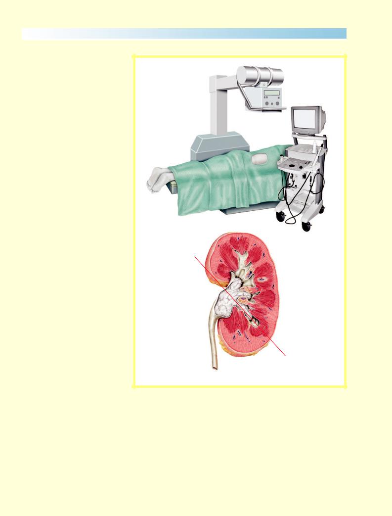
Figure 2
Fig. 2a: Operating room setting with the patient placed on the fluoroscopy desk, monitors for fluoroscopy, US and instruments. If the renal CS is unobstructed, a balloon occlusion catheter is initially placed, with the patient in the lithotomy position to facilitate the puncture. The balloon has to be blocked just below the PUJ. A Foley catheter is then placed and fixed together with the occlusion catheter with tape. The patient is placed prone on an X-ray table with all pressure points padded. The CS is opacified and slightly dilated by retrograde dye instillation. Fig. 2b: The ideal approach to the stone is achieved by a transpapillary puncture with straight access to the renal pelvis.
a
b
© 2 0 0 7 T H E A U T H O R S |
2 1 5 |
J O U R N A L C O M P I L A T I O N © 2 0 0 7 B J U I N T E R N A T I O N A L |
K N O L L E T A L .
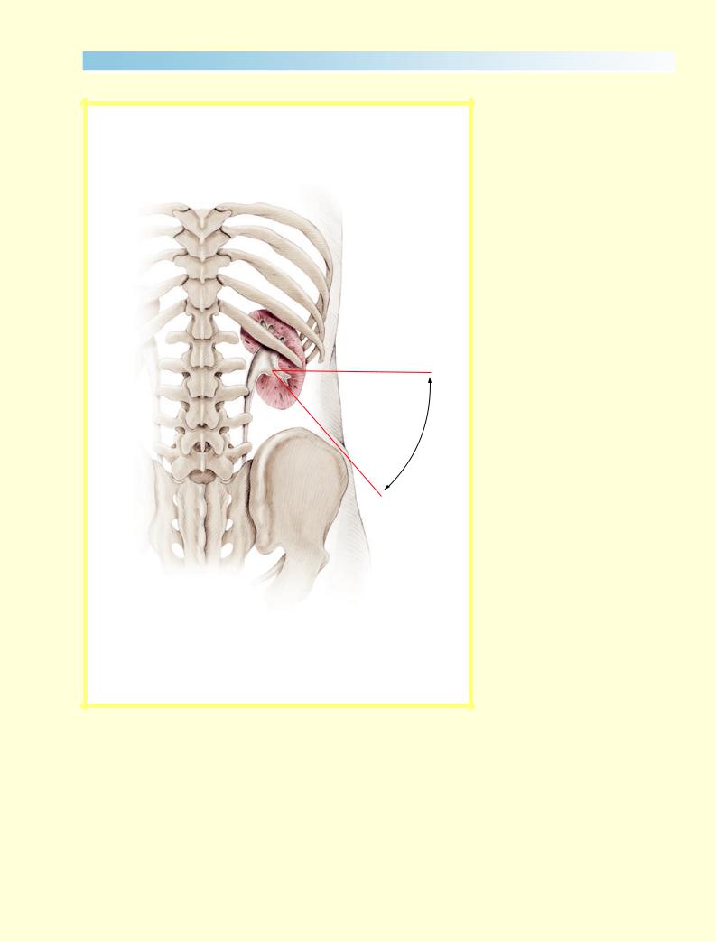
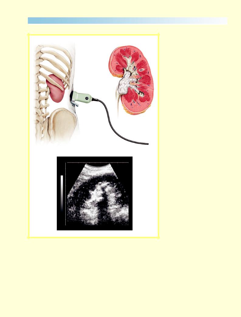
Figure 3
Area of access: The first step in preoperative planning of the procedure is to identify the ideal target calyx and to obtain threedimensional knowledge of the kidney and the stone. It is essential to understand the anatomical location of the kidney. It is located anterior to the psoas muscle, between the 12th thoracic vertebral body and second/third lumbar vertebral body. Both kidneys are within the retroperitoneum at ≈30° posterior to the frontal plane of the body. Access to the kidney is always established individually according to the particular anatomy. Frequently, the ribs or the iliac crest limit the space for access. In these cases, the area has to be shifted a few degrees (caudally or cranially 10–20°).
2 1 6 |
© 2 0 0 7 T H E A U T H O R S |
J O U R N A L C O M P I L A T I O N © 2 0 0 7 B J U I N T E R N A T I O N A L |
S U R G E R Y I L L U S T R A T E D
Figure 4
Determination of the target calyx and puncture plane, puncture site and puncture direction: (i) The long axis of the target calyx is identified by fluoroscopy (Fig. 4a); (ii) the plane between the calyceal axis and its perpendicular projection on the patients back is the ideal puncture plane (Fig. 4b); indicated by blue line in corresponding illustrations.
a
b
© 2 0 0 7 T H E A U T H O R S |
2 1 7 |
J O U R N A L C O M P I L A T I O N © 2 0 0 7 B J U I N T E R N A T I O N A L |
K N O L L E T A L .
Figure 5
US of the kidney.
a |
b |
c
2 1 8 |
© 2 0 0 7 T H E A U T H O R S |
J O U R N A L C O M P I L A T I O N © 2 0 0 7 B J U I N T E R N A T I O N A L |

Figure 6
Determination of the puncture site and direction; within the puncture plane the puncture site is determined so that the puncture direction from that site is as close as possible identical to the extension of the long axis of the target calyx to the skin. This is achieved by moving the scanner head laterally on the predefined puncture plane while keeping the scanning plane within the predefined puncture plane. The image will change until the access calyx points directly towards the scanner head. An electronically generated line marks the puncture path from the needle guidance adapter to the stone. Puncture from this site will guarantee optimal access to the stone. At this moment the direction and depth to reach the CS can be determined. This is the least traumatic access because it establishes a direct and nearly avascular straight path through the parenchyma and the calyx to the renal pelvis.
S U R G E R Y I L L U S T R A T E D
b
a
c
© 2 0 0 7 T H E A U T H O R S |
2 1 9 |
J O U R N A L C O M P I L A T I O N © 2 0 0 7 B J U I N T E R N A T I O N A L |

K N O L L E T A L .
1 |
|
1 |
|
1 |
|
1 |
2 |
3 |
2 |
2 |
3 |
5 |
2 |
|
4 |
|
|
4 |
|
|
|
|
|
|
|
|
a |
|
|
b |
|
1 |
|
1 |
1 |
1 |
|
|
5 |
2 |
2 |
|
2 |
2 |
5 |
|
|
|
|
||
|
|
3 |
|
|
|
|
|
|
c |
d |
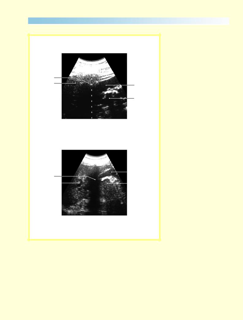
Figure 7
How US helps to avoid mistakes [9]; US allows easy identification of the surrounding organs (lung, liver, spleen, intestine or liver) that are not visible in fluoroscopy.
(a)The US image of a stone-bearing kidney viewed from the 10th intercostal space; (1) surface 10th and 11th rib; (2) back-shadow of the ribs; (3) kidney parenchyma; (4) bright echo of stone with back-shadow.
(b)During inspiration diffuse reflections (5) caused by air in the lung obscures the view of the kidney, which moves caudally.
(c, d) During deep inspiration the intercostal space is completely filled by the lung, indicating that this access should be avoided, as it will lead into the lung or thoracic cavity.
2 2 0 |
© 2 0 0 7 T H E A U T H O R S |
J O U R N A L C O M P I L A T I O N © 2 0 0 7 B J U I N T E R N A T I O N A L |
S U R G E R Y I L L U S T R A T E D
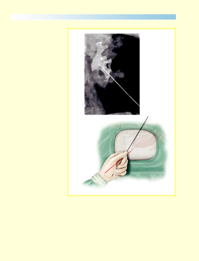
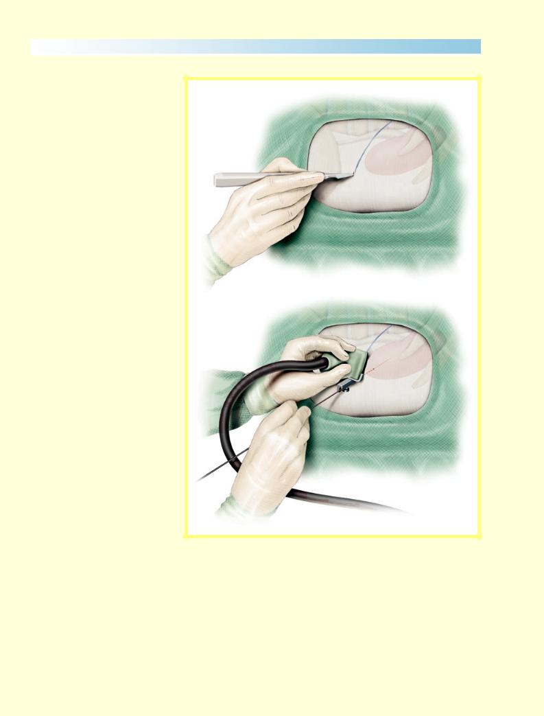
Figure 8
Puncture: A small incision is made at the selected puncture site after the scanner is temporally removed. Two types of puncture are possible, either: (A) puncture with needle guidance; or (B) freehand puncture.
Using the needle guide adapter might be advantageous for the beginner, but frequently the needle will be deflected from the predefined path by different tissue consistencies. Freehand puncture allows easier detection and correction of the needle’s direction. If a correction of the needle direction is necessary it must be moved outside the kidney, sometimes even out of the skin. Immediately before and during the puncture, needle advancement should be monitored by short intermittent fluoroscopy pulses to assess any deflection of the needle and to make sure the target calyx is indeed scanned. To reduce X-ray exposure of the physician, field reduction of the fluoroscopy should be used. The needle can be followed by US until it reaches the calyx. The puncture is usually made with a hollow open needle (with no obturator). When the CS is accessed, clear urine will flow out of the open needle.
In those cases where the stone completely moulds the target calyx, no urine will flow through the needle. Minimal amounts of dye must be injected to assure the optimum position of the needle tip. All further steps are then done under fluoroscopic control.
Once correct access to the CS is ascertained by dye injection, a floppy-tip J-guidewire is passed into the CS. With the aforementioned precautions followed, the guidewire will usually slip straight forward into the renal pelvis.
The access calyx leads to the renal pelvis like the urethra to the bladder.
a
b
© 2 0 0 7 T H E A U T H O R S |
2 2 1 |
J O U R N A L C O M P I L A T I O N © 2 0 0 7 B J U I N T E R N A T I O N A L |
K N O L L E T A L .
Figure 9
(a) US image of the colon filled with gas (3) close to the lower pole of the kidney (1) containing a stone (2).
(b) US image of upper renal pole close to the spleen (4), back-shadow of the 11th rib (3), renal parenchyma (1) stone (2).
3
3
1
2
a
3 |
|
|
|
|
1 |
|
|
|
|
||||
|
|
|
|
|
|
|
4 |
|
|
|
|
2 |
|
|
|
|
|
|||
b
2 2 2 |
© 2 0 0 7 T H E A U T H O R S |
J O U R N A L C O M P I L A T I O N © 2 0 0 7 B J U I N T E R N A T I O N A L |