

4 курс / Лучевая диагностика / ЛУЧЕВАЯ_ДИАГНОСТИКА_И_ЛУЧЕВАЯ_ТЕРАПИЯ
.pdfCompensatory hypertrophy
Unilateral smooth, large kidney that is normal in all respects except for its size and the thickness of the renal parenchyma. The pelvocalyceal system and ureter may appear distended (high urinary flow rate). Response to congenital absence, surgical removal, or disease of the contralateral kidney.
Chronic pyelonephritis
Clubbed, dilated calyces are caused by retraction of papillae and most frequently involve the poles. Depressed cortical scars typically develop over involved calyces.
Renal cyst
This common benign entity is found in approximately 30 % of all elderly patients. It often occurs bilaterally and can be associated with hepatic cysts. Simple renal cysts have a thin, smooth margin, have no internal echo on US, and are of water density on CT (less than 20 Hounsfield Units). They are predominantly found in the renal cortex, but can also protrude into the renal hilum, in which case they have to be distinguished from a dilated renal pelvis (figure 5.25–5.27).
Parapelvic renal cysts located in the renal sinus fat can also create the impression of hydronephrosis but are easily discernible from the actual renal pelvis on the excretory phase of a contrast-enhanced CT scan.
A  B
B 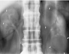
C 
Figure 5.25 (A) Chronic atrophic pyelonephritis. Focal reduction in parenchymal thickness involving the upper pole of the right kidney.
(B)Renal cysts. Nephrotomogram demonstrates bilateral renal cysts (arrows).
(C)Benign renal cyst. Nonenhancing left renal mass (c) with a sharply marginated
border and a thin wall
61

Simple renal cyst
Focal contour expansion of the kidney outline on the nephrogram. The cortical margin appears as a very thin, smooth radiopaque rim about the bulging lucent cyst (beak sign). A thickened wall suggests bleeding into a cyst, cyst infection, or a malignant lesion. When a simple cyst is completely embedded in the kidney parenchyma, the thin rim and beak are absent, and the renal size and contour are normal. A renal cyst causes focal displacement of adjacent portions of the pelvocalyceal system, with the collecting structures remaining smooth and attenuated rather than shaggy and obliterated as with a malignant neoplasm.
A  B
B 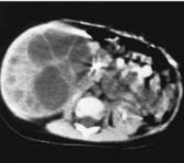 C
C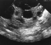
Figure 5.26 Segmental multicystic dysplastic kidney involving only the medial portion of the right kidney. (A) Excretory urogram shows a large multiloculated renal mass displacing the opacified collecting system (arrowhead) over the spine.
(B)Enhanced CT scan confirms the intrarenal multiloculated mass.
(C)Multicystic dysplastic kidney. Sagittal sonogram demonstrates the cystic kidney. Note the absence of communication between the cystic structures
A 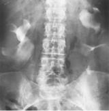 B
B 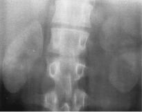 C
C 
Figure 5.27 (A) Adult polycystic kidney disease. Excretory urogram shows marked multifocal enlargement of both kidneys, focal displacement of the collecting structures, and normal opacification.
(B) Chronic glomerulonephritis. Plain film tomogram shows bilateral small, smooth kidneys with diffuse fine calcification in the renal parenchyma.
(C) Renal hamartoma. Arteriography shows the mass to be hypervascular
Acute glomerulonephritides
Bilateral large kidneys (may be of normal size) with global parenchymal thickening and smooth contours. The nephrogram is homogeneously faint or normal. The pelvocalyceal system is normal, though opacification is often faint (ultrasound is the most efficacious test to show that the calyces are not dilated and thus not obstructed). If the disease progresses to a chronic stage (especially in the poststreptococcal type), the kidneys become bilaterally small with smooth contours.
62
Renal hamartoma
A combined excretory urogram and inferior vena cavagram shows a large mass in the lower pole of the right kidney with displacement but no invasion of the pelvocalyceal system and inferior vena cava. Arteriography shows the mass to be hypervascular (the radiographic appearance is indistinguishable from that of renal cell carcinoma) (figure 5.27).
Renal cell carcinoma
Eightypercentofallsolidrenaltumorsarerenalcellcarcinomas(figure5.28–5.29).
A B
B
C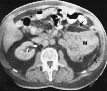 D
D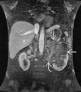
Figure 5.28 (A) Renal cell carcinoma. Upward displacement of the right kidney and distortion of the collecting system by the large lower pole mass.
(B) In another patient, left renal arteriogram demonstrates a large hypervascular mass with striking enlargement of capsular vessels.
(C) Renal cell carcinoma. Large mass (M) of the left kidney with thickening of Gerota's fascia (arrows).
(D) Coronal T1 MR scan shows a large infiltrative and exophytic mass that invades the renal sinus (arrows), filling both the renal pelvis and the renal vein
They are predominantly found in the elderly. Any solid lesion in the kidney represents a malignant neoplasm until proven otherwise, unless there are definite benign features such as internal fat (figure 5.29). Suspicion of renal cell carcinoma always warrants surgery, as tissue biopsy is contraindicated. Renal cell carcinoma is often very vascular and can contain areas of calcification and necrosis; occasionally it can be predominantly cystic. There is a tendency for vascular invasion by the tumor, growing into renal veins, and sometimes also extending into the inferior vena cava, even up to the right atrium.
63

A  B
B 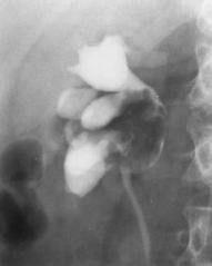
C 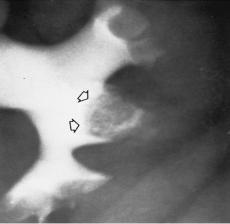
D 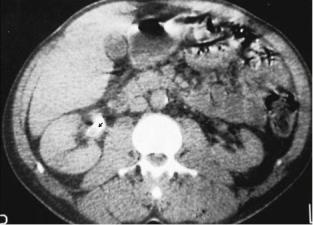 E
E 
Figure 5.29 Transitional cell carcinoma of the renal pelvis in different patients.
(A) A small filling defect (arrow) in the renal pelvis simulates a blood clot, stone, fungus ball, or sloughed papilla.
(B)A huge mass fills virtually all the renal pelvis.
(C)A small filling defect occupies an interpolar calyx (arrows). Although the defect might at first be mistaken for a large but otherwise normal papilla, the many small contrast stipples and the suggestively irregular border make its neoplastic nature evident.
(D)Transitional cell carcinoma. Filling defect (arrow) in the opacified renal pelvis.
(E)Transitional cell carcinoma of the midureter. Irregular stricture with proximal
ureteral and pelvocalyceal dilatation
64
Cystitis (figure 5.30)
A 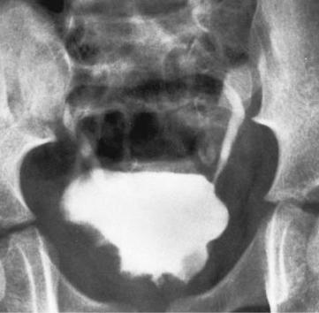 B
B 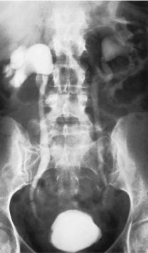
C D
D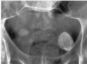
Figure 5.30 (A) Cystitis. Irregular, lobulated filling defects (representing intense mucosal edema) at the base of the bladder.
(B) Radiation cystitis causing ureteral obstruction. After radiotherapy for cervical cancer, an excretory urogram shows the bladder wall to be thickened and bladder opacity to be reduced. Narrowing of the distal ureters causes bilateral hydronephrosis. Bladder calculi. (C) Excretory urogram demonstrates a large stone (arrows)
in a left-sided bladder diverticulum. (D) Plain radiograph of the pelvis shows
the laminated stone and multiple smaller calculi that were obscured by contrast material in the right-sided bladder diverticula on the contrast-filled view
Unilateral or bilateral obstruction of the distal ureters. In acute cystitis, there is compression of the intramural portion of the ureters by edema and inflammation. In chronic cystitis, the ureterovesical junction is obstructed by fibrosis or an inflammatory mass (figure 5.30).
65

A B
B 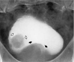
C D
D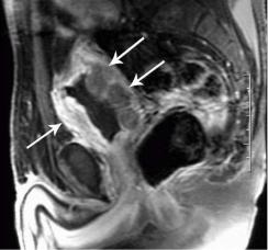 Figure 5.31 Transitional cell carcinoma.
(A)Large, irregular filling defect (arrows) in the bladder.
(B)In another patient, the irregular tumor (open arrows) is associated with a large filling defect(closedarrows),representingbenignprostatichypertrophy,atthebaseofthebladder.
(C)Squamous cell carcinoma. CT cystogram shows an irregular mass involving the lateral wall of the bladder. Note the loss of trabecular structure in the bones and fatty infiltration of the muscles in this paraplegic patient.
(D)Squamous cell carcinoma. Sagittal enhanced and fat-suppressed T1-weighted MR image shows thickening of the anterior and posterior walls of the bladder (arrows),
which represented chronic inflammatory changes with diffuse invasive malignancy.
Urodynamic studies provide accurate and objective information on the pathophysiology of the lower urinary tract in patients with symptoms suggesting dysfunction of the bladder and/or urethra. The basis of urodynamics is the recording of pressure within the bladder (the cystometrogram) or urethra (the urethral profile) and the flow of urine (during voiding (the flow rate). The relationship between pressure and flow has been studied for 50 years but refinements in techniques and advances in technology, such as the addition of ultrasound and real-time imaging, have made urodynamics an accurate science. Urodynamic techniques included: cystodynamogram, intravenous urodynamogram, videocystometrography.
66
Urethra, imaging methods (figure 5.32)
a |
b |
c |
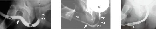
Figure 5.32 Normal male urethra. (a) Retrograde urethrogram (RUG).
(b) Voiding cystourethrogram (VCUG). The penile urethra (PU) extends from the urethral meatus to the suspensory ligament of the penis (straight arrows) at the penoscrotal junction. The bulbous urethra (BU) extends from the penoscrotal junction to the urogenital diaphragm (curvedarrows),markedbythetipoftheconeontheRUGandtheslightnarrowingofurethral caliber on the VCUG. The membranous urethra (curved arrows) is only 1 cm in length and is entirely within the muscle of the urogenital diaphragm. On a RUG, the membranous urethra extendsbetweenthetipoftheconeandtheverumontanum.Theverumontanum(arrowheads) is a nodular structure that produces a filling defect on the urethrograms by bulging into the prostaticurethra.Theprostaticurethraextendsfromtheinferioraspectoftheverumontanum
to the base of the bladder (B).
(c) The stones (arrowhead) lie in a segment of an anterior urethral stricture
The urethra is studied by retrograde and voiding urethrography. The retrograde urethrogram is a simple study of the anterior male urethra. Contrast medium is injected into the anterior urethra by means of a syringe or catheter that occludes the meatal orifice. Films are exposed in the right posterior oblique projection.
Voiding cystourethrography is performed by filling the bladder with contrast via a catheter. The catheter is removed, and films are obtained while the patient urinates into a basin on the fluoroscopy table.
Imaging methods of characteristic
The choices for imaging of the pelvicaliceal system and ureters have expanded. An excretory urogram , also called an intravenous pyelogram (IVP), has been the traditional choice. This study is performed by obtaining a series of radiographs at various times and in various projections following IV administration of contrast agent. The images of the collecting system are routinely of high quality and remain the gold standard. However, detection of lesions in the renal parenchyma is limited. With the development of multidetector CT (MDCT) and continued improvement in MR, we now have available the CT-IVP and the MR-IVP, which combine optimal imaging of the renal parenchyma with satisfactory images of the collecting system and ureters. MR urography can be performed without the use of IV contrast utilizing T2 weighting to provide visualization of urine-filled structures. Poor renal function or high-grade obstruction limits the use of IV contrast agents because of poor concentration of contrast within the collecting system. When a percutaneous nephrostomy catheter has been placed in the collecting system, antegrade pyelography is an additional choice. US is the imaging method of choice for screening for hydronephrosis but is limited in its ability to demonstrate small uroepithelial tumors. CT, routinely performed without IV or oral contrast agents, has supplanted plain radiographs and IVP in the diagnosis of renal stones in the kidneys and ureters.
Scintigraphy has provided a unique tool for the noninvasive evaluation of renal pathophysiology, and the past two decades have witnessed a rapid increase in the scope and number of radionuclide renal studies.
67

6. Radiation therapy
Introduction
Millions cancer patients receive radiation therapy each year, either alone or in conjunction with surgery, chemotherapy or other forms of cancer therapy. Other terms for radiation therapy include radiotherapy or irradiation. Radiation therapy is useful in cases where surgical removal of the cancer is not possible or when surgery might debilitate the patient (for example, when tumors that are located close to the spinal cord). Together with image guided treatment planning, radiation therapy is a powerful tool in the treatment of cancer, particularly when the cancer is detected at an early stage.
Radiation treatment (radiology therapy) principles and methods. Equipment, types used ionizing irradiations
Radiotherapy or radiation treatment is the use of x-rays, electrons or gamma rays to treat cancer. Radiation can cure or control cancer by inhibiting the cancer cells from dividing or reproducing. About fifty or sixty percent of patients with cancer will require radiation at sometime during their lifetime. Radiation is a effective form of treatment for patients.
Radiation therapy uses high-energy of ionizing radiation to stop cancer cells from dividing. During radiation therapy, ionizing radiation deposit energy in the area being treated, damaging the genetic material of cells and making it impossible for these cells to divide. Although radiation damages both cancer cells and normal cells, the normal cells are usually able to repair themselves and function properly.
A rad is the scientific unit of measure of radiation energy dose. A patient who receives radiation therapy as a treatment for cancer will receive several thousand rads over a very short period of time (weeks or months). For example, modern mammography systems used to take x-ray images of the breast use approximately 0,1 to 0,2 rad dose per x-ray.
Radiation therapy is commonly applied to the cancerous tumor because of its ability to control cell growth. Ionizing radiation works of exposed tissue leading to cellular death. Ionizing radiation passes through tissues and dislodges electrons from atoms. Ionization, in turn, can cause cell death or a genetic change. To spare normal tissues (such as skin or organs which radiation must pass through in order to treat the tumor), shaped radiation beams are aimed from several angles of exposure to intersect at the tumor, providing a much larger absorbed dose there than in the surrounding, healthy tissue.
Radiation therapy may be used to treat localized solid tumors, such as cancers of the skin, head and neck, brain, breast, prostate and cervix, can also be used to treat leukemia and lymphoma. The goal of radiation treatment can be:
Radical (or curative) — radiation can be a very effective treatment for prostate cancer.
Palliative, that is, to alleviate or reduce symptoms (a pain).
Symptomatic — elimination symptoms (compression vein cava).
68
There are basically two types of radiation treatment:
1.External (distant) radiation therapy.
2.Brachytherapy, or radiation at a short distance (contact radiation therapy). A patient may receive one or the other, or assotiated of both. The combined
of an irradiation with operation or chemotherapy is possible. The complex therapy included operation-, chemoand radiotreatment.
Types of radiation used to treat cancer
Ionizing radiation can be divided into 2 major types:
photons (x-rays and gamma rays);
particle radiation (electrons, protons, neutrons, alpha particles, and beta particles).
Some types of ionizing radiation have more energy than others. The higher the energy, the more deeply the radiation can penetrate into the tissues. The way a certain type of radiation behaves is important in planning radiation treatments. The radiation oncologist selects the type and energy of radiation that is most suitable for each patient's cancer.
The more common types of radiation used for cancer treatment:
High-energy photons come from radioactive sources such as cobalt, cesium, or a machine called a linear accelerator.
Electron beams produced by a linear accelerator are used for tumors close to a body surface since they penetrate less into deeper tissues.
Protons are a newer form of treatment. Protons are parts of atoms that cause little damage to tissues they pass through but are very effective in killing cells at the end of their path. This means that proton beam radiation may be able to deliver more radiation to the cancer while reducing side effects of nearby normal tissues. Proton beam radiation therapy requires highly specialized equipment and is currently only available in a few medical centers.
Neutrons are used for some cancers of the head, neck, and prostate. They can sometimes be effective when other forms of radiation therapy don’t work.
The periods of radiation treatment included:
1. Preradiation (before treatment, treatment planning).
2. Treatment (sessions).
3. After treatment a period.
I. Treatment planning
The objective of treatment planning is to determine the configuration and parameters of beams that will lead to an optimum dose distribution. The parameters include the number and directions of proton beams. Computerised dose planning systems are used to construct an isodose distribution with beams of appropriate energy, size, weighting, gantry angle and wedge to give a homogeneous result over the target volume. In a complex procedure called dosimetry, computer programs are used to determine how much radiation the surrounding normal structures would be exposed to in order to deliver the prescribed dose to the cancer.
69

The doctor and dosimetrist will work together to determine the amount of radiation patient will receive and the best way to aim it at the cancer, based on the size of the tumor, how sensitive the tumor is to radiation, and the ability of the normal tissue in the area to tolerate the radiation.
Steps in radiation treatment planning:
Consultation: At the beginning, the patient will be seen by the radiation oncologist for a consultation. During the consultation, the radiation oncologist will perform a history and a physical examination. He will review all the pertinent data and all of the investigations that have been performed. He may also request other tests or consultations to be made.
Simulation: After the consultation, the radiation oncologist will formulate a treatment plan. Here, the patient comes to the radiation department and lies down on a table under a machine, called a simulator. Various immobilization devices may be necessary, such as a head rest or a face mask, in order to make sure the patient is positioned correctly and in the same way for each treatment. There will be various markings that will be made on the skin and various x-rays will be taken.
During simulation, the area of the patient's body to be treated is marked directly on the patient's skin with markers. Very tiny permanent marks, or tattoos are sometimes made to ensure that daily treatments are delivered accurately. These are also useful for future treatment planning sessions and treatment updates. CT scans may have to be taken in order that the computers can calculate and prescribe the dose distribution of the radiation.
The radiotherapy simulator is the mechanical analog of an isocentric therapy unit. It serves several functions, but its primary purpose is to help establish the optimal beam and setup parameters for a patient before the first treatment. These parameters may include the gantry angle, field size, target-to-skin distance and treatment couch position for each beam. The patient setup position, skin marks, and immobilization devices can also be determined during the treatment simulation.
Radiation physicists and dosimetrists use computer models to plan the treatment. This computerized treatment planning allows the treatment to be delivered more accurately, both in terms of the "beam shape" that is achieved with precise collimator settings and in terms of the angles and directions used to deliver the radiation to the cancer. By using the computer to help, the radiation oncologist can deliver the radiation precisely to cancer, delivering a minimal amount of radiation to healthy tissues.
The radiation treatment planning models usually use a three-dimensional (3D) re-creation of the patient's anatomy. Images of the patient are acquired from the CT or MR scanner and then sent by network or computer disk to the radiation treatment planning computer.
II. Radiology treatment (of treatment sessions are similar and follow this procedure):
1. Radiation treatments are divided over many sessions encompassing several weeks. Because of this, very tiny marks, or tattoos, are made on the patient's skin to ensure the accuracy of treatment.
70