

Sensory Organ Replacement and Repair - Gerald E. Miller
.pdfSensory Organ Replacement
and Repair

Copyright © 2006 by Morgan & Claypool
All rights reserved. No part of this publication may be reproduced, stored in a retrieval system, or transmitted in any form or by any means—electronic, mechanical, photocopy, recording, or any other except for brief quotations in printed reviews, without the prior permission of the publisher.
Sensory Organ Replacement and Repair
Gerald E. Miller
www.morganclaypool.com
ISBN: 1598290622 |
paperback |
Miller |
1598290630 |
ebook |
Miller |
DOI: 10.2200/S00022ED1V01Y200604BME003
A Publication in the Morgan & Claypool Publishers’ series
SYNTHESIS LECTURES ON BIOMEDICAL ENGINEERING
Lecture #3
First Edition
Printed in the United States of America

Sensory Organ Replacement
and Repair
Gerald E. Miller
Virginia Commonwealth University
SYNTHESIS LECTURES ON BIOMEDICAL ENGINEERING #3
M
&C
Mor gan & Cl aypool Publishers

iv
ABSTRACT
The senses of human hearing and sight are often taken for granted by many individuals until they are lost or adversely affected. Millions of individuals suffer from partial or total hearing loss and millions of others have impaired vision. The technologies associated with augmenting these two human senses range from simple hearing aids to complex cochlear implants, and from (now commonplace) intraocular lenses to complex artificial corneas. The areas of human hearing and human sight will be described in detail with the associated array of technologies also described.
KEYWORDS
Human hearing, Audiology, Hearing Aids, Cochlear Implants, Human vision, Intraocular lens, Cataract surgery, Artificial Cornea, Corneal Transplant

v
Contents
1. Sensory Organ Replacement and Repair . . . . . . . . . . . . . . . . . . . . . . . . . . . . . . . . . . . . . . . . 1
1.1 Hearing AIDS . . . . . . . . . . . . . . . . . . . . . . . . . . . . . . . . . . . . . . . . . . . . . . . . . . . . . . . . . . . 1 1.1.1 Anatomy of the Ear and Human Hearing . . . . . . . . . . . . . . . . . . . . . . . . . . . 1 1.1.2 Hearing Loss . . . . . . . . . . . . . . . . . . . . . . . . . . . . . . . . . . . . . . . . . . . . . . . . . . . . . 7 1.1.3 Hearing Aid Technologies . . . . . . . . . . . . . . . . . . . . . . . . . . . . . . . . . . . . . . . . 12
1.2 Middle Ear Replacement . . . . . . . . . . . . . . . . . . . . . . . . . . . . . . . . . . . . . . . . . . . . . . . . 19 1.2.1 Introduction . . . . . . . . . . . . . . . . . . . . . . . . . . . . . . . . . . . . . . . . . . . . . . . . . . . . 19 1.2.2 Technology and Replacement Components . . . . . . . . . . . . . . . . . . . . . . . . . 20 1.2.3 History of Ossicle Surgery and Replacement . . . . . . . . . . . . . . . . . . . . . . . 22
1.3 Cochlear Implant . . . . . . . . . . . . . . . . . . . . . . . . . . . . . . . . . . . . . . . . . . . . . . . . . . . . . . . 24 1.3.1 Introduction . . . . . . . . . . . . . . . . . . . . . . . . . . . . . . . . . . . . . . . . . . . . . . . . . . . . . 24 1.3.2 Cochlear Implant Components and Surgery . . . . . . . . . . . . . . . . . . . . . . . . 25 1.3.3 Current Devices and Cochlear Implant Companies. . . . . . . . . . . . . . . . . .31 1.3.4 Issues Associated with Implant Use . . . . . . . . . . . . . . . . . . . . . . . . . . . . . . . . 33 1.3.5 History of the Cochlear Implant . . . . . . . . . . . . . . . . . . . . . . . . . . . . . . . . . . 34 1.3.6 Regulatory Issues for Cochlear Implants and Recent Research . . . . . . . . 35
1.4 Intraocular Lens . . . . . . . . . . . . . . . . . . . . . . . . . . . . . . . . . . . . . . . . . . . . . . . . . . . . . . . . 36 1.4.1 Anatomy of the Eye . . . . . . . . . . . . . . . . . . . . . . . . . . . . . . . . . . . . . . . . . . . . . . 36 1.4.2 Cataracts and Their Determination . . . . . . . . . . . . . . . . . . . . . . . . . . . . . . . . 37 1.4.3 The IOL and Implantation Surgery . . . . . . . . . . . . . . . . . . . . . . . . . . . . . . . 41 1.4.4 History of the IOL and Current Research . . . . . . . . . . . . . . . . . . . . . . . . . . 47
1.5 Artificial and Replacement Cornea . . . . . . . . . . . . . . . . . . . . . . . . . . . . . . . . . . . . . . . . 48 1.5.1 Corneal Transplant . . . . . . . . . . . . . . . . . . . . . . . . . . . . . . . . . . . . . . . . . . . . . . 49

1
Sensory Organ Replacement
and Repair
The senses of human hearing and sight are often taken for granted by many individuals until they are lost or adversely affected. Millions of individuals suffer from partial or total hearing loss and millions of others have impaired vision. The technologies associated with augmenting these two human senses range from simple hearing aids to complex cochlear implants, and from (now commonplace) intraocular lenses (IOLs) to complex artificial corneas. The areas of human hearing and human sight will be described in detail with the associated array of technologies also described.
1 HEARING AIDS
1.1Anatomy of the Ear and Human Hearing
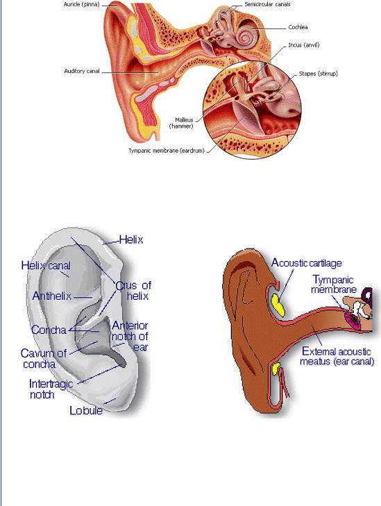
The human ear is a complex arrangement of mechanical and neurological components that are designed to interpret a multifaceted sound pressure waveform into its individual frequency components and then reconstitute these components together into what we recognize as blended sound. The human ear is shown in Figure 1.
The ear is composed of three sections: the outer ear (also known as the ear canal), the middle ear, and the inner ear. The outer ear is shown in greater detail in Figure 2.
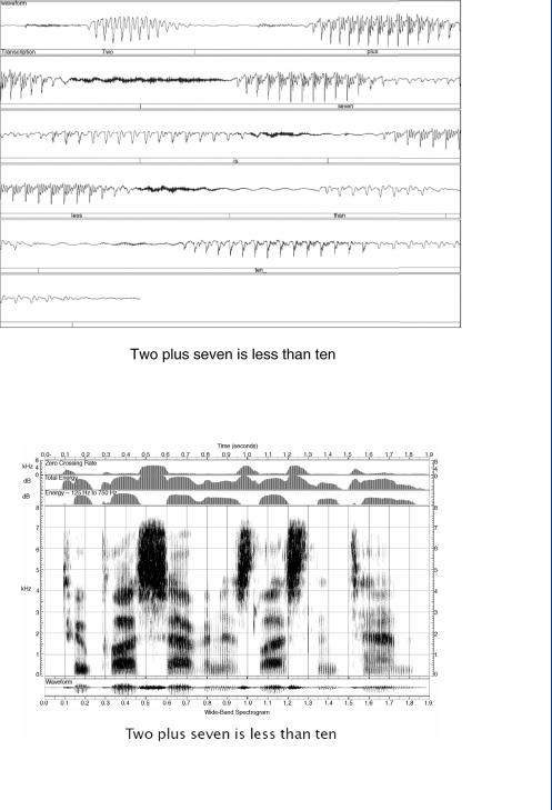
The outer ear is designed to channel sound from a complete hemisphere into a megaphone-shaped channel that is wide at the outside end and funnels down toward the inside end. As was noted above, most sounds are complex waveforms consisting of pressure waves of varying amplitudes and frequencies mixed together into sounds and words that we recognize in our daily lives. A typical sound waveform of spoken words is shown in Figure 3 with the frequency spectrum of those words shown in Figure 4.
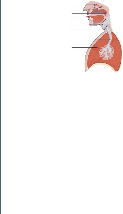
The array of pressure waveforms (Figures 3 and 4) results from a series of anatomical elements including the trachea, lungs, vocal cords, tongue, and mouth, as is seen in Figure 5.
2 SENSORY ORGAN REPLACEMENT AND REPAIR
FIGURE 1: Anatomy of the human ear.
FIGURE 2: The outer ear.
HEARING AIDS 3
FIGURE 3: Speech waveform of spoken words indicating a breadth of amplitudes and frequencies.
FIGURE 4: Frequency spectrum of spoken words indicating a range of embedded frequencies.
4 SENSORY ORGAN REPLACEMENT AND REPAIR
Nasal cavity
Hard palate
Soft palate (Velum)
Tongue
Jaw
Thyroid cartilage
Vocal folds
Trachea
Lung
FIGURE 5: Anatomical structures associated with speech production.
The complex waveforms associated with speech and many sounds are channeled into the outer ear and, as noted above, funneled toward the inside, which culminates with the tympanic membrane, also known as the eardrum. This membrane vibrates with the same complex arrangement as the complex sound waveforms that enter the ear canal.
From the tympanic membrane, the vibrations associated with the sound pressure waveform are then transmitted into the middle ear, which consists of three tiny bones called the ossicles (the malleus, incus, and stapes). These three bones are sometimes given the colloquial names hammer, anvil, and stirrups, which describe their shapes. They amplify the sound and send it through the entrance to the inner ear (oval window) and into the fluid-filled hearing organ known as the cochlea. The middle ear appears to be an overly complex structure of three bones that connect one membrane (the tympanic membrane or eardrum) to the oval window leading to the inner ear. However, there are two important elements to the middle ear. Firstly, the bones not only transmit the complex sounds from the tympanic membrane to the oval window, but also amplify the pressure due to the area difference between the two membranes and the size of the bones that attach to these membranes. In this fashion, the middle ear is similar to an impedance-matching device. Secondly, the middle ear is responsible for balance. If there is a significant pressure difference between the two ears, then one’s balance is adversely affected.
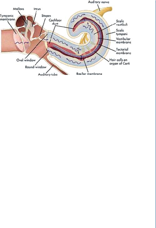
The sound from the middle ear is transmitted into the inner ear known as the cochlea. It is here that the complex sound waveforms of varying frequencies and amplitudes are separated into individual components. This is accomplished by means of the resonant frequency for a material based upon its geometry. Thicker materials vibrate naturally at lower frequencies, while thinner
HEARING AIDS 5
FIGURE 6: Human cochlea with hair cells and auditory nerve.
materials vibrate at higher frequencies. This is why bass speakers are larger than treble speakers in a stereo system. The cochlea is configured as a coiled triangular tissue with a wide thicker end and a narrow thinner end with a continual taper along its length. As complex sound enters into the cochlea, each relevant section of the cochlea would vibrate at its resonant frequency based upon its thickness. Thus, the complex sound is then separated into its individual frequency elements. The cochlea is thus a reverse auditory sound mixer. Along the cochlea, there are thousands of hair cells that are connected to tiny nerves. As the various sections of the cochlea vibrate at each resonant frequency at an amplitude dependent on the incoming sound amplitude (at that frequency), so do the attached hair cells. These hair cells excite their associated nerves. These nerves are then connected en masse to the auditory nerve or eighth cranial nerve, which sends the recombined electrical signal to the brain. The brain translates these impulses into what we experience as sound. It is unusual that the cochlea separates sounds into their individual components, converts them from vibrations into electrical signals, and then reconstitutes them into a blended sound, similar to its original construct. Figure 6 indicates the cochlea with hair cells leading to the auditory nerve.
The wide variety of sounds that we routinely hear vary from a whisper to a roar with pressure variations covering several orders of magnitude. To provide some uniform measurement system, a logarithmic sound scale has been established in units known as decibels. A decibel scale for many common sounds is shown in Figure 7.
The frequency range of human hearing is 20–20 000 Hz with the lowest end being more of a vibratory feeling than true auditory hearing. The range of human hearing as compared to other species is shown in Figure 8.