

Molecular and Cellular Signaling - Martin Beckerman
.pdf5.12 Hsp 90 Chaperones Help Maintain Signal Transduction Pathways |
103 |
FIGURE 5.4. A GroEL-GroES Anfinsen cage for protein folding: Fourteen subunits are arranged into two rinds of seven units each. Each chain contains equatorial, intermediate, and apical domains. Polypeptide chains are inserted into the cage, undergo a round of mechanical manipulations (i.e., the chains are “annealed” to remove unfavorable bondings), and are ejected. Several rounds of these operations may take place.
14 identical subunits; the eukaryotic Hsp 60 molecule has 16 identical subunits. As shown in Figure 5.4 the subunits are arranged into a pair of rings; in the case of GroEL each ring contains 7 identical subunits. Cochaperonins help in this activity. Group I co-chaperonins form a lid over the barrel and assist in the folding.
5.12Hsp 90 Chaperones Help Maintain Signal Transduction Pathways
The second main group of molecular chaperones consists of the Hsp70 and Hsp90 families. The Hsp70 family of chaperones performs a variety of tasks in the prokaryotes. DnaK is the outstanding bacterial Hsp70 family member. It regulates the heat shock stress response and maintains proteins in their physiologically viable configurations. Eukaryotic polypeptide chains are some 30–40% larger on the average than their prokaryotic counterparts. Eukarotic Hsp70 chaperones bind newly synthesized proteins. They assist in transporting these proteins across organelle membranes and into the endoplasmic reticulum (ER) and mitochondria. They also assist in the insertion of nascent membrane proteins into membranes.
Members of the Hsp90 family are involved in maintaining the functional integrity of proteins involved in intracellular signaling. These chaperones often work in association with Hsp70 family members and several other small ancillary proteins to prevent aggregation and mediate refolding. The Hsp90 chaperones form complexes with signal molecules, then help in their translocation to the correct subcellular compartment and into association with other elements of the signal pathway where they operate.
104 5. Protein Folding and Binding
Members of the Hsp90 family of molecular chaperones are among the most abundant proteins in the cell. They account for 1–2% of the total cellular protein even under non-stressful conditions. Hsp90 acts later in the folding process than the other chaperones. In normal cells, Hsp90 family members bind to a selective set of proteins operating in the signaling pathways that regulate cell differentiation and embryonic development. The proteins targeted by Hsp90 typically have low energy conformations that are only marginally stable. These signaling proteins have difficulty remaining in their physiologically competent states and require the assistance of Hsp90 to stabilize them. For example, in the absence of Hsp90, steroid hormone receptors are unable to bind their ligands and DNA. Likewise, bereft of Hsp90, tyrosine kinases such as c-Src lose their ability to function as kinases and to be acted upon by their regulators.
5.13 Proteins: Dynamic, Flexible, and Ready to Change
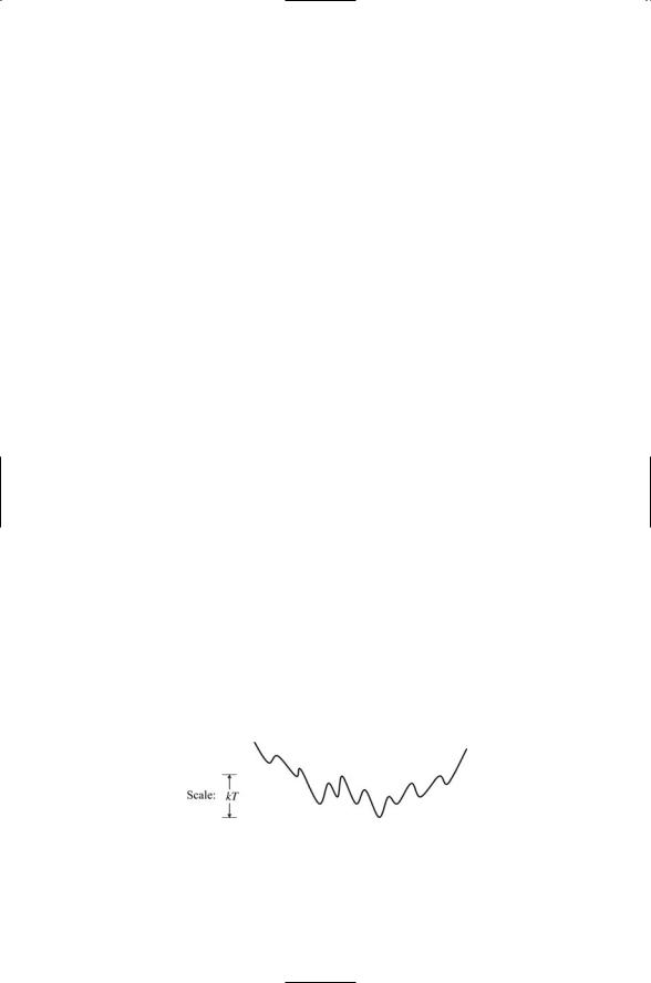
Proteins are not static but instead are dynamic structures that exhibit considerable flexibility, and readily adopt new shapes. Proteins are dynamic entities. If one examines the energy landscape in the vicinity of the protein’s native state one finds that there is an ensemble of low-lying states and the proteins is continually undergoing transitions from one state to another. This population of states and the barriers separating them play an important role in substrate recognition and catalysis. An example of such as ensemble of low-lying conformational states is presented in Figure 5.5. In this figure, none of the barriers are particularly high compared to the thermal energy kT. In this situation, the proteins will easily undergo transitions from one state to another, but will spend more time in the lowest energy state than in any of the others. The protein whose energy landscape is depicted in Figure 5.5 is quite flexible. It is able to move back and forth among a set of different shapes. The energy landscape of a protein that is completely rigid would be far sparser if it lacked low-lying states that were easy to get to.
FIGURE 5.5. Low-lying ensemble of native and nearby state: These states determine the binding properties of the proteins. Different ligands acting as environmental factors select one or more of these preexisting states when they bind the protein.

References and Further Reading |
105 |
The energy landscapes are influenced by environmental factors such as temperature, pH, ionic concentration, and binding events. Catalysts make use of protein motions to increase the rate of reactions. Environment factors, most notably charged groups such as acids, bases, metal ions, and dipoles belonging to the catalyst generate a shift in the energy landscape.
Two kinds of shifts are possible, kinetic and thermodynamic. In a kinetic shift, the energy barrier separating two conformational states is lowered, making possible transitions from a higher energy state to a lower energy state. The initial kinetic barrier is high compared to the thermal (kinetic) energy factor kT, and the system may be trapped in the higher, or metastable, state prior to the kinetic shift.
In a thermodynamic shift (the situation illustrated in Figure 5.5) the barrier between two states remains the same but the relative energies are modified so that a formerly higher energy state is now a lower energy state. The barriers in this scenario low so there are no kinetic barriers, and transitions among the many states can be frequent. In either case, a catalyst generates a shift in the population of conformations towards those that favor the reaction being catalyzed. This latter utilization of protein flexibility is often referred to in the literature as stabilizing the transition state in the case of catalysis, and as the induced fit model of surface complementarity in the case of binding. The key point is that protein flexibility underlies the ability of a protein to bind to another molecule—and to entire groups of proteins of differing shape and size—with the appropriate degree of specificity. In all of these situations the ligand may be thought of as selecting out one or more states from the ensemble of preexisting low-lying states.
References and Further Reading
Anfinsen Nobel Prize Lecture
Anfinsen CB [1973]. Principles that guide the folding of protein chains. Science, 181: 223–230.
Motions of Proteins
Cavanagh J, and Akke M [2000]. May the driving force be with you—Whatever it is. Nature Struct. Biol., 7: 11–13.
Feher VA, and Cavanagh J [1999]. Millisecond-timescale motions contribute to the function of the bacterial response regulator protein Spo0F. Nature, 400: 289–293.
Forman-Kay JD [1999]. The “dynamics” in the thermodynamics of binding. Nature Struct. Biol., 6: 1086–1087.
Frauenfelder H, Sligar SG, and Wolynes PG [1991]. The energy landscapes and motions of proteins. Science, 254: 1598–1603.
Kay LE, et al. [1996]. Correlation between dynamics and high affinity binding in an SH2 domain interaction. Biochem., 35: 361–368.

106 5. Protein Folding and Binding
Kern D, et al. [1999]. Structure of a transiently phosphorylated switch in bacterial signal transduction. Nature, 402: 894–898.
Lee AL, Kinnear SA, and Wand AJ [2000]. Redistribution and loss of side chain entropy upon formation of a calmodulin-peptide complex. Nature Struct. Biol., 7: 72–77.
Stock A [1999]. Relating dynamics to function. Nature, 400: 221–222.
Zidek L, Novotny MV, and Stone MJ [1999]. Increased protein backbone conformational entropy upon hydrophobic ligand binding. Nature Struct. Biol., 6: 1118– 1121.
Protein Folding: The Energy Landscape Picture
Bryngelson JD, et al. [1995]. Funnels, pathways, and the energy landscape of protein folding: A synthesis. Proteins: Structure, Function and Genetics, 21: 167–195.
Chan HS, and Dill KA [1998]. Protein folding in the landscape perspective: Chevron plots and non-Arrhenius kinetics. Proteins: Structure, Function and Genetics, 30: 2–33.
Dill KA, and Chan HS [1997]. From Levinthal to pathways to funnels, Nature Structure Biology, 4: 10–19.
Leopold PE, Montal M, and Onuchic JN [1992]. Protein folding funnels: A kinetic approach to the sequence-structure relationship. Proc. Natl. Acad. Sci. USA, 89: 8721–8725.
Onuchic JN, et al. [1995]. Toward an outline of the topography of a realistic proteinfolding funnel. Proc. Natl. Acad. Sci. USA, 92: 3626–3630.
Sali A, Shakhnovich E, and Karplus M [1994]. How does a protein fold? Nature, 369: 248–251.
Molecular Chaperones and Protein Folding in the Cell
Hartl FU, and Hayer-Hartl M [2002]. Molecular chaperones in the cytosol: From nascent chain to folded proteins. Science, 295: 1852–1858.
Pratt WB [1998]. The Hsp90-based chaperone system: Involvement in signal transduction from a variety of hormone and growth factor receptors. Proc. Soc. Exp. Biol. Med., 217: 420–434.
Rutherford SL, and Lindquist S [1998]. Hsp90 as a capacitor for morphological evolution. Nature, 396: 226–342.
Sauer FG, et al. [2000]. Chaperone-assisted pilus assembly and bacterial attachment.
Curr. Opin. Struct. Biol., 10: 548–556.
Binding Mechanisms
DeLano WL, et al. [2000]. Convergent solutions to binding at a protein-protein interface. Science, 287: 1279–1283.
Freire E [1999]. The propagation of binding interactions to remote sites in proteins: Analysis of the binding of the monoclonal antibody D1.3 to lysozyme. Proc. Natl. Acad. Sci. USA, 96: 10118–10122.
Hilser VJ, et al. [1998]. The structural distribution of cooperative interactions in proteins: Analysis of the native state ensemble. Proc. Natl. Acad. Sci. USA, 95: 9903–9908.

Problems 107
Kumar S, et al. [2000]. Folding and binding cassettes: Dynamic landscapes and population shifts. Protein Sci., 9: 10–19.
Ma B, et al. [2002]. Multiple diverse ligands binding at a single protein site: A matter of pre-existing populations. Protein Sci., 11: 184–197.
Teague S [2003]. Implications of protein flexibility for drug design. Nature Rev. Drug Dis., 2: 527–541.
Problems
5.1Entropy, density of states, and probabilities. Consider a system of N atoms, each of which is placed in one of M bins. The atoms are labeled by the bins into which they are placed, but are otherwise indistinguishable from one another. That is, n1 atoms are placed in bin 1; n2 atoms are placed in bin 2, and so on up to nM atoms in bin M. The quantities
pi = ni  N
N
represent the probabilities of finding an atom in a particular bin. The number of ways a specific configuration can be realized (where “configuration” means finding n1 atoms in bin 1, n2 atoms in bin 2, and so on) is, from elementary probability theory, given by the multinomial coefficient
W({n1 , n2 |
, . . . , nM }) = |
|
N! |
|
|
|
. |
||
n1n2 |
|
|||
|
|
◊ ◊ ◊ nM |
||
The relationship between number of states k, actually coeffs, and entropy S was first established by Ludwig Boltzmann and Max Planck. Their results are usually presented in the form:
S = k ln W({ni }),
N
and sometimes in the form
M
S= -k pi ln pi .
i =1
Show that the two forms are equivalent; that is,
Ê k ˆ |
M |
|||
ln W({ni }) = -k pi ln pi . |
||||
|
|
|
||
Ë N ¯ |
||||
i =1 |
||||
5.2Frustration and rugged energy landscapes. Rugged energy landscapes are formed whenever there is a lack of good low energy conformations. Situations of this type arise when the system of atoms

108 5. Protein Folding and Binding
FIGURE for Problem 5.2. Nonfrustrated and frustrated squares: Atoms (vertices) are connected by bonds (sides) with bond (coupling) strengths as indicated. (a) Nonfrustrated square in which all couplings are positive. (b) Frustrated square in which there is one negative coupling and three positive couplings.
and bonds connecting them becomes frustrated. The term frustration is intended to convey the inability of the atomic system to fold itself in such as way that all bond orientations lead to good low energy states. Instead, for any spatial orientation of bonds, some interactions will be positive and others will be negative, and in place of a few deep low energy states there will be a large number of less deep low energy states.
The notion of frustration can be illustrated using a mini system consisting of four spin 1/2 atoms arranged in a pair of squares as shown above. Each vertex in a square contains an atom whose spin value is either +1 or -1. The energy of the systems is the sum of four interactions of the form Jsisj, where si and sj are the spin values of atoms at neighboring vertices connected by bonds, and J is the bond strength. In other words, the energy E is given by the expression
E = -ÂJij si s j ,
and the sum is over the four pairs of neighboring vertices. There are 16 possible states of this system: 2 ¥ 2 ¥ 2 ¥ 2. The nonfrustrated square on the left possesses two good (deep) low-energy states—one where all spins are +1 and one where all spins are -1, each with energy -4 J. All other states have higher energies. (a) Tabulate the distribution of states and then compare this distribution to that for the frustrated square shown in the right hand part of the figure. (b) What happens if the top and bottom couplings are negative while the two side couplings remain positive?
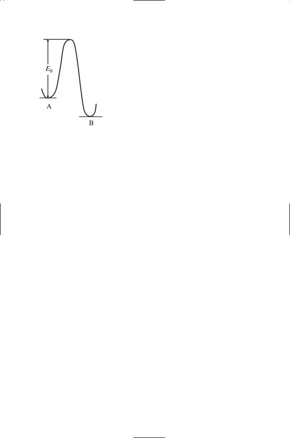
5.3Transition rates and their dependence on barrier height. The transition rate for passage over a barrier from state A to state B is given by the Arrhenius formula:
v = v0 e -E0 kT .
Problems 109
FIGURE for Problem 5.3. Transition over a barrier from one state to another.
In this expression, v is the transition rate, which is equal to the product of a factor v0 that gives the number of attempts being made on the barrier (the attempt frequency) and the Boltzmann factor (i.e., the exponential term) describing the probability for success for any given attempt. Assuming no change in the attempt frequency, how much less likely is a transition for the case where barrier height is a factor of 3 greater than the thermal energy kT as compared to a situation where the barrier height is half that of the thermal energy?

6
Stress and Pheromone Responses in Yeast
Unicellular organisms such as the budding yeast Saccharomyces cerevisiae have to cope with continually changing environments and are subject to a variety of environmental and physiological stresses. These include unfavorable temperatures that produce heat shock, osmotic stresses that disrupt the water balance, carbon source deprivation that leads to starvation, and oxidative stresses. Yeast cells respond to these and other unfavorable conditions by altering their patterns of gene expression and by adjusting their rates of protein synthesis. They may respond in a rather general way to a stressful situation, or they may respond in a far more specific manner to a particular stress condition.
The signaling routes that ultimately produce the alterations in gene expression and protein synthesis begin at the plasma membrane with signal reception. Signal receptors embedded in the plasma membrane sense changes in the local environment and receive chemical messages sent by other cells. The end points of the signaling routes are control points in the fixed infrastructure. It is at these control points that the signals are converted into cellular response. Some signaling routes, especially those found in bacteria, are fairly short with at most a few elements lying between sensing and contact elements. Other signal routes are far longer. In these situations, the routes from cell membrane to the contact points in the cell interior often involve several intermediate signaling elements and control points where several signals converge. Whether short or long, a set of signaling elements is activated in a sequential fashion, from cell surface molecules to intracellular signal elements to one or more final contact elements. The ensemble of signaling elements activated in this way is referred to as forming a signaling pathway.
The different kinds of signaling elements that make up the typical eukaryotic signaling pathway will be introduced in the first part of this chapter. Examples will then be given in the second part of the chapter of how these elements come together to form stress and pheromone signaling pathways in yeast. The short signaling routes found in bacteria will be explored in Chapter 7, and an important set of signaling intermediaries,
111
112 6. Stress and Pheromone Responses in Yeast
commonly referred to as second messengers, will be explored in the chapter after that, Chapter 8.
6.1 How Signaling Begins
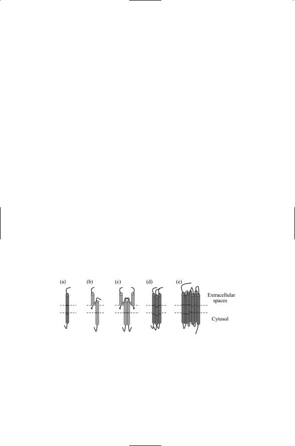
Signaling begins at the plasma membrane with receptor-ligand binding and signal transduction. Transmembrane receptors function as sensors of environmental stresses and as receivers of chemical messages. They transmit, and, in the process, convert the signals from an external, outside-the-cell form to an internal, inside-the-cell one that can be understood and further processed. This process is called signal transduction. Several kinds of transmembrane receptors are presented in Figure 6.1. In each example, part of the receptor lies in the extracellular spaces enabling it to bind to ligands. There are also one or more transmembrane segments, portions of the polypeptide chain that either pass once through the plasma membrane or wind back and forth several times. Lastly, there is a cytoplasmic segment that permits the receptor to make contact with signaling molecules inside the cell, thereby allowing the receptor to transduce a signal.
The notion of signal transduction is a central one. The ligand itself is not passed into the cell. Rather, a different molecule becomes activated and conveys the message inside the cell. This conversion process can happen several times inside the cell with one protein contacting a second one, which then contacts and activates a third molecule, and so on until the final signaling element reaches a control point where the signal is converted into a cellular response. In each of these transfers, one signal molecule replaces another in a sequential manner to carry the signal. Thus, each step involves transduction, the conversion of a message from one form to another.
FIGURE 6.1. Signal receptors: Each receptor has an extracellular region, a transmembrane segment, and an intracellular (cytosol) portion. (a) Single-chain, singlepass receptor; (b) two-chain, single-pass receptor; (c) dimeric arrangement of two-chain, single-pass receptors; (d) trimeric arrangement of single-chain, singlepass receptors; and (e) single-chain, seven-pass receptor. The N-terminal is usually located on the extracellular side and the C-terminal on the cytoplasmic side in the single-chain and seven-pass receptors. In the double-chain receptors, the uppermost side is N-terminal and the lower side is C-terminal.

6.2 Signaling Complexes Form in Response to Receptor-Ligand Binding |
113 |
Receptors can be grouped into families according to their topology, structure, and the kinds of ligands they bind. The first two examples presented in Figure 6.1 are single-pass receptors. In Figure 6.1(b), two chains are used rather than a single chain as in Figure 6.1(a). One chain lies entirely in the extracellular space and is responsible for ligand binding. The other chain is covalently linked to the first (through disulfide bonds). It contains transmembrane and cytoplasmic segments and transduces the signal into the cell.
Receptors form associations with other receptors. One way for a signal to be conveyed into the cell is through the formation and stabilization of receptor complexes. The complex may consist of a pair of receptors, as in Figure 6.1(c), simultaneously bound to a single ligand (1 : 2 stoichiometry) or to a pair of ligands (2 : 2 stoichiometry). Alternatively a receptor trimer may form as in Figure 6.1(d) bound to, for example, three ligands. Higherorder complexes may form and these complexes may even involve a mix of different receptors. The last example, presented in Figure 6.1(e), is for a seven-pass, single chain receptor. This is the topology of the largest family of receptors found in the body—the G protein-coupled receptors (GPCRs). They sense and transduce sensations such as light and odors, and cell-to- cell hormonal and neuromodulatory signals. They are also the targets of about half of all the drugs commercially produced.
6.2Signaling Complexes Form in Response to Receptor-Ligand Binding
Receptor-ligand binding stimulates the assembly of signaling complexes at and just below the plasma membrane. Ligand binding provokes changes in the environment around the cytoplasmic portion of the receptor(s) leading to formation of these complexes. These changes may take one or more forms. In some receptor families, ligand binding stabilizes the formation of receptor dimers. The receptors possess an intrinsic kinase activity, which is turned on when the two chains are brought into close proximity to one another. The ensuing phosphorylation opens up docking sites for cytoplasmic signaling proteins that, in turn, seed the formation of the signaling complex. In families such as the GPCRs, ligand binding triggers a changes in conformation that are sufficient to activate nearby cytoplasmic G- proteins leading to activation of signaling intermediates—second messen- gers—and the formation of signaling complexes.
Proteins functioning as anchor, scaffold, and adapter proteins help organize these signaling complexes. As depicted in Figure 6.2, anchor proteins provide platforms for proteins to attach in close proximity to receptors and other upstream signaling elements. Anchor proteins attach to the plasma membrane and to membranes of organelles. They allow two or more sig-