

Molecular and Cellular Signaling - Martin Beckerman
.pdf114 6. Stress and Pheromone Responses in Yeast
FIGURE 6.2. Assembly of signaling complexes: (a) Signal complex organized by exposure of a docking site by a receptor aided by the utilization of an adapter protein; (b) signal complex organized by a cytoskeleton-associated scaffold protein; and (c) signal complex organized by a membrane-associated anchor protein.
naling proteins to position themselves in close proximity to one another and to their membrane-associated substrates. Scaffold proteins operate in a similar fashion to anchor proteins. They too provide platforms for attachment of multiple proteins. They attach to components of the actin cytoskeleton, or alternatively, to microtubules. These attachment sites are usually located near the plasma membrane and play an important role in relaying signals from the plasma membrane to downstream control sites. The third group of nonenzymatic intermediaries is the adapter proteins. These proteins contain protein-protein interaction domains and serve as intermediaries that allow two proteins that would otherwise not be able to communicate to do so.
In order to form a complex, proteins diffuse over to and collect at the docking sites. The proteins are said to be “recruited” to the collection point. The process of recruitment is mostly a passive one driven by diffusion of proteins localized in the vicinity of docking sites opened up by receptor-ligand binding. The diffusion is largely planar; that is, it is a twodimensional rather than a three-dimensional process because all signaling elements are located at and just below the plasma membrane. The adapters, anchors, and scaffolds localize the necessary signaling elements close to one another at and just below the plasma membrane. Because the proteins are localized nearby and because the process is a two-dimensional one, it is fairly rapid.
6.3 Role of Protein Kinases, Phosphatases, and GTPases |
115 |
6.3 Role of Protein Kinases, Phosphatases, and GTPases
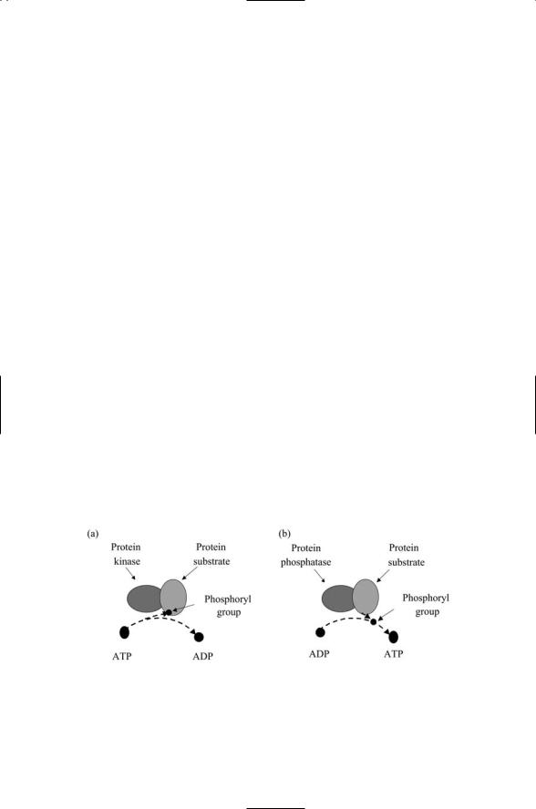
Protein kinases, phosphatases, and GTPases convey signals from the cell surface to downstream cytoplasmic effectors. The organization of signaling complexes by anchors, scaffolds, and adapters is illustrated in Figure 6.2. In each example, proteins designated in a generic sense as protein “a,” or protein “b,” and so on are recruited to the complex. The proteins so indicated are mostly enzymes. Two different kinds of enzymes predominate— protein kinases and protein phosphatases, and these are the primary intracellular signal transducers. Protein kinases are large proteins that catalyze the transfer of phosphoryl groups to target proteins using ATP as a donor. Protein phosphatases do the opposite: They catalyze the removal of phosphoryl groups using ADT as an acceptor. These two operations are illustrated in Figure 6.3. The substrates that accept and lose phosphoryl groups are often kinases themselves, so that one kinase activates another, which then activates a third kinase, and so on.
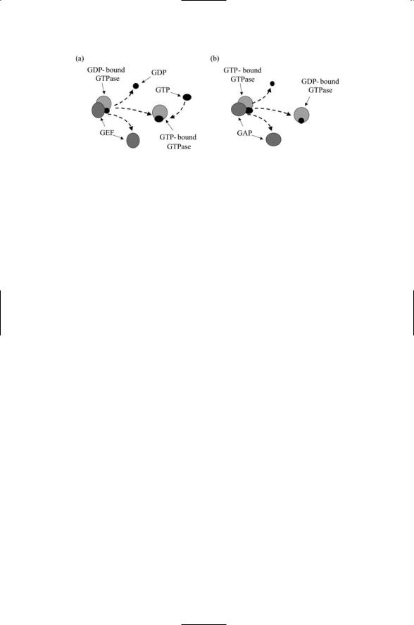
Protein kinases and phosphatases are not the only categories of enzymes that participate in signaling. Enzymes called GTPases also contribute to signaling, as do a variety of proteolytic enzymes, or proteases. GTPases are enzymes that are activated when they bind guanosine triphosphate (GTP) and are deactivated when they bind guanosine diphosphate (GDP). Two sets of ancillary enzymes catalyse GDP/GTP binding and release from the GTPases. As illustrated in Figure 6.4, a guanine nucleotide exchange factor (GEF) catalyzes the dissociation of GDP from the GTPase. GTP is an abundant cytosolic protein and in response to the actions of the GEF it binds the GTPase, thereby activating it. The GTPase-activating protein (GAP) accelerates the enzymatic actions of the GTPase. It also speeds up the hydrolysis of the bound GTP molecule, leaving a bound GDP molecule in its place, resulting in the inactivation of the GTPase.
FIGURE 6.3. Enzymatic actions of protein kinases and protein phosphatases: (a) A protein kinase catalyzes the transfer of a phosphoryl group to its substrate using ATP as a donor. (b) A protein phosphatase catalyzes the removal of a phosphoryl group from its substrate using ADP as an acceptor.
116 6. Stress and Pheromone Responses in Yeast
FIGURE 6.4. Catalytic activities of GTPase-associated factors: (a) A GEF catalyzes the removal of GDP from a GTPase, which then binds to a GTP molecule. (b) A GAP catalyzes the hydrolysis of the GTP molecule bound to the GTPase, leaving a bound GDP molecule.
6.4 Role of Proteolytic Enzymes
Proteolytic enzymes transform, activate, and deactivate signaling elements. Proteolytic processing is yet another kind of posttranslational modification that has an important role in signaling. Several different kinds of proteolytic enzymes, or proteases, are active in the cell. Some of the them catalyze the cleavage of membrane-associated proteins either on the extracellular side or the intracellular side of the plasma membrane. The cleavage of a tether that anchors a signaling protein on the outside of the cell converts that protein into a diffusible ligand, thereby greatly extending its range of influence. The intracellular cleavage of the protein either anchored to or embedded in the plasma membrane converts that protein into a messenger that can relay messages into the cell interior, allowing it to function not only as an upstream signaling element but also as a downstream one. These operations are illustrated in Figure 6.5(a) and (b).
The enzymes involved in transducing signals are localized where they are needed in inactive forms. When an initiating event takes place, such as the arrival of an extracellular ligand that binds a receptor located in the plasma membrane, the proteins that the signaling pathway comprises are activated. A series of events take place with elements upstream activating substrates downstream until the last signaling elements contact the fixed infrastructure at a control point.
The signaling elements must be carefully controlled to prevent unwanted signaling. The signals that convert the signaling molecules from an inactive to an active form are of several kinds, and these often work in concert to “turn on” signaling when and only when appropriate activating signals are present. One way of accomplishing this is to immobilize a signaling protein through binding it to a scaffolding protein. If the scaffold protein is proteolytically processed, the signaling protein will be freed up and can convey

6.5 End Points Are Contact Points to Fixed Infrastructure |
117 |
FIGURE 6.5. Signaling activities of proteolytic enzymes: (a) A protease cleaves the tether, detaching the ligand from the outer leaflet of the plasma membrane allowing it to function as a diffusible ligand. (b) A protease cleaves the polypeptide chain just below the plasma membrane, allowing the cytoplasmic portion of the protein to function as a downstream signaling element. (c) A protease degrades a scaffold protein, freeing up the signaling protein to convey a signal to its downstream substrate. (d) A protease degrades a signaling protein shortening its lifetime and terminating signaling.
a message as shown in Figure 6.5(c). Proteolytic processing can do the opposite and turn off signaling. If a signaling protein rather than a scaffold is tagged for proteolytic processing as indicated in Figure 6.5(d), the signal protein’s lifetime will be reduced and its signaling activity will be damped down.
6.5End Points Are Contact Points to Fixed Infrastructure
The end points of signaling pathways are control points that make contact with the fixed infrastructure. The furthest downstream signaling elements in the signaling pathways make contact with one or more components of the fixed infrastructure. The components being contacted may be a receptor or ion channel embedded in a membrane, or they may belong to the cytoskeleton. They may belong to an intracellular transport organelle, or to the mitochondria, or to the transcription apparatus in the nucleus, or to the translation machinery situated in the endoplasmic reticulum.

118 6. Stress and Pheromone Responses in Yeast
The most commonly occurring end points are those that contact and regulate the transcription machinery. Proteins that convey regulatory instructions to the transcription machinery are called transcription factors.These are the last or furthest downstream signaling elements in the various stress and pheromone pathways. These proteins bind to specific DNA sequences located in noncoding regions of genes and as a consequence influence transcription either positively or negatively. In the former case by makig it easier to carry out transcription and in the latter case by making it more difficult to do so.The noncoding gene regulatory regions contain binding sites for elements of the fixed infrastructure to bind and also sites for regulatory proteins to attach. The control regions are known as promoters and the sequence-specific control points where transcription factors bind and come together to regulate transcription are known as responsive elements (REs).
6.6 Transcription Factors Combine to Alter Genes
In the budding yeast, several types of stress response can be evoked. Those elements of a stress response that are common to all stresses are handled by the yeast general stress response, while additional elements needed to treat certain kinds of stresses are handled by a set of specific stress responses. Organisms such as the yeast respond to stresses by remodeling their pattern of gene expression, upregulating some genes and downregulating others. Experiments performed using DNA microarrays show that changes in the patterns of gene expression are global, often involving substantial fractions of the entire genome. The control points are organized in a way that makes possible these stress responses. Genes that require coexpression because their products must all be present at the same time and work together possess a common set of responsive elements to which the transcription factors may bind. That is, multiple copies of each of these responsive elements are distributed among promoters throughout the genome. Certain combinations of responsive elements are encountered in genes that are cotranscribed in response to specific stress and pheremone signals; other combinations are often encountered in genes coexpressed in response to a different set of stress signals and some are encountered in almost all stress responses. By this means a small number of transcription factors can coordinate and control a major remodeling of the cellular infrastructure.
Transcription factors activated by stresses act in a combinatorial fashion to trigger genome wide alterations in gene expression. In eukaryotes, these noncoding DNA sequences almost always contain binding sites for more than one transcription factor. The transcription factors jointly determine whether and how strongly to initiate transcription. The process whereby several transcription factors regulate transcription is known as combinatorial control. This term signifies one of the key aspects of the process. Tran-

6.7 Protein Kinases Are Key Signal Transducers |
119 |
scription factors can come together in a variety of ways, acting in different combinations of twos, threes and so on. A small number of transcription factors can therefore generate many different patterns of gene expression in response to stresses and other environmental changes.
Combinatorial control is a synergistic process. In combinatorial control the effect produced by two positively acting transcription factors is far greater than the sum of the effects of each individual in the pair. This can happen in several ways. One way of producing an effect of this sort is for one of the partners to relieve an autoinhibitory interaction present in its partner that prevents the partner from acting. Alternatively, one of the partners may act on the DNA substrate to expose or better prepare a binding site for the other partner. These interactions are all cooperative with the first interaction making it easier for the second interaction to occur.
In the remainder of this chapter, the operation of signaling pathways activated by yeast cells in response to stresses will be examined. The yeast stress pathways illustrate how the various kinds of signaling proteins come together to form a pathway. Before examining these pathways the different categories of signaling elements will be looked at in more detail.
6.7 Protein Kinases Are Key Signal Transducers
Protein kinases are central regulators of cellular physiology. These proteins catalyze the transfer of a gamma-phosphoryl group from an ATP molecule (usually bound to Mg2+ ion) to a hydroxyl group on the side chain of specific residues in target proteins. In other words they are phosphotransferases that use ATP (and sometimes GTP) as a donor and specific residues in target proteins as acceptors. Their ability to function as enzymes is due to the presence of a catalytic domain of some 200 to 300 amino acids. The kinase domain, along with one or more regulatory domains, comprise the kinase. Some protein kinases encode their catalytic domains in separate subunits; in others, the catalytic and regulatory domains are all part of a single chain molecule.
The protein kinases can be divided into two large groups. In the first group, are the protein kinases that target hydroxyl groups on serine/threonine residues. These are called serine/threonine (ser/thr) kinases. In the second group are the protein kinases that target hydroxyl groups on tyrosine residues, and these are called tyrosine (tyr) kinases. The proteins in each major grouping can be further placed into a number of families that are highly conserved across all eukaryotes. Listed in Table 6.1 are five prominent groups of serine/threonine kinases, organized according to similarities in kinase domain structure, substrate specificity, and method of regulation/activation. These kinases are encountered in eukaryotes ranging from yeast to man. Tyrosine kinases are more restricted in their distribution. They are largely absent from unicellular organisms and appear to have emerged

120 |
6. Stress and Pheromone Responses in Yeast |
|
TABLE 6.1. Serine/threonine kinases of eukaryotes. |
||
|
|
|
Group |
Family |
Comments |
|
|
|
AGC |
Protein kinase A (PKA)/Protein |
Cyclic nucleotide-regulated; basic |
|
kinase G (PKG) |
amino acid-directed |
|
Protein kinase C (PKC) |
Lipid-, calcium-regulated |
|
Protein kinase B (PKB), Akt, RAC |
Lipid-regulated |
|
G protein-coupled receptor kinase |
Lipid-, calcium-regulated |
|
(GRK) |
|
|
Ribosomal S6 kinase (S6K) |
Lipid-regulated |
|
Phosphoinositide-dependent protein |
Lipid-regulated |
|
kinase-1 (PDK1) |
Ca2+/calmodulin-regulated; Basic |
CaMK |
CaM kinase (CaMK) |
|
|
|
amino acid-directed |
|
AMP-dependent protein kinase |
Activated when AMP:ATP is |
|
(AMPK) |
elevated; basic amino acid-directed |
CMGC |
Cyclin-dependent kinase (CDK) |
Proline-directed |
|
Mitogen-activated protein kinase |
Substrate of MEKs; proline-directed |
|
(MAPK) |
|
|
Glycogen synthase kinase-3 (GSK-3) |
Principal substrate of PKB; dual |
|
|
specificity; proline-directed |
|
Casein kinase-2 (CK2) |
Uses both ATP, GTP; Acidic amino |
|
|
acid-directed; dual specificity |
MEK |
MAP/ERK kinase (MEK) |
Dual specificity; highly specific |
|
MAP/ERK kinase kinase (MEKK) |
Highly specific |
|
MAP/ERK kinase kinase kinase |
Highly specific |
|
(MEKKK) |
|
PIKK |
ATM/ATR; DNA-PKcs |
Glutamine-directed |
|
TOR |
Proline-directed |
|
|
|
in response to the need for increased cell-to-cell signaling and control in multicellular organisms. Tyrosine kinases will be examined in detail in later chapters.
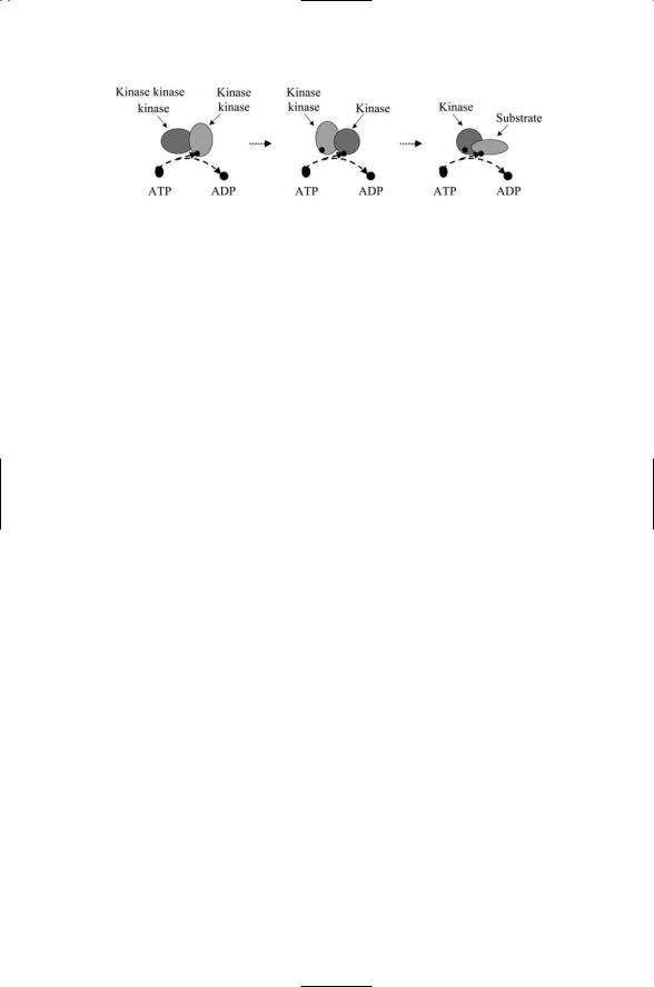
Serine/threonine kinases have many features in common. The most important of these similarities from the signaling perspective is their activation through phosphorylation. The kinases become catalytically active only when crucial residues in a region of their catalytic domain known as the activation loop becomes phosphorylated. In many, if not most instances one kinase activates a second kinase, and these kinases, activated sequentially in an upstream to downstream direction, form the core of the signaling pathway. Many of the entries in Table 6.1 can be arranged into pathways according to their actions on one another. That is, these kinases catalyze the transfer of phosphoryl groups to other kinases. Many of these are called by the same name as the downstream kinase, with the addition of another kinase in the name. An illustration of a kinase signaling cascade whose components are named in this manner is presented in Figure 6.6. Some common examples of this naming convention are ERK kinase kinases (ERKKs),
AMP kinase kinases (AMPKKs), and CaM kinase kinases (CAMKKs). Other kinases that act on kinases have their own distinct names. These
6.8 Kinases Often Require Second Messenger Costimulation |
121 |
FIGURE 6.6. A kinase signaling cascade: A kinase kinase kinase phosphorylates a kinase kinase, which in turn phosphorylates a kinase, which then phosphorylates its substrate. In some cascades, the kinases are localized in close proximity to one another by scaffolding proteins; in others, the upstream element must diffuse to where the downstream element is tethered.
kinases typically can act on more than one kind of target kinase; that is, they have broader substrate specificity than the first group of rather narrowly acting kinase kinases, and consequently have their own name. An example of this kind of broader acting kinase kinase is phosphoinositide dependent kinase (PDK). This signaling molecule acts upstream of many, if not most members of the Protein kinase A, G, and C (AGC) group.
6.8Kinases Often Require Second Messenger Costimulation
Phosphorylation by itself may not be sufficient to activate a kinase. Many of the serine/threonine kinases require the presence of a second messenger molecule. Second messengers are signaling intermediates connecting events taking place at the plasma membrane with the intracellular signaling that eventually converts the signal into a cellular response. Second messengers are small molecules. There are three main kinds—cAMP, lipids, and calcium. These intermediates are sent into the cytoplasm in response to receptor binding. Members of the AGC group and the CaM kinase family members are regulated in this way. Binding of a second messenger to the kinase, and also to the kinase kinase, precedes activation of the signaling pathways by phosphorylation.
The first of the second messengers mentioned above, cyclic adenosine monophosphate (cAMP), is generated from ATP by the enzyme adenylate cyclase embedded in the plasma membrane. Catalytic and regulatory sites are located within the cytoplasmic loops and termini of this doubleclustered, multipass molecule (depicted in Figure 8.9). The production of cAMP is triggered by signals relayed from the cytosolic surface of plasma membrane-bound receptors, and other intracellular signaling elements located in its near vicinity. Cyclic AMP binds to protein kinase A and this kinase is the sole cellular mediator of cAMP stimulated signaling.

122 6. Stress and Pheromone Responses in Yeast
Lipid second messengers regulate the activity of a number of kinase families belonging to the AGC group. The kinases of the AGC group are activated in a multistep manner. First, there is an initial phosphotransfer process that induces the migration of the protein kinases to the plasma membrane where subsequent phosphotransfers serve to activate the catalytic properties of the kinase. Prominent agents of the second messenger system are lipid kinases and phospholipase C that generate the second messengers, such as diacylglycerol (DAG) that activates protein kinase C, and other lipid second messengers that trigger the release of Ca2+ from intracellular stores. The lipid second messenger generating system and AGC kinase activation will be examined in detail in Chapter 8.
The third major kind of second messenger is intracellular calcium. As noted in Table 6.1 calcium is a regulator of the CaM kinases. It activates the kinase by first binding to calmodulin; then the calcium-calmodulin complex binds and activates the protein kinase and its upstream kinase kinases. Calcium is not only stored and released from intracellular stores located in the endoplasmic reticulum, but also enters the cells through ion channels found in excitable cells such as neurons. Calcium signaling and CaM kinases, along with many of the AGC kinases, are key agents of neural signaling and for these reasons the CaM and associated signaling pathways are called learning pathways.
6.9Flanking Residues Direct Phosphorylation of Target Residues
A third common theme in kinase actions pertains to the establishment of substrate specificity. This issue was briefly touched upon in the naming of kinases depending on whether they had a narrow or broad specificity. The key notion is that specificity is determined not only by the presence or absence of serine and threonine residues in a potential target protein, but also by the identity of the residues that flank the potential acceptor of the phosphoryl group. The flanking residues, that is, the amino acid residues immediately preceding or following the target site, help determine whether a particular target residue can be phosphorylated or not.
The most general observation that can be made is that kinases belonging to the AGC and CaMK groups tend to be basic amino acid-directed while those belonging to the CDK, MAPK, GSK3, CK2, and cyclin dependent kinase-like kinase (CMGC) group are mostly proline directed. In more detail, kinases belonging to the AGC group are more likely to catalyze the transfer of phosphoryl groups to S/T residues lying near basic amino acids such as Lys and Arg. The GRKs are an exception to this preference, as they prefer flanking acidic residues. A similar observation holds for the CaMK group. These kinases too prefer basic amino acid environments. In contrast, members of the CMGC group are mostly proline-directed, phosphorylat-

6.11 Protein Phosphatases Are Prominent Components |
123 |
ing S/T residues lying within proline-rich regions. The main exceptions to this rule are the CK2s. These kinases prefer the flanking amino acids to be acidic. Finally, phosphoinositide 3-kinase related kinases (PIKKs) are either glutamine or proline directed, as listed in Table 6.1.
6.10 Docking Sites and Substrate Specificity
One of the challenges to the reliable operation of cellular control layers is how to ensure that the kinases phosphorylate the correct substrate at the right time. The cell is crowded with many proteins, and each of these proteins likely to contain many potential phosphorylation sites. This selection problem is solved, but only in part, by the presence of specific flanking residues. It turns out that the amino acids lying either just before or just after the target serines and threonines are not the only amino acid residues involved in kinase-substrate recognition. Additional residues located well away from the target phosphorylation sites confer additional substrate specificity. These sites are referred to as docking motifs or docking domains.
The presence of docking sites on kinase and substrate increases the substrate specificity. These sites are typically located far from the catalytic site of the kinase and far from the phosphoacceptor site of the substrate. An example of a kinase family that uses this form of matching is the MAP kinase family. In this family, the docking sites are located in a C-terminal domain of the kinase. The docking site is characterized by the presence of several acidic amino acids, and is matched to a cluster of basic amino acids in the matching docking sites used by upstream activators, deactivators, and downstream substrates. Another family of kinases, the glycogen synthase kinase-3s (GSK-3s), uses a different docking strategy. In this family the presence or absence of a phosphorylated serine located four residues from the substrate target serine is a crucial determinant of whether the kinase can dock at the substrate. A crucial arginine residue serves as the kinase docking site, and the complementary matching of the arginine residue in the kinase to the phosphorylated, or “primed,” site in the substrate stabilizes the kinase in a catalytically active conformation.
6.11Protein Phosphatases Are Prominent Components of Signaling Pathways
Reversible phosphorylation, the covalent attachment and removal of phosphoryl groups to and from proteins, is the most widespread method of regulation protein activity in the cell. In addition to a various families of protein kinases there are several families of protein phosphatases. Protein kinases catalyze the transfer of phosphoryl groups to target proteins using