

Molecular and Cellular Signaling - Martin Beckerman
.pdfProblems 185
PTEN
Yamada KM, and Araki M [2001]. Tumor suppressor PTEN: Modulator of cell signaling, growth, migration and apoptosis. J. Cell. Sci., 114: 2375–2382.
Anchoring Proteins
Colledge M, and Scott JD [1999]. AKAPs: From structure to function. Trends Cell Biol., 9: 216–221.
Feliciello A, Gottesman ME, and Avvedimento EV [2001]. The biological function of A-kinase anchoring proteins. J. Mol. Biol., 308: 99–114.
Mochly-Rosen D, and Gordon AS [1998]. Anchoring proteins for protein kinase C: A means for isozyme selectivity. FASEB J., 12: 35–42.
Protein Kinases B and C Signaling
Dudek H, et al. [1997]. Regulation of neuronal survival by the serine-threonine protein kinase Akt. Science, 275: 661–665.
Newton AC, and Johnson JE [1998]. Protein kinase C: A paradigm for regulation of protein function by two membrane-targeting modules. Biochim. Biophys. Acta, 1376: 155–172.
Ron D, and Kazanietz MG [1999]. New insights into the regulation of protein kinase C and novel phorbel ester receptors. FASEB J., 13: 1658–1676.
Shepherd PR, Withers DJ, and Siddle K [1998]. Phosphoinositide 3-kinase: The key switch mechanism in insulin signaling. Biochem. J., 333: 471–490.
Vanhaesebroeck B, and Alessi DR [2000]. The PI3K-PDK1 connection: More than just a road to PKB. Biochem. J., 346: 561–576.
Protein Kinases
Huse M, and Kuriyan J [2002]. The conformational plasticity of protein kinases. Cell, 109: 275–282.
Taylor SS, et al. [1999]. Catalytic subunit of cyclic AMP-dependent protein kinase: Structure and dynamics of the active site cleft. Pharmacol. Ther., 82: 133–141.
Problems
8.1As discussed in the chapter, biological membranes can be described as fluids in which lipids and proteins freely diffuse within the plane of the membrane. Singer and Nicholson [Singer SJ and Nicholson GL [1972]. The fluid mosaic model of the structure of cell membranes. Science, 175: 720–731] presented the basic features of this picture in a 1972 paper that appeared in Science. The membranes are not homogeneous structures but rather are organized into domains, each characterized by a somewhat different mix of lipids and proteins. A typical value for the
diffusion coefficient for lateral diffusion in the plane of the lipid bilayer in the fluid phase is D = 3 ¥ 10-8 cm2/s. When the cholesterol content is

186 8. Organization of Signal Complexes by Lipids, Calcium, & Cyclic AMP
increased to high values the diffusion coefficient decreases to D = 2 ¥ 10-9 cm2/s. How far will the lipids diffuse in 1 s in the fluid phase?
8.2Proteins diffuse more slowly than lipids. Some proteins, especially those involved in signaling, are not free to diffuse at all. These proteins are immobilized through interactions with each other and with the cytoskeleton elements. Protein diffusion coefficients are consequently quite variable, but in those instances where the proteins can diffuse
freely within a domain, diffusion coefficients ranging from D = 4 ¥ 10-9 cm2/s to D = 2 ¥ 10-10 cm2/s are observed. Given these numbers, how does the viscosity of the lipid bilayer compare to the viscosity of water?

9
Signaling by Cells of the Immune System
The human body has three super signaling and control systems—the immune system, the endocrine system, and the nervous system. Each system has a myriad of cells extending throughout the body and specialized for signaling. Cells of the immune system communicate using cytokines; cells of the endocrine system send out hormones and growth factors, and the cells of the nervous system utilize neurotransmitters and neuromodulators. The immune system, the subject of this chapter, consists of several organs, colonies of leukocytes, and large numbers of extracellular messengers. The immune system’s job is to identify and destroy pathogens, entities that enter the body, establish themselves in a specific locale, or niche, multiply, cause damage, and exit. Leukocytes, the white blood cells, are highly motile, shortlived cells that move through the cardiovascular and lymphatic systems into damaged tissues. The leukocytes kill bacterial, protozoan, fungal, and multicellular pathogens; they destroy cells infected with viruses and bacteria, and eliminate tumor cells.
Leukocytes are highly mobile cells that can migrate from blood into tissues and back again into blood. They respond to infections by attacking and destroying the causative agents, or pathogens. They carry out inflammatory responses, innate immune responses, and adaptive immune responses. The inflammatory response to an infection involves the triggering of physiological responses such as fever and pain, redness and swelling, and a buildup of white blood cells, the leukocytes, at the infection site. A local environment is formed that promotes migration of leukocytes to the infection site, the destruction of the invasive agents, and the repair of damaged tissues. It takes several days for the adaptive immune response to develop, and during that the inflammatory response contains the infection.
The innate immune response is a phylogenetically ancient form of defense by multicellular organisms against pathogens. It involves recognition by host cell surface receptors of molecules situated on the outer surface of pathogens. Such molecules are characteristic of the pathogen. Because the receptors are encoded by the host genome and thus innate, the recognition response is termed an innate immune response. Lipopolysaccharides
187

188 9. Signaling by Cells of the Immune System
(LPS) are an example of molecules that are recognized by receptors expressed on cells of host multicellular organisms. They are prominent components of the outer membrane of gram-negative bacteria such as
Escherichia coli.
The adaptive immune response is unique to vertebrates. It involves the production of antibodies and enables the host to respond to pathogens that have eluded the innate immune response, and to pathogens have not been encountered before. There are two basic situations: Pathogens may reside outside of the host cells, or they may hide within cells of the body. Antibodies are receptors that recognize and bind antigens, foreign substances uniquely derived from the pathogens. Antigens (antibody generators) may be molecules or structures located on the surface of pathogens, toxins secreted by a pathogen, foreign RNA, or any other molecule identified by the host as not belonging to the host (not self). In situations where the pathogens hide within the cell the solution used by the immune system is to present peptide fragments of the pathogen, i.e., antigens, on the surface of the host cells where they be seen and can trigger immune responses. Antigens that are encountered extracellularly are taken up by leukocytes specialized for their ingestion, and these are presented on the cell surface of the leukocytes.
This chapter will begin with a review of the different kinds of leukocytes found in the body and the signaling proteins—the cytokines—used by them to communicate with one another. The signaling pathways used by each of the five classes of cytokines will then be examined. The chapter will conclude with an examination of signaling through the T cell receptor.
9.1 Leukocytes Mediate Immune Responses
An immune response involves detection, the marshalling and movement of resources (cells) between tissue and blood, creation of a protected environment, production of leukocytes needed for fighting the infection, and destruction of the invaders and diseased cells. Colonies of leukocytes are formed from hematopoietic (blood-forming) stem cells in several developmental stages in a number of locations in the body—in bone marrow, in lymphoid tissue, in the circulating blood, and in body tissues. There are two main categories of leukocytes: Cells that migrate in and out of the lymphatic system and mature and differentiate there are categorized as lymphocytes. These cells are the key players in the adaptive immune response. Members of the second group of cells differentiate and mature in bone marrow. These myeloid cells are key mediators of inflammation and innate immune responses and are categorized as in Table 9.1 as myeloid-derived phagocytes/ granulocytes.
Dendritic cells and mast cells are distributed throughout the body, serving as sentinels whose purpose is to detect the presence of pathogens. As

9.1 Leukocytes Mediate Immune Responses |
189 |
TABLE 9.1. Leukocytes: Cells of the immune system.
Leukocyte |
Function |
Lymphocytes |
|
Dendritic cells |
Antigen-presenting cells (APCs); sentinels |
|
distributed throughout the body |
B cells |
Produce antibodies, mediate adaptive immune |
|
responses; antigen-presenting cells |
T cells |
Mediate adaptive immune responses; help B |
|
cells respond to antigens |
NK cells and cytotoxic T cells |
Kill virally infected and cancerous cells |
Myeloid-derived phagocytes/granulocytes |
|
Basophils |
Circulate in the bloodstream; mediate |
|
inflammatory responses and recruit leukocytes |
Eosinophils |
Reside in submucosal tissues; kill multicellular |
|
parasites |
Mast cells |
Sentinels broadly distributed throughout the |
|
body; initiate inflammatory and allergic |
|
responses and recruit luekocytes |
Macrophages |
Antigen-presenting cells; engulf and digest |
|
bacteria, fungi, dead/dying cells |
Neutrophils |
Engulf and digest viruses, bacteria, protozoa, |
|
fungi, viral infected cells, and tumor cells |
|
|
already noted, antigen presentation is the way cells respond to pathogens such as viruses that hide within host cells, thereby eluding direct detection by the sentinels. The immune system deals with these pathogens by shipping and displaying antigens on the external surfaces of antigen-presenting cells (APCs), where T cells can recognize them through ligand-receptor binding. In their role as sentinels, dendritic cells are able to take foreign materials from the extracellular spaces and display them on their surfaces leading to activation of T cells. They along with B cells and macrophages are called professional APCs. All nucleated cells in the body are capable of displaying peptides derived internally in the cytosol from invading pathogens. This kind of activity is constitutive so that in the absence of pathogens only self-molecules are displayed on the surface. These surface display and recognition processes are the essence of selfversus non-self recognition. If not dealt with in the treatment of patients, they lead to the rejection of tissue grafts and organ transplants.
Many of the leukocytes listed in Table 9.1 contain vesicles filled with histamines, hormones, and other inflammatory agents; with cytokines for signaling; and with enzymes that mediate the destruction of pathogens. They release the contents of their vesicles (granules) in response to the appropriate signals, and as a result they are called granulocytes, while leukocytes that are able to engulf pathogens are referred to in Table 9.1 as phagocytes.

190 9. Signaling by Cells of the Immune System
9.2 Leukocytes Signal One Another Using Cytokines
Leukocytes continually send and receive messages from one another, acting in many ways like a highly mobile nervous system whose job it is to identify and destroy pathogens. Cytokines are small, secreted molecules, usually less than 30 kDa in mass. They are synthesized and secreted by leukocytes, most commonly macrophages and T cells. They convey messages to other leukocytes and to nonhematopoietic cells such as neurons. The cytokine signals instruct leukocytes to grow, differentiate, mature, migrate, and die. The signals stimulate antiviral and antitumor activities, and stimulate and regulate the three kinds of immune responses.
Cytokines, like many proteins belonging to the control layer, are pleiotropic, or multifunctional, in their actions. The specific physiological response elicited by a given cytokine is dependent upon cellular context. Different responses are produced in different cellular environments, that is, in cells expressing different mixes of proteins. Furthermore, the specific effect of a cytokine on a leukocyte not only depends upon the type of leukocyte but also upon the leukocyte’s physiological state. For example, it will depend upon the cell’s state of maturation.
Cytokines often act in a redundant manner in which several different cytokines produce the same physiological response in a cell. Several different cytokines are usually produced at the same time and they act synergistically. The receipt of messages conveyed by cytokines often triggers another burst of cytokine signals by the recipient thereby creating a cascade of signaling events. Cytokines convey signals in a targeted fashion. They usually operate over short distances and have short half-lives. However, they can convey signals over long distances too. For example, they can send messages to bone marrow to instruct cells to make more leukocytes. The overall result of the cytokine signaling is to rapidly activate and recruit cells of the immune system in response to the onset of an infection.
Interleukins are cytokines secreted mostly by T cells. After T cells, monocytes (and macrophages) are the most prolific interleukin conversationalists. The messages are received by other leukocytes, but especially by B and T cells, and instruct the recipients to growth and proliferate and to differentiate. A representative sampling of the interleukins is presented in Table 9.2, while a more extensive but compressed listing of cytokines is presented Table 9.3. As can be seen from an examination of the entries in Table 9.2, T cells send out multiple cytokines. These signaling proteins work synergistically with one another in cell-dependent ways to induce a variety of changes in the recipients.
Although colonies of different leukocytes are present at all times, their numbers are increased when an infection occurs, and then their numbers are reduced once the infection has been treated. Many of these cells are maintained in an immature state awaiting signals that instruct them to differentiate into specific, more mature forms. This aspect is reflected in the

9.2 Leukocytes Signal One Another Using Cytokines |
191 |
TABLE 9.2. A sampling of leukocyte-to-leukocyte signaling proteins (interleukins).
Interleukin |
Sending cell |
Receiving cell |
Instructions |
IL-1 |
Monocytes and DCs |
Th cells |
Proliferation |
IL-2 |
Th1 cells |
Activated B, T cells |
Growth and proliferation; Ig |
|
|
|
production |
IL-3 |
Th cells |
Stem cells |
Growth and differentiation |
IL-4 |
Th2 cells |
Activated B, T cells |
Proliferation and differentiation; |
|
|
|
promotes Th2 differentiation; |
|
|
|
Ig production |
IL-5 |
Th2 cells |
Activated B cells |
Maturation, proliferation and |
|
|
|
differentiation; Ig production |
IL-6 |
Monocytes and other |
Activated B cells |
Proliferation and differentiation; |
|
cells |
|
Ig production |
IL-7 |
Stromal cells |
Stem cells |
Growth factor for pre-T, B cells |
IL-9 |
T cells |
T cells |
Growth |
IL-10 |
B, T cells, monocytes |
Monocytes, Th cells |
Inhibits cytokine production |
IL-11 |
Bone marrow stromal |
B cells |
Growth and proliferation |
|
cells |
|
|
IL-12 |
Monocytes and other |
Th1 cells |
Promotes Th1 differentiation |
|
APCs |
|
|
|
|
|
|
TABLE 9.3. Signaling molecules of the immune system.
Family and representative members |
Main role(s) |
Toll/IL-1: IL-1, TLR1-10, Toll |
Mediates the innate immune response to |
|
bacterial pathogens; mediates |
|
inflammatory responses and stimulates |
|
lymphocytes |
Tumor necrosis factor (TNF): FasL, TNFa, |
Mediates adaptive immune responses of |
TRAIL, Apo3L |
growth, proliferation, and death |
|
(apoptosis) |
Hematopoietic (Class I cytokine receptors): |
Key mediators of the adaptive immune |
Prolactin, EPO, GH, GM-CSF, TPO, IL-2, |
response; regulates and coordinates |
IL-3, IL-4, IL-5, IL-6, IL-7, IL-9, IL-11, |
leukocyte activities; functions as |
IL-12 |
leukocyte-specific growth hormones, |
|
promoting growth and differentiation |
Interferon/IL-10 (Class II cytokine receptors): Mediates antiviral and antitumor responses,
IFNa, IFNb, IFNg, IL-10 |
and promotes the adaptive immune |
|
response |
Chemokine: IL-8, Rantes, MIP-1 |
Leukocyte chemoattractants |
|
|
table by the frequent presence of differentiation in the list of instructions conveyed by the interleukins. Yet another kind of instruction conveyed by interleukins is to express a specific set of cell surface molecules, for example, those belonging to the immunoglobulin (Ig) family of antigen-binding receptors.
192 9. Signaling by Cells of the Immune System
9.3APC and Naïve T Cell Signals Guide Differentiation into Helper T Cells
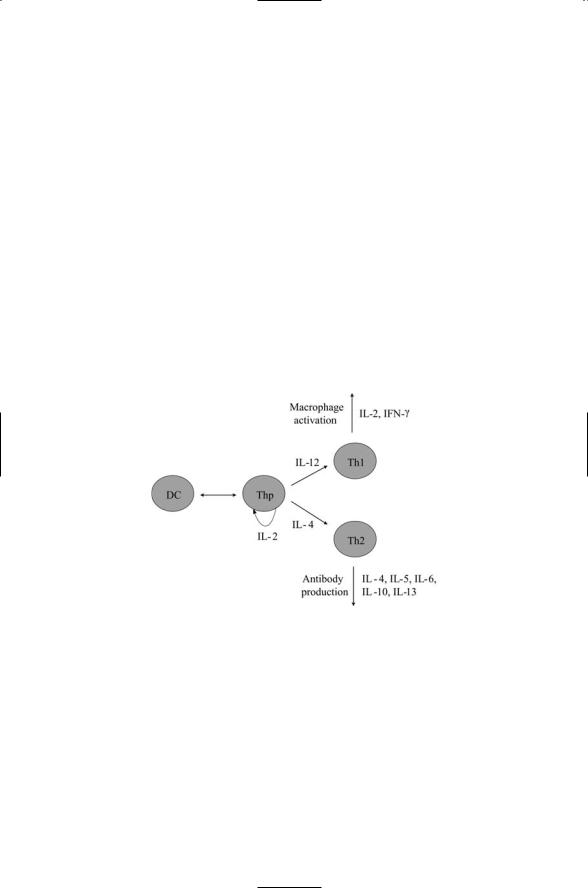
Interactions between dendritic cells and naïve T cells leading to differentiation into helper T cells is illustrative of interleukin signaling. Immature dendritic cells serve as sentinels in the body. Their morphology is specialized for capturing antigens from their surrounding. When they encounter an antigen denditic cells migrate to the secondary lymphoid organs. They differentiate into antigen-presenting dendritic cells, and interact with naïve T cells thereby stimulating their differentiation into either T1 helper cells or T2 helper cells (Figure 9.1). T1 and T2 helper cells perform different immune functions. T1 helper cells secrete cytokines that stimulate actions by macrophages in inflammatory responses. T2 helper cells secrete a mix of cytokines that triggers the differentiation of B cells into antibody-releasing plasma cells.
FIGURE 9.1. Helper T cell development: The first steps in the specification involve interactions between the immature dendritic cells and the pathogen they have encountered. These interactions determine the precise nature of the receptor complexes expressed on the surface of the antigen-presenting dendritic cells and the type of cytokines secreted when they interact with the naïve T cells in the secondary lymphoid tissue. Differentiation is triggered by interactions between the dendritic cell (DC) and a naïve T cell (Thp). The Thp expresses IL-2, which, acting in an autocrine manner on itself, leads to its differentiation into either a Th1 cell or a Th2 cell. IL-12 production stimulates differentiation into Th1 cells and inhibits formation of Th2 cells. IL-4 production stimulates differentiation into Th2 cells and inhibits formation of Th1 cells.

9.4 Five Families of Cytokines and Cytokine Receptors |
193 |
9.4 Five Families of Cytokines and Cytokine Receptors
There are five families of cytokines and cytokine receptors. The five families, representative members, and their most prominent activities are listed in Table 9.3. The first family in the table is the Toll/IL-1 family, named for the interleukin-1 (IL-1) cytokine, the family’s founding member. IL-1 is primarily sent from macrophages to lymphocytes stimulating lymphocyte activation. When received by granulocytes, IL-1 promotes inflammatory responses. The Toll receptors are key mediators of the innate immune response. Their ligands are not cytokines but instead are molecular markers found on the surfaces of pathogens.
The next family of cytokines is the tumor necrosis factor (TNF) family. The tumor necrosis factor superfamily consists of a group of ligands and receptors that coordinate and regulate growth, proliferation, and survival of leukocytes. They coordinate the development of lymphoid organs and temporary inflammatory structures, an essential part of the adaptive immune response. One group of these proteins conveys death signals that instruct cells to suicide, or die by apoptosis (discussed in Chapter 15). A key function of these signals is homeostasis, the maintenance of stable (baseline) populations and the preservation of a balance between different subpopulations of cells ready to respond to future infections.
Hematopoietin receptors are sometimes referred to as Class I cytokine receptors and the interferon/IL-10 receptors as Class II cytokine receptors. The hematopoietins are a family of more than 20 different signal molecules that are especially prominent in lymphocyte signaling. As already mentioned, interleukins, secreted by macrophages, T cells, and bone marrow stromal cells stimulate growth, proliferation, and differentiation of several cell types. In response to these signals, stem cells differentiate into progenitor B and T cells; B cells differentiate into antibody-secreting plasma cells and B cells and macrophages express core components of their antigenspecific responses.
Interferons mediate antiviral responses by alerting other leukocytes that a virus attack is underway and by upregulating genes whose products have antiviral activities. In addition, interferons act as antitumor agents by downregulating genes important for cell proliferation, and they regulate the expression of many genes essential for the adaptive immune response.
Lastly, leukocytes are guided to the site of an infection by chemokines, a family of small, 7- to 15-kDa protein chemoattractants. These molecules are secreted by several different kinds of cells at an infection site in order to recruit leukocytes from blood to sites of infection in tissue. Neutrophils, monocytes, fibroblasts, endothelial cells, and epithelial cells secrete chemokines. The chemokines form chemical concentration gradients that guide the migration of monocytes, neutrophils, eosinophils, and lymphocytes, and they arrest their movement at the appropriate location.
194 9. Signaling by Cells of the Immune System
9.5 Role of NF-kB/Rel in Adaptive Immune Responses
The NF-kB/Rel signaling module plays an important role in the inflammatory, innate, and adaptive immune responses. The signaling pathway activated in response to Toll receptor binding has several features in common with the pathway activated in response to tumor necrosis factor receptor binding. Both utilize signaling through the NF-kB signaling module; both convey signals through MAP kinase modules, and both use adapters belonging to the TRAF family to link upstream receptor binding to downstream intracellular signaling.
NF-kB proteins are transcription factors that regulate genes for cytokines, chemokines, adhesion molecules, antimicrobial peptides, and other factors important for immune responses. In the absence of activating signals these proteins form an inactive cytosolic module with their IkB inhibitors, and a set of IKK regulatory kinases.The IKK proteins consist of two catalytic subunits, IKKa and IKKb, and a regulatory subunit, IKKg, also called NF-kB essential modulator (NEMO). Both of the IKK catalytic subunits are competent to phosphorylate IkB proteins.The IKKs are themselves activated by upstream kinases, and are the key point of convergence of a variety of regulatory events triggered by extracellular signals and intracellular stresses.
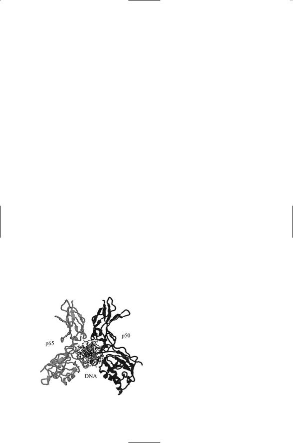
There are five members of the NF-kB family of proteins: NF-kB1 (p50/p105), NF-kB2 (p52/p100), c-Rel, RelB, and RelA (p65). In the absence of activating signals, these proteins are sequestered in inactive forms in the cytosol by inhibitory IkB factors. When the repressive effects of the IkBs are lifted, the NF-kBs form homoand heterodimers, the most common combination being p65/p50, and translocate to the nucleus where they function as transcription factors (Figure 9.2).
The NF-kB1 and NF-kB2 proteins are formed and sequestered in the cytosol as p105 and p100 precursors. They are proteolytically processed to make the functional p50 and p52 forms. The C-terminus portion that is
FIGURE 9.2. Binding of a NF-kB heterodimer to DNA: Shown is a p65/p50 NF-kB heterodimer (ribbon model) bound to an IFNb enhancer (ball-and- stick representation) viewed looking down along the DNA axis. The figure was prepared using Protein Explorer with atomic coordinates deposited in the PDB under accession numbers 1le5.