

Molecular and Cellular Signaling - Martin Beckerman
.pdf3.3 Protein Structure via X-Ray Crystallography |
51 |
FIGURE 3.4. Scattering of X-rays from a series of planes in a crystal: A beam of X-rays impinges on a series of crystal planes. Some of the light is scattered back (reflected) from each of the planes while the remainder of the light is transmitted. The optimal situation where the angle of incidence equals the angle of reflection is depicted in the figure.
W.H. Bragg and his son W.L. Bragg formulated the basic theory of relating diffraction patterns to atomic positions in a crystal in 1912–1913. They noted that a three-dimensional crystal could be viewed as a set of equidistant parallel planes. The conditions for maximal constructive interference are twofold. First, the scattering from each plane must be a specular, or mirror, reflection in which the angle of incidence equals the angle of reflection. This situation is depicted in Figure 3.4. Second, the X-ray wavelength l, distance between parallel planes d, and angle of reflection q, obey the relationship known as Bragg’s law:
2d sin q = nl. |
(3.4) |
In Eq. (3.4), n is an integer that can take on the values 1, 2, 3, and so on. When n = 1, the spots of light are known as first order reflections, and when n = 2, they are called second order reflections. First order reflections are more intense than second order reflections, and similarly for third and higher order contributions.
The light spots are produced when the light waves arrive at the detector in phase with one another and thus constructively interfere. In an X-ray diffraction experiment, the intensities and positions of the light spots are recorded. The diffraction patterns are converted into electron density maps through application of a mathematical operation known as a Fourier transform. Several tens of thousands of reflections are collected in a typical X-ray diffraction experiment. Computer programs, taking as input the resulting electron density map and knowledge of the primary sequence, are

52 3. Exploring Protein Structure and Function
FIGURE 3.5. X-ray crystallography: A unit cell is shown containing the molecule to be studied. It is replicated many times in the crystal sample. X-rays scattered from the atoms in the crystal produce a complex three-dimensional pattern of light and dark spots in the detector called reflections. The patterns of light and dark spots are a consequence of the way the waves from different atoms in different locations in the unit cell interfere with one another. A typical diffraction pattern produced in one-dimension from a pair of atoms is depicted in the figure. As can be seen, there is a series of peaks where the waves constructively interfere, and each peak is separated from the next by a trough where the waves from the two atoms destructively interfere. These reflections are then converted into an electron density map. A typical portion of an electron density map is depicted. The map, a contour plot, shows peaks where the electron density is high and valleys or open areas where the electron density is low. In the model-building portion of the data analysis, the threedimensional structure of the molecule is deduced using computer programs.
used deduce the three-dimensional arrangement of atoms in the protein (Figure 3.5).
X-ray crystallographic data have been used to deduce the atomic structure of more than 16,000 proteins to date. In 1953, Crick and Watson deduced the double helix structure of DNA using the X-ray crystallographic data of Franklin and Wilkins. This discovery was followed by those of Perutz and Kendrew, who used X-ray crystallography to deduce the atomic structure of hemoglobin and myoglobin in the period from 1953 to
1960. These were the first two proteins to be so described in atomic detail.
Since that time a rapidly increasing number of proteins have had their atomic structure solved. The three-dimensional coordinates (x, y, z) for each atom in the protein are deposited in the Protein Data Bank (PDB) and made available to the research community. The PDB repository was estab-

3.5 Determining Protein Structure Through NMR |
53 |
lished at Brookhaven National Laboratory in Long Island, and at several mirror sites throughout the world. Of the more than 16,000 structures for proteins and peptides that have been deposited in the PDB to date, 14,000 were determined using X-ray crystallography and 2000 using nuclear magnetic resonance (NMR) spectroscopy.
3.4Membrane Protein 3-D Structure via Electron and Cryoelectron Crystallography
The overall limitation of X-ray crystallography is the growing of purified crystals with sufficient numbers of molecules. This outcome is exceptionally difficult to achieve in the case of large and complex protein molecules such as those embedded in membranes that pass back and forth through the lipid bilayer several times. These proteins have a hydrophobic band that tends to destabilize them when they are removed from the lipid bilayer and solubilized. In electron crystallography, both the native environment and the biological activity of the membrane proteins are preserved.
The central idea in electron crystallography is similar to that which drives X-ray crystallography. By growing a crystal containing a large number of the molecules of interest one can greatly amplify the crystal’s weak signals. In this approach, two-dimensional membrane crystals are prepared. Electron diffraction patterns are then acquired using high-resolution cryoelectron microscopes. This method has been used to study light-harvesting complexes that function as antennae of solar energy in plants, and to investigate the atomic structure of porins of gram-negative bacteria. The method was first applied to the study of bacteriorhodopsin, the light-driven proton pump located in the purple membrane of Halobacteria and, more recently, to the analysis of the three-dimensional structure of the acetylcholine receptor, a neurotransmitter-gated ion channel located in nerve-muscle synaptic membranes.
3.5 Determining Protein Structure Through NMR
Nuclear magnetic resonance (NMR) spectroscopy is the second major experimental method used to determine the three-dimensional atomic structure of proteins. In this method, one utilizes pulses of electromagnetic radiation in the radiofrequency (RF) range of the EM spectrum. NMR pulse frequencies are typically in the range 300 to 600 MHz. These frequencies correspond to wavelengths of 100 to 50 cm, and the corresponding
RF energies are 10 orders of magnitude weaker that the X-ray energies. Photons of these energies are used to induce transitions between nuclear (proton and neutron) spin states. The subsequent relaxation of the nuclear spin distributions back to their equilibrium distribution is sensitive both to
54 3. Exploring Protein Structure and Function
the electron distribution surrounding the nucleus and to neighboring nuclear spins. As a consequence, one can deduce atomic structure of a protein from an analysis of its NMR spectra.
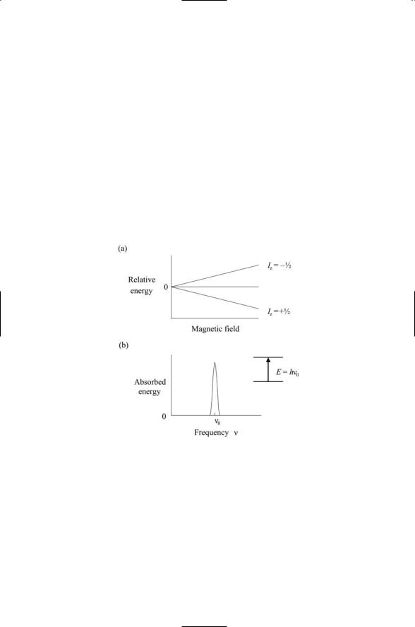
Electrons, protons, and neutrons have an intrinsic angular momentum, or spin. Because they have a spin angular momentum, they have a magnetic dipole moment and can interact with an external magnetic field as depicted in Figure 3.6. Like electrons, protons and neutrons have a spin of 1/2. Nuclei with an odd number of neutrons and an even number of protons, or alternatively, an odd number of protons and an even number of neutrons will have a net spin of 1/2. Spin 1/2 nuclei encountered in biomolecules are 1H, 13C, 15N, 19F, and 31P. This is not the only nonzero spin value possible. Nuclei with an odd number of proteins and also an odd number of neutrons, like 2H, have a spin of 1. In Figure 3.6, spin is indicated by Iz.
FIGURE 3.6. Splitting of energy levels of a spin 1/2 particle by an external magnetic field: (a) In the absence of a magnetic field, the energies of the spin up and spin down states of the spin 1/2 particle are the same. The energy levels are no longer the same in the presence of a magnetic field. The energy of the particle whose spin is aligned parallel to the magnetic field, the spin +1/2 particle, is lower than the particle whose spin is aligned against the external magnetic field, that is, the spin -1/2 particle. The energy levels are said to be split by the magnetic field. (b) If the spin 1/2 particles are exposed to a source of electromagnetic energy, while in the magnetic field, there will be a sharp absorption peak when the frequency of the electromagnetic wave exactly matches the frequency of the photon needed to trigger a transition from the lower to the higher energy level.
3.5 Determining Protein Structure Through NMR |
55 |
The negatively charged electron clouds surrounding nuclei reduce the magnitude of the magnetic field experienced by the spin 1/2 protons and neutrons residing in the atomic nucleus. This effect is called shielding. Each atomic species has a uniquely different electron cloud, and thus different atoms will undergo different line splittings in the presence of the same external magnetic field. Nearby atoms will influence the energy splitting through the shielding effects, as well. Atoms such as oxygen that are strongly electronegative will have a far greater influence on neighboring atoms than will, for example, carbon atoms.
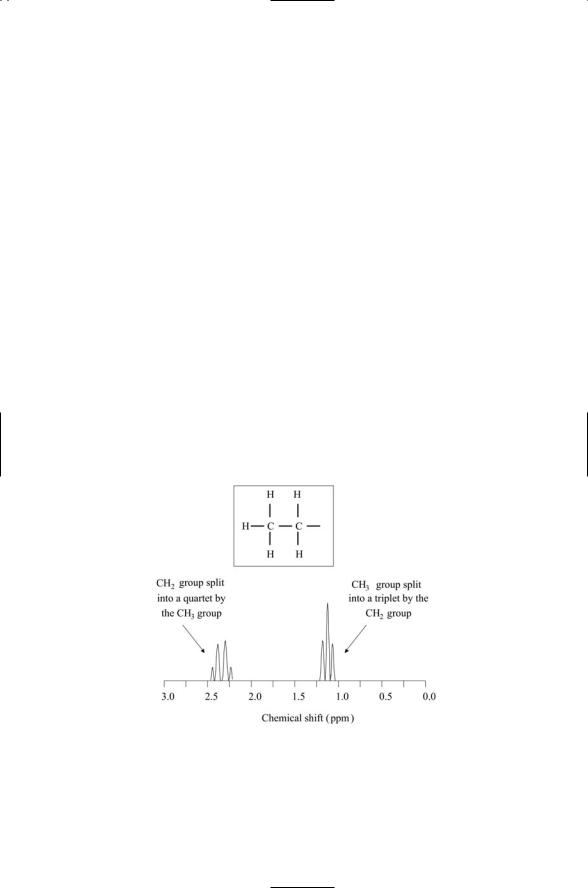
Shielding by the atom’s electron cloud and those of its neighbors give rise to several different observable effects. The electron clouds will alter the locations of the peaks; they will create clusters of peaks in place of single isolated peaks and produce variations in the peak heights. The creation of clusters of peaks arises through interactions of the spins (magnetic fields) of the atoms in one group with the spins (magnetic fields) of atoms in neighboring groups. In more detail, interactions between spins split the energy levels, and the transitions between these energy levels appear as the distinct peaks in the plot of chemical shifts. An example of this, for the case of interactions between a methyl group and a methylene group, is presented in Figure 3.7. If the methyl group were isolated from its neighbors, there would be a single hydrogen peak, since each of the three hydrogen atoms sees the same environment. The presence of a nearby CH2 group induces the splitting of the single peak into three peaks, and similarly, the presence
FIGURE 3.7. Chemical shifts of hydrogen atoms in a CH3CH2 molecular environment: The arrangement of atoms in the two groups is depicted in the insert. The chemical shifts are plotted with respect to a reference shift, taken as the zero of the axis, and are given in units of parts per million (ppm).

56 3. Exploring Protein Structure and Function
of the nearby CH3 group induces the splitting of the single peak for CH2 into four peaks. Variation in peak heights is not arbitrary, but instead is a direct consequence of the relative contribution to each peak from different spin-spin interactions (see Problem 3.4).
3.6Intrinsic Magnetic Dipole Moment of Protons and Neutrons
Protons and neutrons, like electrons, have an intrinsic magnetic dipole moment. The magnitude of the magnetic moment for a proton is
mp = 2.79 mN ,
and for a neutron it is
mn = -1.91mN .
The quantity mN is known as the nuclear magneton. It is defined in a manner analogous to the electron dipole moment as
mN = eh , |
(3.5) |
2Mp |
|
- p
where e is the unit charge, h is Planck’s constant divided by 2 , Mp is the proton rest mass, and c is the speed of light. Since the mass of a proton is 1837 times the mass of an electron, the magnetic dipole moment of a proton or neutron is 1/1000 that of an electron.
A free proton at rest in a uniform static magnetic field B0 oriented along the z-axis can occupy one of two spin states. It can either be in a spin up state (Iz = +1/2) or a spin down state (Iz = -1/2). The energy associated with lower energy spin up state is
E+ = -mp B0 ,
and that of the higher energy spin down state is
E- = mp B0 .
If an RF pulse of electromagnetic energy is applied that matches the transition energy, or energy difference, between these two states, the spin can be flipped from the lower energy state to the higher. The proton will then relax back to its lower energy state. The resonant frequency of the RF pulse that triggers the spin transition is
n0 |
= 2mp B0 . |
(3.6) |
|
h |
|
Nuclear magnetic resonance of protons in bulk matter was first demonstrated in 1946 by Felix Bloch and, independently, by Edward M. Purcell.

3.7 Using Protein Fluorescence to Probe Control Layer |
57 |
Their efforts followed earlier work by Isidor I. Rabi, who discovered the NMR effect in molecular beam experiments in 1937. Norman F. Ramsey, working in Rabi’s laboratory, acquired the first radiofrequency (RF) spectra the following year, and developed the chemical shift theory in 1949. These initial studies have evolved into a powerful technique used to study biomolecules in solution, and to a diagnostic tool, magnetic resonance imaging (MRI), used in medicine.
3.7 Using Protein Fluorescence to Probe Control Layer
Fluorescent proteins can be used as sensitive probes of the movements and interactions of control layer elements. Proteins that fluoresce emit light at a characteristic emission wavelength, lem, shortly after absorbing light at their characteristic excitation wavelength, lex. The wavelengths at which both absorption and emission occur fall in the short to middle portion of the visual spectrum. An energy level diagram, a schematic depiction of the energies of the low-lying electron orbitals of the protein’s fluorophore, is presented in Figure 3.3. As shown in the figure, there is a ground state band consisting of number of closely spaced energy levels, and an excited state band, also consisting of a cluster of energy levels. Levels within the excited state band are populated when light is absorbed. The emission of light corresponds to electronic transitions from the excited state band back down to the ground state band. The energies of absorbed and emitted photons differ slightly. The emitted photon has a greater wavelength, reflecting its slightly lower energy.
Green fluorescent protein (GFP) and other naturally fluorescent molecules are found in organisms ranging from the bioluminescent jellyfish
Aequorea victoria to the non-bioluminescent coral Discosoma striata. The ability of GFP to fluoresce is due to the presence of a fluorophore consisting of a sequence of three amino acid residues—Ser 65-Tyr 66-Gly 67. This internal sequence is post-translationally modified to a ring structure and the tyrosine is oxidized. These two changes convert the trio of amino acid residues into a fluorescing center. The natural form, or wild type, of GFP has an excitation maximum at 395 nm, a secondary excitation maximum at 470 nm, and an emission peak at 509 nm. These peaks correspond to green light. The protein extracted from Discosoma striata fluoresces in the red range, and is named dsRed. In addition to these naturally occurring fluorescent proteins a number of variants have been created with altered properties that improve their utility as markers. The excitation and emission peaks for several fluorescent protein variants are presented in Table 3.3.
The DNA sequence for the 28 kDa green fluorescent protein has been determined. To make a fusion protein, the complementary DNA, or cDNA, of the protein of interest is inserted into a vector along with the DNA for the GFP. When the combined gene is expressed in a transfected cell (the

58 3. Exploring Protein Structure and Function
TABLE 3.3. Spectral properties of green fluorescent protein variants.
GFP variant |
Color |
lex (nm) |
lem (nm) |
eBFP |
blue |
380 |
440 |
eCFP |
cyan |
434 |
476 |
eGFP |
green |
488 |
509 |
eYFP |
yellow |
514 |
527 |
dsRed |
red |
558 |
583 |
|
|
|
|
cell into which the vector was inserted) the resulting fusion protein will contain the GFP covalently attached to the study protein, in a way that does not interfere with the normal operation of that protein. The fusion protein functions as a fluorescent reporter. When excited either by a laser or by an incandescent lamp, the protein will emit light and thus report its presence. Fusion proteins made in this manner have been used to study how signaling proteins move about the living cell. They have been employed to visualize the subcellular compartmentalization of the signaling molecules, and to view how the proteins move from one locale to another, in and out of organelles, and back and forth to the plasma membrane.
In Aequorea victoria, GFP operates in close association with another protein, aquorin, a naturally luminescent protein that emits blue light. Energy is transferred in a radiationless manner from the fluorophore of aquorin to the fluorophore of GFP. The GFP absorbs the blue light and in turn emits green light. The process of radiationless energy transfer from one fluorophore to another is called fluorescence resonance energy transfer
(FRET). This process can occur when the energy level differences involved in emission from one fluorophore overlap the energy level differences involved in excitation of the second fluorophore. That is, FRET can occur if the donor emission spectrum overlaps the acceptor excitation spectrum.
3.8 Exploring Signaling with FRET
Fluorescence resonance energy transfer can be used to explore signaling within the living cell. Fluorescence resonance energy transfer and related techniques can be used to study protein-protein interactions. In this approach, one fluorophore is fused to the first of two proteins and the other to the second protein. When the first protein is well separated from the second, the first protein will emit light at its characteristic emission wavelength upon stimulation. If the two fluorophores satisfy the conditions for FRET, there will be a shift in wavelength of the emitted light from that of

3.8 Exploring Signaling with FRET 59
the first protein (the donor) to that of the second (the acceptor) when the two proteins some into contact. These processes can then be studied with light microscopy. The nature of the emitted light is sensitive to relative orientation of the two fluorophores and to their spatial separation. These dependencies can be exploited in the design of the fusion proteins, so that the emission spectra reflect conformational changes and properties of the protein-protein interactions.
The principle behind the use of FRET for studying protein-protein interactions can be applied in a variety of ways to the study of signaling. As was the case for protein-protein interactions, two particles are brought into close proximity, one fused to a fluorescent donor and the other to a fluorescent acceptor. The sought after signaling interactions are then studied and quantified by measuring the changes in FRET efficiency. When the two fluorophores are far apart the efficiency is low, but this changes in the presence of the interaction of interest. This is the basis for studying post-translational modifications such as proteolytic cleavage and phosphorylation; the movement of small signaling intermediaries such as cyclic adenosine monophosphate (cAMP), Ca2+, and cytochrome c; and it has been used to study the trafficking of proteins as they are secreted from the cell.
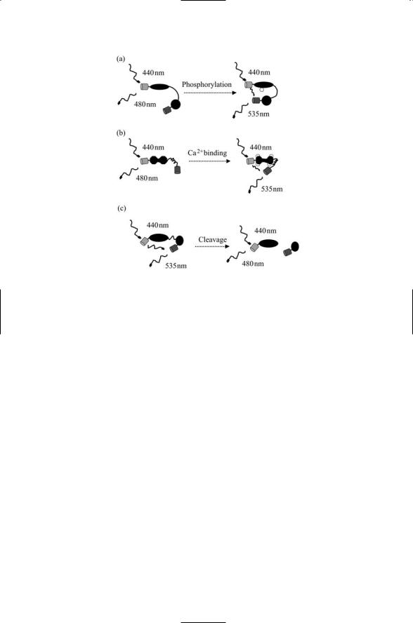
Several examples illustrating how the FRET principle is applied are presented in Figure 3.8. In the first example, that of phosphorylation by a protein kinase, a phosphorylation reporter is created. This (the reporter) protein contains an amino acid sequence that is typically phosphorylated by the kinase of interest. When the sequence is phosphorylated by the kinase a second domain in the reporter protein recognizes and binds to the phosphorylated sequence. The second domain contains a fluorescent protein that acts as the acceptor, while the first domain contains the donor. When the protein is phosphorylated by the kinase and the second domain binds it, the fluorescent acceptor is brought closer to the donor and the
FRET efficiency increases.
A similar procedure is followed for proteins called cameleons, which report the presence of calcium, an exceptionally important small signaling molecule. In this case, calmodulin (CaM), a calcium-binding protein, is used in constructing the reporter. Again a donor and acceptor are appended; the donor is appended to CaM and the acceptor to a peptide known as M13 that binds tightly to CaM what the latter binds calcium. The third example presented in Figure 3.8 represents the converse situation, that of proteolysis. To study proteolysis, a protein is created with donor and acceptor fluorescent proteins. The two regions are connected to one another by an amino acid sequence that is characteristically cleaved by the proteolytic enzyme
(protease) of interest. When the reporter protein is cleaved by the proteolytic protein, FRET ceases as the two fluorescent proteins drift apart and no longer interact.
60 3. Exploring Protein Structure and Function
FIGURE 3.8. Fluorescence resonance energy transfer (FRET) used to explore intracellular signaling: (a) Phosphorylation—A laser emits radiation at 440 nm, triggering emission from a CFP at 480 nm in the left hand panel. The phosphorylation recognition domain connected to the first domain by a flexible linker binds the phosphorylated protein following kinase activity in the right hand panel. These changes result in FRET and an increased emission from the YFP at 535 nm. (b) Calcium binding—In the absence of calcium, a calcium-binding protein, calmodulin, is in a confirmation that loosely tethers an M13 peptide. When it binds four calcium ions, it assumes a dumbbell-like shape that induces a tight binding of the M13 peptide resulting in FRET and increased emission at 535 nm. (c) Proteolysis—In the absence of the protease, the two domains remain in close proximity and emit radiation at 535 nm. When the protease cleaves the linker, the two portions of the protein move apart and no longer exhibit FRET.
3.9 Exploring Protein Structure with Circular Dichroism
Another important property of biomolecules is their chirality, or handedness. All amino acids used to make proteins are left handed (L-amino acids) and all sugars used in DNA and RNA are right handed (D-sugars). This is not accidental. A consistent handedness on the part of protein-forming amino acids is essential for the proper folding of the polypeptide chains into compact three-dimensional shapes. Not all molecules possess a handedness. For this to occur a central atom surrounded by four different atoms or atomic groups must be present. The alpha carbon in the amino acids serves as the chiral center, and all amino acids with the exception of glycine satisfy