

Dankwardt A. - Immunochemical assays in pesticide analysis(en)
.pdfIMMUNOCHEMICAL ASSAYS IN PESTICIDE ANALYSIS
Immunochemical Assays in
Pesticide Analysis
Andrea Dankwardt
Sension GmbH, Augsburg, Germany
1 |
Introduction |
1 |
|
2 |
Antibodies |
2 |
|
|
2.1 |
Antibody Structure |
2 |
|
2.2 |
Antibody Production |
3 |
|
2.3 |
Immunogens |
3 |
3 Immunoassay Types |
5 |
|
3.1 |
Assay Formats |
5 |
3.2 |
Labels |
5 |
3.3 |
Solid Phases |
6 |
4 Properties of Competitive Immunoassays |
6 |
|
4.1 |
Dose ± Response Curve |
6 |
4.2 |
Quality Control |
7 |
4.3 |
Cross-reactivity |
8 |
4.4 |
Sample Preparation and Matrix |
|
|
Effects |
8 |
5 |
Application to Environmental Samples |
9 |
|
|
5.1 |
Water |
9 |
|
5.2 |
Soil |
13 |
|
5.3 |
Food |
13 |
|
5.4 |
Biomonitoring |
14 |
6 |
New Developments |
14 |
|
Acknowledgments |
16 |
||
Abbreviations and Acronyms |
16 |
||
Related Articles |
16 |
||
References |
16 |
||
|
|
|
|
Immunochemical assays (immunoassays, IAs) are biochemical assays which work according to the law of mass action. They are based on the recognition of an antigen (Ag) or a hapten by antibodies (Abs). Abs are serum glycoproteins of the immunoglobulin (Ig) class and are produced by the vertebrate immune system against foreign material of high molecular mass. The result of the binding reaction between the Ab and an analyte is usually made visible by means of enzymatic, chemiluminescent, fluorescent or radioactive markers. According to the label used IAs can be classified into enzyme immunoassays (EIAs), radioimmunoassays (RIAs), fluorescence immunoassays (FIAs) or chemiluminescent immunoassays (CLIAs). The measuring range of most IAs for pesticides is in the parts
1
per trillion to lower parts per billion range. A lot of samples can be analyzed within a short time, while only low sample volumes are necessary. In many cases (water, some liquid food samples) no extraction step and no cleanup are necessary. Not all assays are completely specific to one single compound. Cross-reactivities of the Abs with haptens similar to the analyte can be observed. In some cases, matrix effects may occur, especially with soil or colored food extracts. Therefore, validation of the assays for the matrix of interest should be carried out. As IAs are usually targeted at a single analyte or a group of analytes, multianalyte approaches using Ab arrays or a combination of immunochemical techniques with liquid chromatography (LC) are pursued.
1 INTRODUCTION
Interest in immunochemical assays for the determination of pesticides has been steadily increasing. IAs are now commonly applied for the analysis of contaminants in water, soil, food and body fluids..1 ± 9/ The first immunological experiments had already been carried out as early as the late eighteenth century when Edward Jenner, an English physician, used cowpox to prevent infection with smallpox. Based on these studies Louis Pasteur developed the use of attenuated strains of microorganisms for successful vaccinations. Emile Roux and Alexandre Yersin then found that immunity is caused by soluble compounds of microorganisms, which they called toxins. These toxins induce specific compounds in the immunized animal, which were named ``antitoxins'' by Emil von Behring and Shibasaburo Kitasato (1890) and are now called Abs. The Ab ``generating'' compounds are known as Ags.
Around the turn of the century it was shown that Abs are not only produced against microorganisms and their toxins, but also by other substances such as milk, protein or plant-derived toxins. Paul Ehrlich was the first to carry out quantitative studies on Ag ± Ab interactions. The great interest in this field led to the first book on immunochemistry, published by Svante Arrhenius in 1907..10/ Karl Landsteiner also belongs to the pioneers in immunochemistry. He systematically used small artificial molecules, which he called haptens, coupled to a carrier molecule for immunization. In 1923 Heidelberger and co-workers found polysaccharides to be antigenic as well.
Studies by Rodney Porter (1959) and Gerald Edelman (1961) have provided the chemical structure of the Ab molecule. The enormous variety of Abs was explained by Frank McFarlane Burnet in 1957, based on a hypothesis of Niels Jerne dating from 1955, the now widely accepted clonal-selection theory. It describes each Ab-producing
Encyclopedia of Analytical Chemistry
Edited by Robert A. Meyers. © John Wiley & Sons Ltd, Chichester. ISBN 0471 97670 9
2
cell as carrying on its surface only one type of Ab as a receptor. The binding of a respective Ag to this receptor leads to a clonal expansion of this cell and to the maturation of Ab-producing cells.
Immunochemical methods have their origin in the medical field. The first IA, a RIA for the quantification of insulin in serum, was described by Yalow and Berson..11/ Later, radiolabels were replaced by enzymes in EIAs by Engvall and Perlmann.12/ and Van Weeman and Schuurs..13/ Since then radiolabels have obtained broad application in medical diagnostics and environmental analysis.
IAs belong to the most common methodology in the field of immunoanalysis. Even though Abs are (still) produced by a biological process, IAs are nevertheless chemical analytical procedures. The basic principle applying to all immunoreactions is based upon the law of mass action. In the equilibrium reaction between a free Ag or a hapten, such as a pesticide, and the Ab forming the hapten ± Ab complex HAb (Dbound hapten), represented
by Equation (1), |
|
H C Ab D HAb |
.1/ |
the affinity constant K determines the concentration ratio between the bound hapten and the free reaction partners, Equation (2):
[HAb] |
|
K D [H][Ab] mol 1 |
.2/ |
PESTICIDES
Occupancy of antibody binding sites by analyte
|
|
|
|
|
|
|
|
|
|
|
|
|
|
|
|
|
|
|
|
|
|
|
|
|
|
|
|
|
|
|
Occupied |
Unoccupied |
|
|
|||||||||||||||||||||
|
|
|
|
|
|
|
binding sites |
|
|
||||||||||||||||||
|
|
Competitive |
|
Measurement of |
|
|
|||||||||||||||||||||
|
|
|
|
|
|||||||||||||||||||||||
|
|
assays |
|
unoccupied |
|
|
|||||||||||||||||||||
|
|
|
|
|
|
|
|
|
|
|
binding sites |
|
|
||||||||||||||
(a) Immobilized antibody |
|
|
(b) Immobilized coating |
|
|
||||||||||||||||||||||
|
|
|
|
||||||||||||||||||||||||
|
|
|
|
|
|
|
|
|
|
|
|
|
|
|
conjugate |
|
|
||||||||||
|
|
|
|
|
|
|
|
|
|
|
|
|
|
|
|
|
|
|
|
|
|
|
|
|
|
|
|
|
|
|
|
|
|
|
|
|
|
|
|
|
|
|
|
|
|
|
|
|
|
|
|
|
|
|
|
|
|
|
|
|
|
|
|
|
|
|
|
|
|
|
|
|
|
|
|
|
|
|
|
|
|
|
|
|
|
|
|
|
|
|
|
|
|
|
|
|
|
|
|
|
|
|
|
|
|
|
|
|
|
|
|
|
|
|
|
|
|
|
|
|
|
|
|
|
|
|
|
|
|
|
|
|
|
|
|
|
|
|
|
Inverse relation between |
Inverse relation between |
antibody-bound tracer and |
coating conjugate-bound |
analyte concentration |
labelled antibody and |
|
analyte concentration |
Antibody |
Immobilized |
Labeled |
|
antibody |
antibody |
||
|
|||
Analyte |
Immobilized |
Labeled |
|
(hapten) |
analyte |
analyte |
A low detection limit (DL) in an IA therefore requires a high affinity of the Ab toward the analyte, which is expressed by a high affinity constant. For further details refer to Hock et al..14/
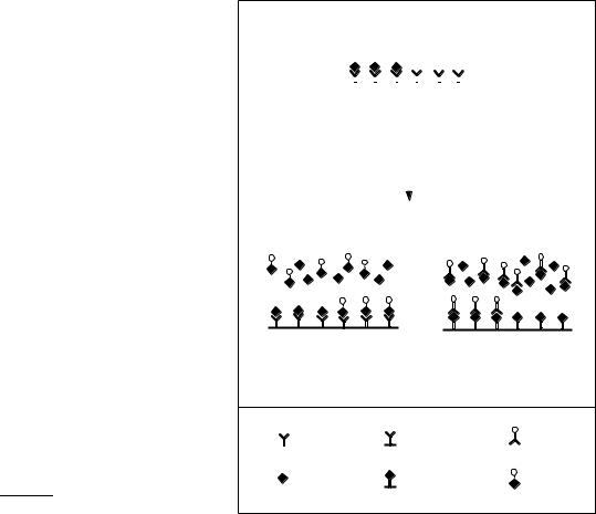
IAs are based upon the measurement of Ab bindingsite occupancy by the analyte (Figure 1). This reflects the analyte concentration in the sample. Since the binding reaction does not produce a signal which can be detected by simple means, various markers, e.g. radioactivity, enzymes, or fluorescence, are employed for the detection of the immunoreaction (see section 3.2). However, more sophisticated techniques like some immunosensing methods do not rely on a label (see section 6).
2 ANTIBODIES
2.1 Antibody Structure
Immunochemical analysis is based upon the specific reaction between an Ab and its corresponding Ag or hapten. Abs are part of the vertebrate defense system (for more details refer to immunology textbooks, for example Golub.15/ and Roitt.16/). They are serum glycoproteins
Figure 1 Principle of the competitive IA. In the first format with immobilized Ab (a) the plates are coated with Ab. Analyte and enzyme-labeled analyte compete for the Ab binding sites. In the second format a hapten ± protein conjugate is immobilized in the solid phase (b). This protein conjugate and the free analyte compete for the binding sites of the Ab in solution. (Reproduced from Dankwardt and Hock.7/ with permission from Food Technology and Biotechnology.)
of the Ig class produced by the immune system against foreign material such as pathogens or xenobiotics, and bind the target substance with high selectivity and affinity. Although there are five distinct classes of Ab in most higher mammals (IgA, IgD, IgE, IgG, IgM) IgG makes up approximately 80% of the total Ig in human serum. Most IAs rely upon IgG as the major Ig.
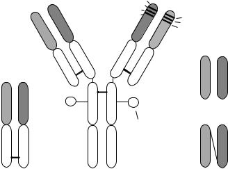
The basic structure of an Ab molecule is shown in Figure 2. It consists of two identical heavy (H) chains and two identical light (L) chains stabilized and linked by interand intrachain disulfide bonds. The H- and L-chains are organized into variable and constant regions. The Ag binding site (combining site) is formed by the association of parts of the variable regions of the H- and L-chains, located at the amino terminal end. The
IMMUNOCHEMICAL ASSAYS IN PESTICIDE ANALYSIS
|
Heavy chain |
FR1 |
Antigen binding site |
|
Light chain |
FR2 |
|
|
|
FR3 |
|
|
||
|
|
CDR1 |
|
|
|
VH |
FR4 |
|
|
|
CDR2 |
|
||
|
VL |
|
|
|
|
|
CDR3 |
|
|
|
|
|
|
|
|
|
CH1 |
|
|
|
|
CL |
VL |
VH |
VL |
VH |
CH2 |
|
|
|
|
Fv |
||
|
|
|
|
|
|
|
Carbohydrate |
|
|
CL |
CH1 |
CH3 |
CL |
VH |
Fab |
Antibody |
scFv |
||
Figure 2 Structure of IgG Abs and their fragments (modified after Hock et al..17/). scFv is the recombinant antibody fragment, a single chain fragment containing only the variable region, FR is the frame region, VL is the variable region of light chain, VH is the variable region of heavy chain, CL is the constant region on the light chain, CH1, CH2, CH3 is a constant region on the heavy chain.
variable regions of both chains are organized into three hypervariable or complementarity determining regions (CDRs) separated by four framework regions. The greatest amino acid sequence variation occurs within the CDRs whereas the framework regions are more conserved. It is assumed that the association of the CDR regions forms the combining site. The lower part of the molecule, the Fc (antibody fragment containing the crystallizable fragment) is responsible for some important biological effector functions such as complement fixation and is not necessary for Ag or hapten binding. It contains the last heavy chain domains. The whole of the Ig molecule or Ab fragments, F(ab)2 and Fab (antibody fragments containing the antigen binding site(s)) can be used in IAs.
A substance that after injection into the body of a vertebrate induces a specific Ab synthesis, is called an Ag. Ags are principally macromolecules, for instance proteins, polysaccharides or nucleic acids. Synthetic polymers also belong to the antigens, i.e. they can be used as, or act as, Ags. Small molecules (haptens) such as pesticides have to be coupled to a macromolecular carrier to elicit an Ab response (see section 2.3). The ability of an Ab molecule to bind an Ag or a hapten specifically is controlled by structural and chemical interactions between the ligand and the Ab at the combining site. The Ag ± Ab interaction is reversible and does not involve formation of covalent bonds..16/ The binding is a result of a variety
3
of interactions such as hydrophobic, ionic, H-bonding, p-p electron interaction, and van der Waals forces. The binding energy (relative affinity of the Ab) increases with the number of specific chemical interactions between the analyte and the amino acid residues in the Ab combining site. Therefore, the selectivity and sensitivity of an IA is controlled by the nature of the Ag ± Ab binding process.
2.2 Antibody Production
Ab production is conveniently carried out in warm blooded animals, e.g. rabbits, sheep, mice or chickens..17/ Polyclonal antibodies (pAbs) are obtained from the serum and comprise a mixture of different Ab populations. Monoclonal antibodies (mAbs) consist of a single monospecific Ab population. These Abs are produced in cell culture by a single hybridoma cell derived from the fusion of B-lymphocytes with myeloma cells..18/ The hybridoma cells can then be propagated almost indefinitely in culture and will continue to produce the Ab of the lymphocyte parent. Since an individual lymphocyte produces only a single Ab type, all of the Ab molecules produced by a hybridoma cell line derived from a single hybrid cell are identical and have the same binding properties. Therefore, the hybridoma technology guarantees the unlimited production of mAbs with constant characteristics..19/ Owing to the great effort involved in mAb production many IAs still employ pAb. A third possibility for creating Abs has emerged, recombinant antibody (rAb) techniques. Here, Ig genes can be cloned, introduced and expressed in inexpensive and relatively simple host systems..20;21/ Although several nonmammalian host systems (yeast, plant and insect cells) have been used to produce rAbs, the most common vehicle is Escherichia coli..22;23/ The main properties of pAbs, mAbs and rAbs are listed in Table 1.
2.3 Immunogens
Most pesticides are of low molecular mass and therefore are not ordinarily antigenic. They have to be coupled to a carrier molecule, usually a protein, in order to induce an Ab response in the vertebrate immune system..14/ The site of coupling to the carrier, the coupling procedure as well as the number of haptens bound to one carrier molecule can be of major importance for the sensitivity and the selectivity of the resulting Ab (for reviews refer to Erlanger,.24;25/ Goodrow et al..26/ and Szurdoki et al..27/).
The protein carriers used in various laboratories include globulin fractions, serum albumins of different species, hemocyanin, ovalbumin, thyroglobulin, and fibrinogen. Also nonproteinaceous carriers have been used
4 |
|
|
PESTICIDES |
Table 1 Properties of polyclonal, monoclonal and recombinant Abs |
|
||
|
|
|
|
Properties |
pAb |
mAb |
rAb |
|
(Ab from serum) |
(Ab from hybridoma cells) |
(Ab produced by gene technology) |
|
|
|
|
Supply |
Limited and variable |
Unlimited production possible |
Unlimited production possible, |
|
|
|
immunization not mandatory |
Uniformity |
Changing properties with |
Constant properties of a mAb |
Constant properties of a rAb, can be |
|
different sera and bleedings |
|
changed by genetic manipulations |
Affinity |
Mixture of Ab with different |
Uniformly high or low, can be |
Uniformly high or low, can be |
|
affinities, affinity often higher |
selected by testing |
selected by testing and can be |
|
with pAb |
|
modified |
Cross-reactivity |
Results from different |
Different, dependent upon the |
Different, dependent upon the |
|
selectivities and low affinity |
individual Ab |
individual Ab, can be modified |
|
interactions |
|
|
Classes and |
Typical spectrum |
One defined isotype |
Different, depending on molecular |
subclasses |
|
|
design |
Demands on Ag |
High purity required for specific |
Impure Ags or mixture of Ags |
Impure Ags or mixture of Ags can |
|
antisera |
can be used for immunization, |
be used for immunization, pure |
|
|
pure Ags necessary for |
Ags necessary for screening, |
|
|
screening |
immunization not mandatory |
Costs |
Low |
High |
Once established, low |
|
|
|
|
such as liposomes or dextran..28;29/ Keyhole limpet hemocyanin (KLH), a protein from mollusks, is often viewed as a superior carrier because it is foreign to the vertebrate immune system..30/
Another important issue concerns the optimal number of haptens bound to the carrier protein (i.e. optimal epitope density). Highly substituted carriers usually lead to the best results. For bovine serum albumin (BSA) molar ratios of 10 : 1 to 20 : 1 (hapten : carrier) are desirable; for larger molecules such as hemocyanin, ratios of 800 : 1 to 1000 : 1 should be obtained..17/ However, very high ratios may reduce immunogenicity because of either the changes in tertiary structure of the protein caused by masking of the essential free amino groups or the removal of critical determinant sites on the carrier by haptenic blocking.
For the production of pAbs the purity of the hapten used to prepare an immunoconjugate should be as high as possible. After synthesis of haptenic substances, closely related substances may be present in small quantities, leading to the production of nonspecific Abs and, consequently, to unwanted cross-reactivities of the antisera. This is not a problem with mAb, because a single cell line producing only one kind of Ab with the desired properties can be selected.
Another important consideration is the point of attachment on the hapten. Ab specificity is directed primarily at the part of the hapten molecule farthest away from the functional group that is linked to the protein carrier..25/ Even better specificity can be obtained with conjugates in which the hapten is coupled to the carrier via a spacer, thereby giving much better exposure of the hapten on the surface of the carrier. The spacer should be attached as far as possible from the unique
determinant structures..26/ Usually C3 ± C6 spacers are used; if the spacer is too long, it may bend back to the carrier and the hapten will not be properly exposed. Strategies for IA hapten design for the triazine, arylurea and chloroacetanilide herbicides have been summarized by Goodrow et al..26/
The functional groups of the hapten govern the selection of the method to be used to conjugate the hapten to the functional groups of the carrier. The functional groups of the protein carrier available for attachment of the haptens are the carboxyl group of the C terminal and of the aspartic and glutamic acid residues, the amino group of the N terminal and the lysine residues, the imidazol and phenolic functions of the histidine and tyrosine residues, respectively, and the sulfhydryl group of cysteine residues. General procedures for the preparation of conjugates can be found in Erlanger..24;25/
After coupling, characterization of the conjugates can be carried out (see Erlanger.25/). Generally, the haptenic groups have an absorbance spectrum that can be differentiated from the protein carrier. Elemental analysis for the chlorine content can be carried out for some triazine conjugates. A more direct procedure is the incorporation of some radioactive hapten in the conjugation procedure. Another approach is quantitating the change in free amino groups as a result of conjugation. A recently applied technique is the determination of hapten density by matrix-assisted ultraviolet laser desorption/ionization mass spectrometry (MALDIMS).31/ and electrospray ionization mass spectrometry (ESIMS)..3/ Application of energy-minimized molecular modeling methods to hapten design will help to choose the best derivatives and conjugation methods for successful Ab production..32/

IMMUNOCHEMICAL ASSAYS IN PESTICIDE ANALYSIS
3 IMMUNOASSAY TYPES
3.1 Assay Formats
For low-molecular-mass analytes (haptens) such as pesticides in solution, competitive tests have to be employed, using limiting Ab concentrations. The tests can be performed as homogeneous assays without separation of the reactants,.33/ but more common are heterogeneous tests where unreacted reagents are removed before evaluation. Two different formats are available, (1) with immobilized Ab (Figure 1a) and (2) with immobilized coating conjugate (Figure 1b). In variant (1) analyte and a labeled analyte (tracer) compete for the free Ab binding sites. After removal of unbound reactants the bound tracer yields a signal that is inversely proportional to the analyte concentration. The variant (2) employs an immobilized hapten-carrier conjugate on the solid phase to which analyte and Ab are added. The Ab binds to the free analyte or to the immobilized hapten according to the concentration of the reactants. If a labeled Ab is used, the amount of Ab bound to the solid phase can be directly determined after a washing step. Alternatively, a secondary labeled Ab may be used to detect the Ab which has bound to the solid phase. The signal is inversely proportional to the amount of free analyte in the sample. Very sensitive competitive IAs have been developed with DLs between 1 and 50 ng L 1, for example for the triazines and urea herbicides..34 ± 36/
An example for a homogeneous assay system is the polarization fluoroimmunoassay (PFIA). PFIA measures the increased polarization of fluorescence when a fluorophore-labeled hapten (tracer) is bound by a specific Ab, and the decreased signal when free analyte competes
Table 2 Enzyme systems commonly used for EIAs
5
with the tracer for binding..37/ While these assays are easy to carry out and very suitable for automation, they usually show a lower sensitivity than EIAs, e.g. for simazine a DL of 5 μg L 1 was observed..37/
Noncompetitive assays can only be applied for high- molecular-mass analytes with more than one antigenic determinant (i.e. Ag) or low-molecular-mass analytes (haptens) bound to a solid phase, exposing the antigenic determinant. They work with an Ab excess. Noncompetitive IAs have been employed for the detection of soil-bound pesticides..38;39/ In this case the soil particles, to which the pesticide residues have bound, form the solid phase, and the residues can be detected by a labeled Ab specific to the analyte.
3.2 Labels
Depending on the label, IAs are classified in different groups. Radioisotopes are used in RIAs, enzymes in enzyme-linked immunosorbent assays (ELISAs) or EIAs, fluorophores in FIAs or PFIAs and chemiluminescent compounds in CLIAs. Additional types of IA exist, but are not very common in pesticide analysis. A more detailed description of these IAs can be found in Gosling..40/
EIAs are most commonly used in pesticide analysis as they avoid the necessity of working with radioactive material and low DLs can be reached. Simple and cheap photometers which give an extremely rapid measurement capability and long-lasting stability of the colored product after the reaction has stopped make EIA superior to fluorimetry or luminometry, even though with these methods lower DLs may be reached..33/ Enzymes
Enzyme |
Source |
Molecular |
pH |
Colorimetric |
Fluorometric |
Luminometric |
|
|
weight |
optimum |
substrates |
substrates |
substrates |
|
|
|
|
|
|
|
Alkaline |
Calf intestine |
100 000 |
9 ± 10 |
p-Nitrophenyl- |
4-Methylumbelliferyl- Adamantyl-1,2- |
|
phospha- |
|
|
|
phosphate |
phosphate |
dioxyethane |
tase |
|
|
|
|
|
Phenylphosphate- |
|
|
|
|
|
|
substituted |
|
|
|
|
o-Nitrophenyl-b-D- |
4-Methylumbelliferyl- |
dioxyethane |
b-Galacto- |
Escherichia coli |
540 000 |
6 ± 8 |
± |
||
sidase |
|
|
|
galactopyranoside |
b-D-galacto- |
|
|
|
|
|
Chlorophenolic |
pyranoside |
|
|
|
|
|
red-b-D-galacto- |
|
|
|
|
|
|
pyranoside |
|
|
Peroxidase |
Horseradish |
40 000 |
5 ± 7 |
2,20-Azino-di(3-ethyl- |
p-Hydroxyphenyl- |
Luminol |
|
|
|
|
benzthiazoline sulfonic |
acetic acid |
|
|
|
|
|
acid-6) (ABTS)/H2O2 |
p-Hydroxyphenyl- |
|
|
|
|
|
3,30-5,50-Tetramethyl- |
propionic acid |
|
benzidine (TMB)/H2O2 o-Phenylendiamine (OPD)/H2 O2
6 |
|
|
|
|
|
|
|
PESTICIDES |
||
|
Table 3 Solid phases used for EIAs |
|
|
|
|
|
|
|
|
|
|
|
|
|
|
|
|
|
|
|
|
|
Material |
Form |
Binding |
|
|
|
Capacity |
|
|
|
|
|
|
|
|
|
|
|
|||
|
Polystyrene |
Microtiter plates, tubes, pins, |
Noncovalent |
250 ± 500 ng cm2 |
|
|
|
|||
|
|
beads |
|
ca. 300 ng cm 2 |
|
|
|
|||
|
Polyethylene |
Tubes |
Noncovalent |
|
|
|
||||
|
Polypropylene |
Microtiter plates |
Noncovalent |
ca. 300 ng cm 2 |
|
|
|
|||
|
Polyvinylchloride and |
Microtiter plates, membranes |
Noncovalent |
ca. 300 ng cm 2 (plates) |
||||||
|
similar |
|
|
ca. 300 ng cm 2 (beads) |
||||||
|
Polycarbonate |
Beads, membranes |
Noncovalent |
|||||||
|
Nitrocellulose |
Microtiter plates, membranes |
Noncovalent |
ca. 100 μg cm 2 |
|
|
|
|||
|
Protein A coated |
Microtiter plates, beads |
Noncovalent |
20 mg mL 1 (for Ab only) |
||||||
|
Activated polymer, with |
Microtiter plates, beads |
Covalent, using |
2 |
|
1013 |
1 |
14 |
reactive |
|
|
amino or carboxyl groups |
|
bifunctional reagents |
|
|
± 2 10 |
|
|||
|
|
|
sites/cm |
|
|
|
||||
|
Magnetic |
Beads |
Depends on the |
Varies |
|
|
|
|
||
|
|
|
surface of the beads |
|
|
|
|
|
|
|
|
|
|
|
|
|
|
|
|
|
|
commonly used as labels in heterogeneous EIA are listed in Table 2.
The following requirements are necessary for the use of an enzyme as a marker:
(1)high specific activity (turnover number) of free enzyme and after labeling,
(2)availability of soluble, purified enzyme at low cost and reproducible quality,
(3)high stability in free and conjugated form under storage and assay conditions,
(4)presence of reactive groups for covalent linkage to hapten,
(5)simple and gentle conjugation methods,
(6)inexpensive and stable nontoxic substrates with formation of stable chromogenic, fluorogenic and/or chemiluminogenic products.
covalent binding. Those solid supports contain amino or carboxy groups on a modified surface through which the immunoreagents can be bound by water-soluble carbodiimides or bifunctional reagents such as glutaraldehyde.
Other solid-phase supports for IAs are membranes. They can be used for dip sticks, which are incubated for a short time in the solution.36/ or for dot blots and immunofiltration tests. Here the reactants are filtered through the membrane..43;44/ The test principle is the same as for the microtiter plate tests but the reaction time is much shorter owing to the high surface area of the membrane and the short distance between reaction partners. Application of remission measurements yields a proportional relationship between analyte and remitted light. By using a pocket reflectometer, this set-up is ideally suited for field-monitoring purposes..45;46/
3.3 Solid Phases
IAs are mainly carried out in 96-well polystyrene, polyethylene, polypropylene or polyvinyl microtiter plates, owing to the easy separation of the reactants in a washing step, but polystyrene tubes, beads or pins are also available (Table 3). The plastic plates are of comparatively low binding capacity and low surface area to volume ratio. High-binding supports include agarose and cellulose. Particulate solid phases are very efficient, because they become scattered throughout the reaction mixture and have a much higher surface area to volume ratio..41/ For example, many chemically different beads are available (e.g. polystyrene, latex, polycarbonate and copolymer beads). Immunological reagents are bound to the beads in a similar manner as they are to microtiter plates. Separation of bound and free reagents occurs by washing and centrifuging. IAs using magnetic beads employ a magnet for the separation step..42/ Abs and Ags may be immobilized to some solid phases via
4PROPERTIES OF COMPETITIVE IMMUNOASSAYS
4.1 Dose ± Response Curve
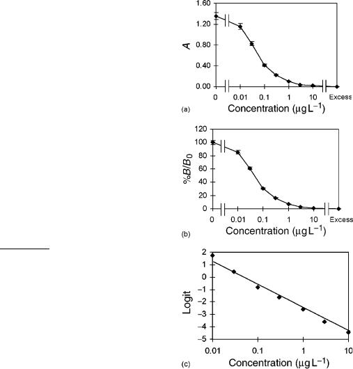
In IAs the signal produced is inversely correlated to the analyte concentration in the sample (Figure 3a). The typical dose ± response curve is of sigmoidal shape when the signal is plotted versus the logarithm of the analyte concentration. A linear range is obtained around the middle of the test (IC50, middle of assay, concentration of analyte that causes 50% inhibition), which should be used for determinations. Within this working range, the change in absorbance is linearly correlated to the analyte concentration. The linear part of the curve is confined by the upper and lower limits of quantification. These are the cut-off values above or below which quantitative results can be obtained with a stated relative precision, or specified degree of confidence in real samples..47/ The experimental errors
IMMUNOCHEMICAL ASSAYS IN PESTICIDE ANALYSIS
increase toward these limits. Consequently, the most precise measurements are obtained in the region close to the middle of the test.
The DL (or least detectable dose) is the smallest concentration of the analyte that produces a signal which can be significantly distinguished from zero for a given sample matrix with a stated degree of confidence. Very often a dose is selected which inhibits 10 ± 20% of enzyme tracer from binding with the Ab or the dose calculated after subtraction of two or three times the standard deviation from the mean measurements of the zero dose signal..3/
Linearization of the calibration curve is useful for many purposes, for instance, for the direct comparison of curves if matrix effects are evaluated. Absorbance curves can be normalized by converting the absorptions to %B=B0 values. These can be expressed as the ratio of bound tracer in the presence of hapten to bound tracer in the absence of hapten and lies between 100% (DA0, the upper asymptote of the curve) and 0% (D AExcess, the lower asymptote) (Figure 3b). They are calculated by Equation (3):
%B |
A AExcess |
|
100 |
.3/ |
|
B0 |
D A0 AExcess |
|
|||
|
|
Linearization can be obtained by various mathematical transformations..47/ Usually, IAs are evaluated with commercial IA programs, often based on logistic models (cf. Rodgers.48/ and Dudley et al..49/), e.g. four-parameter models or the more simple logit-log transformation (twoparameter model, Figure 3c) which can also be carried out with a calculator (s), Equation (4):
logit |
%B |
D ln |
%B=B0 |
.4/ |
B0 |
100 %B=B0 |
4.2 Quality Control
Precision and accuracy of IA are important properties which deserve special attention. The quality and stability of the employed material (microtiter plates, pipettes) and reagents (e.g. Abs, enzyme tracer or buffers), play a crucial role..50/ The long-term stability of reagents has to be ensured, e.g. by freeze-drying of Abs and, if necessary, addition of stabilizing components to the test reagents such as the enzyme tracer..51/
In spite of the simple handling of the assays, expert knowledge is required, especially to recognize and remove incident errors. Therefore, IAs should be performed by trained personnel. The development of simple and rapid assays, such as dip-stick assays or immunofiltration tests reduces the requirement for trained users, but one has still to be aware of potential problems such as interferences from the sample matrix.
The precision of an IA is defined as the extent to which replicate analyses of a sample agree with each other.
7
Figure 3 EIA for the determination of atrazine using pAb.
(a) Absorption curve (means of three determinations standard deviations), (b) B=B0 curve, and (c) logit/log transformation by the two-parameter fit.
The reproducibility is the ability to yield the same results within analyses, between analyses, and between operators. The investigation of the variability of an IA gives valuable information about the consistency of the test. Coefficients of variation (CV) of IA measurements are usually between 10 and 20% for an optimized assay,.52;53/ although more precise results can be obtained..54;55/ Same-day and day-to-day CV of samples have been determined in different matrices..53;56/ Interlaboratory tests of the same IA as that carried out by Hock and the IA Study Group.57/ and Hayes et al..58/ for the investigation of triazines help to evaluate the general applicability of a test. However, several conditions like exact description of the assay including calibration curves, DLs, crossreactivities, a working range close to the middle of the
8
test, enough parallel measurements, etc. must be met (see also AOAC (Association of Official Analytical Chemists) criteria). Meanwhile, standardized procedures for IAs in water are adopted by AOAC International and have been established in Germany as a prenorm..50;58/
A validation of the results obtained by IA should be carried out. To a limited extent this can be done by IA itself. Dilution of the samples as well as spiking of the authentic sample with known amounts of the contaminant can be used to check whether the matrix interferes with the IA..59/ However, spiked samples do not completely mimic real unknown samples. They do not contain potential metabolites of the contaminant nor residues from other compounds which may be present in real samples. Furthermore, spiked samples cannot be a model for aged residues which are more difficult to extract and detect because, for example, they may have bound to soil constituents. Therefore, an IA should also be validated by a different established method like high-performance liquid chromatography (HPLC), gas chromatography (GC) or GC/MS (gas chromatography/mass spectrometry). Many groups have used this approach and have usually obtained correlation coefficients of >0.9..60 ± 63/ Often a slight overestimation of the IA in comparison with HPLC or GC is observed owing to cross-reactivities of the Ab or matrix effects.
|
|
|
|
|
|
|
|
|
|
|
|
PESTICIDES |
|||
|
100 |
|
|
|
|
|
|
|
|
|
|
|
|
|
|
(%) |
80 |
|
|
|
|
|
|
|
|
|
|
|
|
|
|
-reactivity |
|
|
|
|
|
|
|
|
|
|
|
|
|
|
|
60 |
|
|
|
|
|
|
|
|
|
|
|
|
|
|
|
40 |
|
|
|
|
|
|
|
|
|
|
|
|
|
|
|
Cross |
20 |
|
|
|
|
|
|
|
|
|
|
|
|
|
|
|
|
|
|
|
|
|
|
|
|
|
|
|
|
|
|
|
0 |
2 |
3 |
4 |
5 |
1 |
2 |
3 |
4 |
5 |
1 |
2 |
3 |
4 |
5 |
|
1 |
||||||||||||||
|
|
Selective |
Intermediate |
Group-specific |
|||||||||||
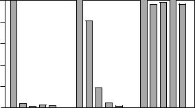
Figure 4 Selective, intermediate and group specific Abs. This example uses Abs which have been produced against hapten 1. Substances 2 ± 5 are assumed to be cross-reacting haptens (modified after Hock et al..14/).
Strong cross-reactivities of an Ab to unexpected metabolites, for example, can produce false positive values. An Ab for alachlor was found to react very strongly to the sulfonic acid metabolite using an alachlor screening kit..69/ This problem could be solved, however, by using solid-phase extraction (SPE) prior to IA and sequential elution of the two compounds with different organic solvents.
4.3 Cross-reactivity
Depending on the conjugate used for immunization and the class of chemicals under investigation, crossreactivities of the Ab with haptens similar to the analyte are frequently observed (see e.g. Hock.14/ and Harrison et al..64/). Therefore, it should be checked which compounds cross-react to what degree with the Ab. This is usually done by comparing the standard curves of the analyte under investigation with similar haptens, using analyte concentrations at 50% of the inhibition curve as the reference. However, cross-reactivity with a certain analyte is not the same over the whole range of a standard curve. Often higher cross-reactivities can be observed at low concentrations of the cross-reacting analyte..3/ Therefore, it has been recommended that cross-reactivities be measured at different concentrations over the range where the assay is suitable..65/
If an Ab is selective for a single compound, it is regarded as monospecific.66/ (Figure 4). An Ab that recognizes several compounds to the same extent (e.g. a group of s-triazines), can be used for the screening of a class of herbicides.67/ (group-specific Ab, Figure 4). If crossreacting compounds are not expected in the samples, because the compounds are not licensed (e.g. propazine in most European countries), a group-specific Ab can also be used for quantitative measurements of one compound..68/
4.4 Sample Preparation and Matrix Effects
Samples can contain compounds in addition to the target analyte, which may interfere with the test. Several groups investigated the influence of ions on EIAs..70 ± 72/ Ruppert et al..70/ observed an inhibition by several anions like azide, which inhibits the peroxidase by binding to the heme group of the enzyme. Most cations did not have an effect except for Ca2C, which leads to an activation of the peroxidase. No interference by different ions such as nitrate, copper, magnesium etc. up to a concentration of 250 ppm was detected in an EIA for pentachlorphenol in water..71/ While ions may inhibit the enzyme used as a label or lead to precipitates by reacting with the buffer components, humic substances present in water or soil extracts may bind nonspecifically to the Ab and thereby interfere with the specific binding of the analyte..73/ These reactions may lead to false positive results. Water samples from forest stands or soil extracts particularly contain a high content of organic compounds such as humic acids (HAs).
Matrix effects in food samples frequently occur owing to colored extracts or to the content of lipids, proteins or polyphenols that may be coextracted during sample preparation..74/ As food samples usually have to be extracted prior to immunochemical analysis, the method of analyte extraction is of great importance. Analytes
IMMUNOCHEMICAL ASSAYS IN PESTICIDE ANALYSIS
that are water soluble and can be efficiently extracted in aqueous buffer will have the most direct extraction method and eliminate the need for organic solvents. However, many pesticides are not readily water soluble and must be extracted with an organic solvent..5/ For the extraction of pesticides from solid foods a variety of solvents have been tested, such as acetone, ether, petroleum ether, methanol, acetonitrile or hexane..75/ Direct analysis of extracts by IA requires the use of solvents that are miscible with water and (at low concentrations) are nondenaturing to proteins such as Ab. IAs are to a certain degree tolerant to a variety of solvents, but each system must be tested to determine which solvent can be accepted and to what extent (for example Hill et al.,.75/ Nugent.76/ and Schneider and Hammock.77/). Usually the extracts are further diluted with water prior to the EIA, but an EIA for parathion was developed, in which the analyte dissolved in hexane could be directly measured in the EIA without prior removal of the hexane. This was achieved by using Ab encapsulated in reverse micelles composed of Aerosol T with aqueous centers..78/ However, a 104-fold decrease in sensitivity was observed.
In some cases a cleanup step is introduced, in which the analyte of interested is separated from the matrix. This can be carried out by C18-columns or immunoaffinity columns..69;79;80/ A very interesting approach is the application of supercritical fluid extraction (SFE) prior to immunoanalysis. These methods generally employ CO2 or CO2 containing various modifiers..5;81/
Some problems with interfering ions can be solved by changing the buffer of the assay system so that no precipitates may be formed..70/ Addition of BSA to the plates prior to the addition of the standard and sample solutions.82/ or to the enzyme tracer.73/ greatly reduces the influence of humic and fulvic acids on the EIA. It may also be helpful to switch to a different batch of Abs or a different assay kit, as different Abs may show different sensitivities to interfering substances. The buffering capacity of the assay buffer should also be checked, as some water or food samples may show relatively low pH values. No effects, however, were observed between pH 3 and 10 by different investigators..56;67;83/
5APPLICATION TO ENVIRONMENTAL SAMPLES
IAs have been developed for many environmental contaminants during the 1990s. A list of several IAs described in the literature can be found in Table 4. Most of them have been developed in laboratories, showing the increasing importance of immunochemical
9
methods in residue analysis. Not all of them are commercially available. Available commercial IAs have been listed in e.g. Dankwardt et al.,.2/ Hennion and Barcelo,.3/ Knopp,.8/ but a lot of movement has been observed in environmental IA markets, leading to the disappearance of IA companies. At the moment IAs for environmental contaminants can be obtained for example from Strategic Diagnostics Inc. (Newark, DE, USA, sells former Ensys, Millipore and Ohmicron kits) and EnviroLogix (Westbrook, MA, USA).
5.1 Water
EIA have been used intensively for the determination of pesticides in surface and rainwater.56;60;69;211 ± 217/ and groundwater..60;69;214;218;219/ A substantial number of these studies were carried out for triazine herbicides..56;60;211;212;214;216;218;219/ This illustrates the widespread occurrence of these herbicides in the aquatic environment. Many groups have used commercial test kits, which allow the investigation of samples without time-consuming Ab production. Thurman et al.,.60/ for example, used a Res-I-Mune kit (ImmunoSystems) for the investigation of triazines in surface and groundwater. The EIA was compared to GC/MS results obtained from samples that were extracted by SPE. Correlation coefficients between 0.91 and 0.95 were obtained after introducing cross-reactivity factors for each of the triazines in order to calculate a sum parameter for the GC. The majority of the samples contained only atrazine (up to 3 μg L 1). Therefore, the EIA results corresponded well with the atrazine concentrations obtained by GC/MS.
Mouvet et al..220/ compared four commercially available test kits and one in-house developed assay for the determination of triazines in surface and groundwater. Operational characteristics, cross-reactivity, sensitivity, CV and agreement with GC/LC (gas chromatography/liquid chromatography) measurements were investigated. DLs were determined between 0.003 and 0.07 μg L 1. Intra-assay CVs were below 7% for all tests, interassay CVs below 20%. Correlation studies between the EIA kits and GC/LC were carried out for samples from different water matrices. Depending on the water source, different levels of significance were observed with different tests. The best results were obtained for surface water, while not all kits showed a good agreement for lysimeter samples.
Apart from the triazines some other pesticides were investigated in water samples, also using commercial test kits. Alachlor was determined in ground and surface water using commercial tests..213/ SPE was carried out prior to EIA to remove interfering substances and to concentrate the analyte. Concentrations of up to 0.8 μg L 1 were observed, and a comparison with GC/MS showed a

10 |
PESTICIDES |
Table 4 Pesticide IAs described in the literature
Pesticides |
Test |
Ab |
|
Range, DL, |
Ref. |
|
|
format |
|
or middle of test (IC50) |
|
||
Herbicides |
|
|
0.2 ± 8 μg L 1 |
|
||
Alachlor |
EIA |
p |
84 |
|||
|
EIA |
p |
0.1 ± 10 μg L 1 |
85 |
||
|
EIA |
p, m |
0.2 ± 8 μg L 1 |
86 |
||
Amitrole |
EIA |
p |
1.7 ± 4200 μg L 1 |
87 |
||
Atrazine |
CLIA |
p |
25 |
± 500 ng L 1 |
88 |
|
|
EIA |
p |
0.5 ± 10 μg L 1 |
89 |
||
|
EIA |
p |
0.01 μg L 1 (DL) |
90 |
||
|
EIA |
m |
0.03 |
± 1 μg L 1 |
35 |
|
|
EIA |
p |
0.2 ± 100 μg L 1 |
64 |
||
|
EIA |
p |
0.011 ± 33 μg L 1 |
91 |
||
|
EIA |
m |
0.05 |
± 3 μg L 1 |
92 |
|
|
EIA |
p, m |
0.1 ± 100 μg L 1 |
93 |
||
|
EIA |
p |
0.5 ± 10 μg L 1 |
94 |
||
|
EIA |
m |
0.05 μg L 1 (DL) |
95 |
||
|
EIA |
m |
0.01 |
± 10 μg L 1 |
77 |
|
|
EIA |
p |
0.03 |
± 3 μg L 1 |
96 |
|
|
EIA |
p |
1 |
± 1000 ng L 1 |
34 |
|
Bentazon |
EIA |
p |
2 |
± 24 μg L 1 |
97 |
|
Bromacil |
EIA |
p |
0.1 ± 160 μg L 1 |
98 |
||
|
EIA |
p |
0.01 |
± 1 μg L 1 |
99 |
|
Chlorodiamino-s-triazine |
EIA |
p |
160 |
± 480 μg L 1 |
100 |
|
Chlorsulfuron |
EIA |
p |
0.1 μg L 1 (DL) |
101 |
||
Clomazone |
EIA |
p |
2 |
± 250 μg L 1 |
102 |
|
|
EIA |
p |
0.5 ± 500 μg L 1 |
103 |
||
Cyanazine |
EIA |
p |
0.035 ± 3 μg L 1 |
104 |
||
|
EIA |
p |
0.5 μg L 1 (DL) |
105 |
||
|
EIA |
p |
0.5 μg L 1 (DL) |
106 |
||
Diethylatrazine |
EIA |
p |
0.01 |
± 100 μg L 1 |
107 |
|
Diclofop-methyl |
EIA |
p |
10 |
± 75 μg L 1 |
108 |
|
2,4-D |
EIA |
p |
50 |
± 5000 μg L 1 |
109 |
|
|
EIA |
m |
2 |
± 20 μg L 1 |
110 |
|
|
RIA |
p |
0.1 ± 10 mg L 1 |
111 |
||
|
EIA |
p |
0.05 |
± 10 mg L 1 |
111 |
|
|
RIA |
p |
5 |
± 250 μg L 1 |
112 |
|
|
PFIA |
m |
0.6 μg L 1 (DL) |
113 |
||
|
RIA |
p |
1 |
± 1000 μg L 1 |
114 |
|
|
EIA |
m |
0.096 μg L 1 (DL) |
115 |
||
Dichlorprop |
PFIA |
p |
0.01 |
± 100 μg mL 1 |
116 |
|
Diuron |
EIA |
m |
2 |
μg L 1 (IC50) |
117 |
|
|
EIA |
p |
0.05 |
± 1 μg L 1 |
118 |
|
Hexazinone |
EIA |
p |
0.22 |
± 17.6 μg L 1 |
119 |
|
Hydroxyatrazine |
EIA |
m |
0.03 |
± 1 μg L 1 |
120 |
|
|
EIA |
m |
0.05 μg L 1 (DL) |
95 |
||
|
EIA |
p |
0.01 |
± 10 μg L 1 |
66 |
|
|
EIA |
p |
3 |
± 300 μg L 1 |
121 |
|
Imazamethabenz |
EIA |
p |
0.5 ± 32 μg L 1 |
122 |
||
Imazaquin |
EIA |
p |
0.45 |
± 25 μg L 1 |
123 |
|
Isoproturon |
EIA |
p |
0.01 |
± 10 μg L 1 |
124 |
|
|
EIA |
m |
20 |
± 250 μg L 1 |
125 |
|
|
EIA |
NA |
0.02 |
± 1 μg L 1 |
126 |
|
Maleic hydrazide |
EIA |
m |
0.01 |
± 11 μg mL 1 |
127 |
|
MCPB |
EIA |
p |
0.03 |
± 0.9 μg L 1 |
128 |
|
Metazachlor |
EIA |
p |
10 |
± 1000 ng L 1 |
129 |
|
(continued overleaf )