

1Paul M Dewick Medicalc Natural / booktext@id88013694placeboie
.pdfMedicinal Natural Products. Paul M Dewick
Copyright 2002 John Wiley & Sons, Ltd
ISBNs: 0471496405 (Hardback); 0471496413 (paperback); 0470846275 (Electronic)
8
CARBOHYDRATES
Some fundamental modifications that occur in carbohydrate metabolism are outlined, then specific examples of monosaccharides, oligosaccharides, and polysaccharides are described. The formation of amino sugars follows, leading to a discussion of aminoglycoside antibiotics. Monograph topics giving more detailed information on medicinal agents include monosaccharides and disaccharides, vitamin C, polysaccharides, aminoglycoside antibiotics based on streptamine and 2-deoxystreptamine, acarbose, lincomycin, and clindamycin.
The main pathways of carbohydrate biosynthesis and degradation comprise an important component of primary metabolism that is essential for all organisms. Carbohydrates are among the most abundant constituents of plants, animals, and microorganisms. Polymeric carbohydrates function as important food reserves, and as structural components in cell walls. Animals and most microorganisms are dependent for their very existence on the carbohydrates produced by plants. Carbohydrates are the first products formed in photosynthesis, and are the products from which plants then synthesize their own food reserves as well as other chemical constituents. These materials then become the foodstuffs of other organisms. Secondary metabolites are also ultimately derived from carbohydrate metabolism, and the relationships of the acetate, shikimate, mevalonate, and deoxyxylulose phosphate pathways to primary metabolism have already been indicated. Many of the medicinally important secondary metabolites described in the earlier chapters have been seen to contain clearly recognizable carbohydrate portions in their structures, e.g. note the frequent occurrence of glycosides. In this chapter, some of the important natural materials which can be grouped together because they are composed entirely or predominantly of basic carbohydrate units are discussed. Because of their widespread use in medicinal preparations, some materials with no inherent biological activity, and which are clearly of primary metabolic status, e.g. sucrose and starch, are also included.
MONOSACCHARIDES
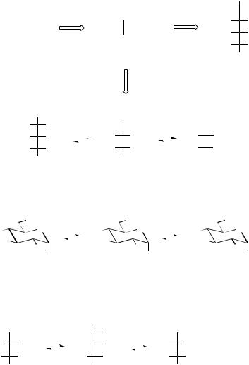
Six-carbon sugars (hexoses) and five-carbon sugars (pentoses) are the most frequently encountered carbohydrate units (monosaccharides) in nature. Photosynthesis produces initially the three-carbon sugar 3-phosphoglyceraldehyde, two molecules of which are used to synthesize glucose 6-phosphate by a sequence which effectively achieves the reverse of the glycolytic reactions (Figure 8.1). Alternatively, by the complex reactions of the Calvin cycle, 3-phosphoglyceraldehyde may be used in the construction of the pentoses ribose 5-phosphate, ribulose 5-phosphate, and xylulose 5-phosphate. These sequences incorporate some of the fundamental reactions which are used in the biochemical manipulation of monosaccharide structures:
• Mutation, |
where repositioning of a phos- |
phate group in the monosaccharide phosphate |
|
molecule, |
e.g. the isomerization of glu- |
cose 6-phosphate and glucose 1-phosphate (Figure 8.2), is achieved via an intermediate diphosphate.
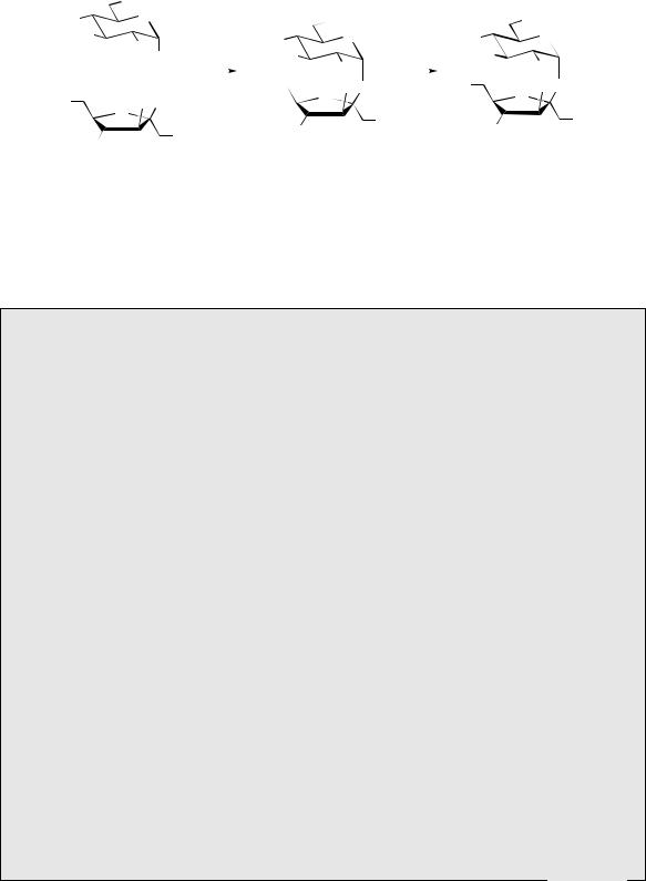
•Epimerization changes the stereochemistry at one of the chiral centres, e.g. the interconversion of ribulose 5-phosphate and xylulose 5- phosphate (Figure 8.3). This reaction involves epimerization adjacent to a carbonyl group and probably proceeds through a common enol tautomer, but some other epimerizations are
466 |
|
|
|
|
|
|
|
|
|
|
|
|
|
|
CARBOHYDRATES |
|
|
|
|
|
|
|
|
|
|
|
||||||||||||
|
|
|
|
|
|
|
|
|
|
|
|
|
|
|
|
|
|
|
|
|
|
|
|
|
|
|
|
|
|
|
|
|
|
|
|
|
CHO |
|
|
|
|
|
|
|
|
|
|
|
|
|
|
|
|
|
|
CHO |
|
|
|
|
|
|
|
H |
OH |
||||||||||||
|
|
|
|
|
|
|
|
|
|
|
|
|
|
|
|
|
|
|
|
x 2 |
|
HO |
H |
|||||||||||||||
PHOTOSYNTHESIS |
|
|
|
|
H |
|
|
|
OH |
|
|
|
|
|||||||||||||||||||||||||
|
|
|
|
|
|
|
|
|
|
|
|
|
|
H |
OH |
|||||||||||||||||||||||
|
|
|
|
|
|
|
|
|
|
|
|
|
|
|
|
|
CH2OP |
|
|
|
|
|
|
|
||||||||||||||
|
|
|
|
|
|
|
|
|
|
|
|
|
|
|
|
|
|
|
|
|
|
|
|
H |
OH |
|||||||||||||
|
|
|
|
|
|
|
|
|
|
|
|
|
|
|
|
|
|
|
|
|
|
|
|
|
|
|
|
|
|
|
|
|
|
|
||||
|
|
|
|
|
|
|
|
|
|
|
|
|
D-3-phosphoglyceraldehyde |
|
|
|
|
|
|
|
|
|
CH2OP |
|||||||||||||||
|
|
|
|
|
|
|
|
|
|
|
|
|
|
|
|
|
|
|
|
|
|
|
|
|
|
|
|
|
|
|
|
|
|
|
|
|
||
|
|
|
|
|
|
|
|
|
|
|
|
|
|
|
|
|
|
|
|
|
|
|
|
|
|
|
|
|
|
|
|
|
|
|
D-glucose 6-P |
|||
|
|
|
|
|
|
|
|
|
|
|
|
|
|
CALVIN |
|
|
|
|
|
|
|
|
|
|
|
|
|
|
|
|
|
|
|
|
|
|
||
|
|
|
|
|
|
|
|
|
|
|
|
|
|
|
CYCLE |
|
|
|
|
|
|
|
|
|
|
|
|
|
|
|
|
|
|
|
|
|
|
|
|
|
|
|
|
|
|
|
|
|
|
|
|
|
|
|
|
|
|
|
|
|
|
|
|
|
|
|
|
||||||||||
|
|
|
CHO |
|
|
|
|
|
CH2OH |
|
|
|
|
CH2OH |
|
|
|
|||||||||||||||||||||
|
|
|
|
|
|
|
|
|
|
|
|
|||||||||||||||||||||||||||
|
|
H |
|
OH |
|
|
|
|
|
|
|
|
O |
|
|
|
|
|
|
|
O |
|
||||||||||||||||
|
|
|
|
|
|
|
|
|
|
|
|
|
|
|
|
|
|
|||||||||||||||||||||
|
|
H |
|
OH |
|
|
|
|
|
|
H |
|
|
|
OH |
|
|
|
|
|
|
|
HO |
|
H |
|
||||||||||||
|
|
|
|
|
|
|
|
|
|
|
|
|
|
|
|
|
|
|
|
|
|
|
|
|
|
|
|
|
|
|
|
|
|
|
|
|
|
|
|
|
H |
|
OH |
|
|
|
|
H |
|
|
|
OH |
|
|
|
|
H |
|
OH |
|
|||||||||||||||||
|
|
|
CH2OP |
|
|
|
|
|
CH2OP |
|
|
|
|
CH2OP |
|
|||||||||||||||||||||||
|
|
D-ribose 5-P |
|
|
|
|
D-ribulose 5-P |
|
|
|
|
D-xylulose 5-P |
|
|||||||||||||||||||||||||
|
|
|
|
|
|
|
|
|
|
|
|
|
|
|
|
Figure 8.1 |
|
|
|
|
|
|
|
|
|
|
|
|||||||||||
6 |
OP |
|
|
|
|
|
|
OP |
|
|
|
|
|
|
|
OH |
|
|||||||||||||||||||||
HO |
|
O |
|
|
HO |
|
|
|
|
O |
|
|
|
|
HO |
|
|
|
O |
|
||||||||||||||||||
|
|
|
|
|
|
|
|
|
|
|
|
|
|
|
|
|
|
|
|
|
|
|
|
|
|
|
|
|
|
|
|
|||||||
HO |
|
|
|
|
|
|
|
|
|
|
|
|
|
|
HO |
|
|
|
|
|
|
|
|
|
|
|
|
|
|
|
HO |
|
|
|
|
1 |
||
|
|
|
|
|
|
|
|
|
|
|
|
|
|
|
|
|
|
|
|
|
|
|
|
|
|
|
|
|
|
|
||||||||
|
|
|
HO |
|
|
|
|
|
|
|
HO |
|
|
|
|
|
|
|
HO |
OP |
||||||||||||||||||
|
|
|
|
OH |
|
|
|
|
|
|
|
|
|
|
OP |
|
|
|
|
|
|
|
|
|
|
|||||||||||||
D-glucose 6-P |
|
|
|
|
D-glucose 1,6-di P |
|
|
|
|
D-glucose 1-P |
|
|||||||||||||||||||||||||||
|
|
|
|
|
|
|
|
|
|
|
|
|
|
|
|
Figure 8.2 |
|
|
|
|
|
|
|
|
|
|
|
|||||||||||
CH2OH |
keto−enol |
|
|
|
CH2OH |
|
enol−keto |
|
CH2OH |
|
|
|
|
|
||||||||||||||||||||||||
tautomerism |
|
|
|
|
tautomerism |
|
|
|
|
|
|
|||||||||||||||||||||||||||
|
|
O |
|
|
|
|
|
|
|
|
|
|
|
|
|
OH |
|
|
|
|
|
|
|
|
|
|
|
|
|
|
|
O |
achieves |
|
||||
|
|
|
|
|
|
|
|
|
|
|
|
|
|
|
|
|
|
|
|
|
|
|
|
|
|
|
|
|
|
|||||||||
H |
OH |
|
|
|
|
|
|
|
|
|
|
|
|
|
OH |
|
|
|
|
|
|
HO |
|
|
|
|
H |
epimerization |
||||||||||
|
|
|
|
|
|
|
|
|
|
|
|
|
|
|
|
|
|
|
||||||||||||||||||||
H |
OH |
|
|
|
|
|
|
|
|
|
H |
|
|
OH |
|
|
|
|
|
|
|
|
H |
|
|
|
|
OH |
|
at position 3 |
||||||||
|
|
|
|
|
|
|
|
|
|
|
|
|
|
|
|
|
|
|
|
|
|
|
|
|||||||||||||||
|
|
|
|
|
|
|
|
|
|
|
|
|
|
|
|
|
|
|
|
|
|
|
|
|
|
|
|
|||||||||||
CH2OP |
|
|
|
|
|
|
|
|
|
|
|
|
CH2OP |
|
|
|
|
|
|
|
|
|
|
|
|
CH2OP |
|
|
|
|
|
|||||||
D-ribulose 5-P |
|
|
|
|
|
|
|
|
|
|
|
|
D-xylulose 5-P |
|
|
|
|
|
||||||||||||||||||||
Figure 8.3
known proceed through oxidation to an intermediate carbonyl, followed by reduction to give the opposite configuration. The substrate for epimerization is often the UDPsugar rather than the monosaccharide phosphate.
•Aldose–ketose interconversions, e.g. glucose 6-phosphate to fructose 6-phosphate (Figure 8.4), also proceed through a common enol intermediate.
•Transfer of C2 and C3 units in reactions catalysed by transketolase and transaldolase respectively modify the chain length of the sugar.
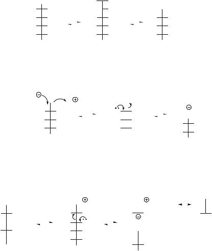
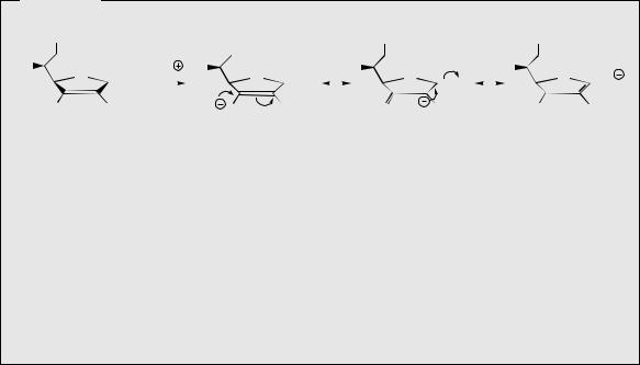
Transketolase removes a two-carbon fragment from ketols such as fructose 6-phosphate (alternatively xylulose 5-phosphate or sedoheptulose 7-phosphate) through the participation of thiamine diphosphate. Nucleophilic attack of the thiamine diphosphate anion on to the carbonyl results in an addition product which then fragments by a reverse aldol reaction, generating the chain-shortened aldose erythrose 4- phosphate, and the two-carbon carbanion unit attached to TPP (Figure 8.5) (compare the role of TPP in the decarboxylation of α-keto
|
|
|
|
|
|
|
|
|
|
|
|
|
|
|
|
|
|
|
MONOSACCHARIDES |
|
|
|
|
|
|
|
|
|
|
|
|
|
|
467 |
||||||||||||||||||||||
|
|
|
|
|
|
|
|
|
|
|
|
keto−enol |
|
|
|
|
|
|
|
|
enol−keto |
|
|
|
|
|
|
|
|
|
|
|
|
|
|
|
|
|
||||||||||||||||||
|
|
|
|
|
|
CHO |
|
tautomerism H |
|
|
|
|
OH |
|
tautomerism |
|
|
CH2OH |
|
achieves |
|
|
|
|||||||||||||||||||||||||||||||||
|
|
|
|
|
|
|
|
|
|
|
||||||||||||||||||||||||||||||||||||||||||||||
|
|
|
|
H |
OH |
|
|
|
|
|
|
|
|
|
|
|
|
|
|
|
OH |
|
|
|
|
|
|
|
|
|
|
|
|
|
|
|
|
O |
|
aldose−ketose |
||||||||||||||||
|
|
|
|
|
|
|
|
|
|
|
|
|
|
|
|
|
|
|
|
|
|
|
|
|
|
|
|
|
|
|
|
|
|
|||||||||||||||||||||||
|
|
|
|
HO |
H |
|
|
|
|
|
|
|
|
HO |
|
|
|
|
H |
|
|
|
|
|
|
|
|
HO |
|
|
|
|
|
H |
|
conversion |
|
|
|
|||||||||||||||||
|
|
|
|
|
|
|
|
|
|
|
|
|
|
|
|
|
|
|
|
|
|
|
|
|
|
|
|
|
|
|
|
|
|
|
|
|
|
|
||||||||||||||||||
|
|
|
|
H |
OH |
|
|
|
|
|
|
|
|
|
H |
|
|
|
|
OH |
|
|
|
|
|
|
|
|
H |
|
|
|
|
|
OH |
|
|
|
|
|
|
|
|
|
|
|||||||||||
|
|
|
|
|
|
|
|
|
|
|
|
|
|
|
|
|
|
|
|
|
|
|
|
|
|
|
|
|
|
|
|
|
|
|
|
|
|
|
|
|||||||||||||||||
|
|
|
|
|
|
|
|
|
|
|
|
|
|
|
|
|
|
|
|
|
|
|
|
|
|
|
|
|
|
|
|
|
|
|
|
|||||||||||||||||||||
|
|
|
|
H |
OH |
|
|
|
|
|
|
|
|
|
H |
|
|
|
|
OH |
|
|
|
|
|
|
|
|
H |
|
|
|
|
|
OH |
|
|
|
|
|
|
|
|
|
|
|||||||||||
|
|
|
|
|
|
CH2OP |
|
|
|
|
|
|
|
|
|
|
|
|
|
CH2OP |
|
|
|
|
|
|
|
|
|
|
|
|
CH2OP |
|
|
|
|
|
|
|
|
|
|
|||||||||||||
|
|
|
|
D-glucose 6-P |
|
|
|
|
|
|
|
|
|
|
|
|
|
|
|
|
|
|
|
|
|
|
D-fructose 6-P |
|
|
|
|
|
|
|
|
|
|
|||||||||||||||||||
|
|
|
|
|
|
|
|
|
|
|
|
|
|
|
|
|
|
|
|
|
|
|
|
|
|
Figure 8.4 |
|
|
|
|
|
|
|
|
|
|
|
|
|
|
|
|
|
|||||||||||||
|
|
|
|
|
|
|
|
|
|
|
|
|
|
|
|
|
|
|
|
|
|
|
|
|
|
reverse aldol reaction |
|
|
|
|
|
|
|
|
|
|
|
|
|
|
C2 unit attached to |
|||||||||||||||
|
|
|
|
|
|
|
CH |
|
OH |
|
|
|
|
|
|
|
|
|
|
|
|
|
|
|
CH2OH |
|
|
|
|
|
|
|
|
CH2OH |
TPP anion; this is |
|||||||||||||||||||||
nucleophilic |
|
TPP |
|
2 |
|
|
|
|
|
|
|
|
|
|
|
|
|
|
|
|
|
|
|
|
|
|
|
|
|
|
|
|
|
|
|
|
|
|
|
|
|
|
|
|
|
|
subsequently |
|||||||||
|
|
|
|
|
O H |
|
|
|
|
|
|
|
|
|
|
|
PPT |
|
C |
|
OH |
|
|
|
|
|
PPT |
|
C |
|
OH |
|||||||||||||||||||||||||
attack of thiamine |
|
|
|
|
|
|
|
transketolase |
|
|
|
|
|
|
|
|
|
|
|
transferred to a |
||||||||||||||||||||||||||||||||||||
|
|
|
|
|
|
|
|
|
|
|
|
|
HO |
|
|
|
|
H |
|
|
|
|
|
|
|
|
|
|
|
|
|
|
|
|||||||||||||||||||||||
diphosphate anion |
|
HO |
|
|
|
|
H |
|
|
|
|
|
|
|
|
|
|
|
|
|
|
|
|
|
|
|
|
different aldose via |
||||||||||||||||||||||||||||
|
|
|
|
|
|
|
|
|
|
|
|
|
|
|
|
|
|
|
|
|
|
|
|
|
|
|
|
CHO |
||||||||||||||||||||||||||||
on to carbonyl |
|
|
H |
|
|
|
|
OH |
|
|
|
|
|
|
|
|
|
|
|
|
H |
|
|
|
|
OH |
|
|
|
|
|
|
|
|
||||||||||||||||||||||
|
|
|
|
|
|
|
|
|
|
|
|
|
|
|
|
|
|
|
|
|
|
|
|
|
|
|
|
|
|
the reverse reaction |
||||||||||||||||||||||||||
|
|
|
|
|
|
|
|
|
|
|
|
|
|
|
|
|
|
|
|
|
|
|
|
|
|
|
|
|
|
|
|
|
|
|
|
|||||||||||||||||||||
|
|
|
|
|
|
|
|
|
|
|
|
|
|
|
|
|
|
|
|
|
|
|
|
|
|
|
|
|
|
|
H |
|
|
|
|
OH |
||||||||||||||||||||
|
|
|
|
|
|
H |
|
|
|
|
OH |
|
|
|
|
|
|
|
|
|
|
|
|
H |
|
|
|
|
OH |
|
|
|
|
|
|
|
|
|
|
|
|
|||||||||||||||
|
|
|
|
|
|
|
|
|
|
|
|
|
|
|
|
|
|
|
|
|
|
|
|
|
|
|
|
|
|
|
H |
|
|
|
|
OH |
|
|
|
|||||||||||||||||
|
|
|
|
|
|
|
CH2OP |
|
|
|
|
|
|
|
|
|
|
|
|
|
|
|
CH2OP |
|
|
|
|
|
|
|
|
|
|
|
|
|||||||||||||||||||||
|
|
|
|
|
|
|
|
|
|
|
|
|
|
|
|
|
|
|
|
|
|
|
|
|
|
|
|
|
|
CH2OP |
|
|
|
|||||||||||||||||||||||
|
|
|
|
|
D-fructose 6-P |
|
|
|
|
|
|
|
|
|
|
|
|
|
|
|
|
|
|
|
|
|
|
|
|
|
|
|
|
|
|
|
|
|
|
|
|
|||||||||||||||
|
|
|
|
|
|
|
|
|
|
|
|
|
|
|
|
|
|
|
|
|
|
|
|
|
|
|
|
|
|
|
|
|
|
|
D-erythrose 4-P |
|
|
|
||||||||||||||||||
|
|
|
|
|
|
|
|
|
|
|
|
|
|
|
|
|
|
|
|
|
|
|
|
|
|
|
|
|
|
|
|
|
|
|
|
|
|
|
|
|
|
|
|
|
|
|
|
|||||||||
|
|
|
|
|
|
|
|
|
|
|
|
|
|
|
|
|
|
|
|
|
|
|
|
|
|
Figure 8.5 |
|
|
|
|
|
|
|
|
|
|
|
|
|
|
|
|
|
|||||||||||||
|
|
|
|
ketose binds |
|
|
reverse aldol reaction |
|
|
|
|
|
|
|
|
|
|
|
|
|
|
|
|
|
|
|
|
|
|
|
|
|
|
|
|
|||||||||||||||||||||
|
|
|
|
|
|
|
|
|
|
|
|
|
|
|
|
|
|
|
|
|
|
|
|
|
|
|
|
|
|
|
|
|
|
|
|
|
|
|
|
|
||||||||||||||||
|
|
|
|
to enzyme via |
|
CH2OH |
|
|
|
|
|
|
|
|
|
|
|
|
|
CH2OH |
|
|
|
|
|
|
|
|
|
|
|
|
|
|
|
|
|
|||||||||||||||||||
CH |
OH |
Schiff base |
|
|
|
|
|
|
|
|
|
|
|
|
|
|
|
|
|
|
|
|
|
|
|
|
|
|
|
|
|
CH2OH |
||||||||||||||||||||||||
2 |
|
|
|
|
|
|
|
|
|
|
|
|
|
|
|
|
|
|
|
|
|
|
|
|
|
|
|
|
|
|
|
|
|
|
|
|
|
|
|
|
|
|
|
|
|
|
|
|
|
|
|
|
|
|||
|
|
|
O |
|
|
|
|
|
|
|
|
|
|
|
|
|
|
|
NH |
|
|
Enz |
|
|
|
|
|
|
|
|
NH |
|
|
Enz |
|
|
|
|
|
|
|
|
NH |
|
Enz |
|||||||||||
HO |
H |
transaldolase |
|
HO |
|
|
|
|
H |
|
|
|
|
|
|
|
|
HO |
|
|
|
|
H |
|
|
|
|
|
|
|
|
|
|
HO |
|
|
H |
|||||||||||||||||||
|
|
|
|
|
|
|
|
|
|
|
|
|
|
|
|
|
|
|
|
|
|
|
|
|
|
|
|
|||||||||||||||||||||||||||||
H |
OH |
|
H |
|
|
|
|
OH |
|
|
|
|
|
|
|
|
|
|
|
|
|
|
|
|
|
|
|
|
|
|
|
C3 unit attached to enzyme as |
||||||||||||||||||||||||
|
|
|
|
|
|
|
|
|
|
|
|
|
|
|
|
|
|
|
|
|
|
|
|
CHO |
|
|
|
|
|
|||||||||||||||||||||||||||
|
|
|
|
|
|
|
|
|
|
|
|
|
|
|
|
|
|
|
|
|
|
|
|
|
|
|
|
|
||||||||||||||||||||||||||||
|
|
|
|
|
|
|
|
|
|
|
|
|
|
|
|
|
|
|
|
|
|
|
|
|
|
|
|
|
||||||||||||||||||||||||||||
H |
OH |
|
|
|
|
|
|
|
|
H |
|
|
|
|
OH |
|
|
|
|
|
|
|
|
|
|
|
|
|
|
|
|
|
enamine; this is subsequently |
|||||||||||||||||||||||
|
|
|
|
|
|
|
|
|
|
|
|
|
|
|
|
|
|
|
|
|
H |
|
|
|
|
OH |
|
|
|
|
|
transferred to a different aldose via |
||||||||||||||||||||||||
H |
OH |
|
|
|
|
|
|
|
|
H |
|
|
|
|
OH |
|
|
|
|
|
|
|
|
|
|
|
|
|
|
|
|
|
|
|||||||||||||||||||||||
|
|
|
|
|
|
|
|
|
|
|
|
|
|
|
|
|
|
|
|
|
H |
|
|
|
|
OH |
|
|
|
|
|
the reverse sequence |
||||||||||||||||||||||||
|
|
|
|
|
|
|
|
|
|
|
|
|
|
|
|
|
|
|
|
|
|
|
|
|
|
|
|
|
|
|
|
|
|
|
|
|
|
|
|
|||||||||||||||||
CH2OP |
|
|
|
|
|
|
|
|
|
|
|
CH2OP |
|
|
|
|
|
|
|
|
|
|
|
|
|
|
|
|
|
|
|
|
|
|
|
|
|
|
|
|
|
|
||||||||||||||
|
|
|
|
|
|
|
|
|
|
|
|
|
|
|
|
|
|
|
|
|
|
|
|
|
|
|
|
|
|
|
|
|
|
|
|
|
|
|
|
|
|
|
|
|
|
|||||||||||
CH2OP
D-sedoheptulose 7-P
D-erythrose 4-P
Figure 8.6
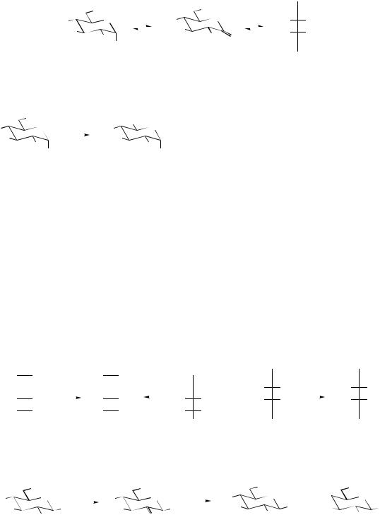
acids, page 21). Then, in what is formally the reverse of this reaction, this carbanion can attack another aldose such as ribose 5- phosphate (alternatively erythrose 4-phosphate or glyceraldehyde 3-phosphate), thus extending its chain length by two carbons. Transaldolase removes a three-carbon fragment from a ketose such as sedoheptulose 7-phosphate (alternatively fructose 6-phosphate) in a reverse aldol reaction, though this requires formation of a Schiff base between the carbonyl group and an active site lysine of the enzyme (Figure 8.6). Again, the reaction is completed by a reversal
of this process, but transferring the C3 carbanion to another aldose such as glyceraldehyde 3- phosphate (alternatively erythrose 4-phosphate or ribose 5-phosphate) and thus increasing its length.
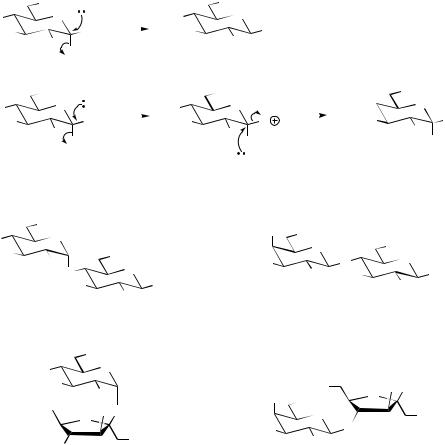
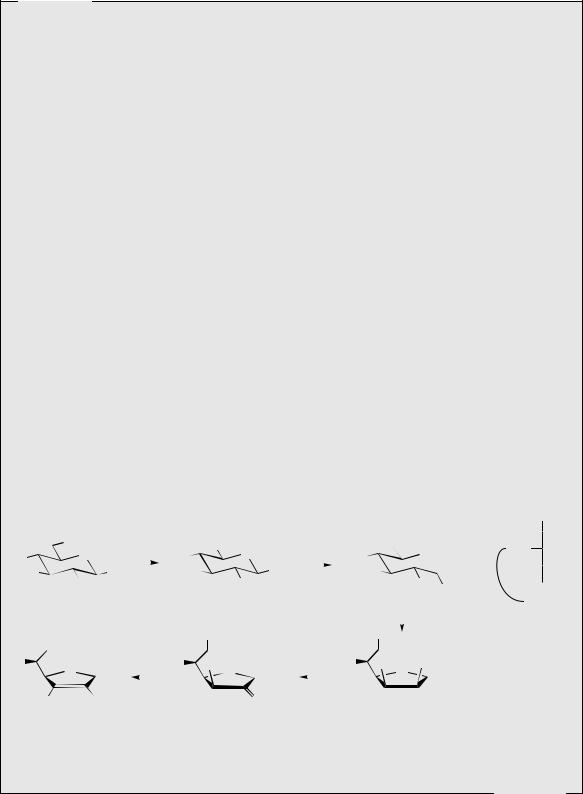
•Oxidation and reduction reactions, typically employing the NAD/NADP nucleotides, alter the oxidation state of the substrate. Oxidation at C-1 converts an aldose into an aldonic acid, e.g. glucose 6-phosphate gives gluconolactone 6-phosphate and then the open-chain gluconic acid 6-phosphate (Figure 8.7). Oxidation at C-6 yields the corresponding uronic acids,
468 |
|
|
|
|
|
|
CARBOHYDRATES |
|
|
||||||
|
|
oxidation at C-1 |
|
hydrolysis |
|
CO2H |
|||||||||
|
|
|
of lactone |
|
|||||||||||
|
OP |
|
|
|
|
|
|
OP |
|
|
|
|
|
H |
OH |
HO |
O |
NADP |
+ |
HO |
O |
|
H2O |
HO |
H |
||||||
|
|
|
|||||||||||||
HO |
1 |
|
|
|
|
HO |
|
|
|
|
|
|
H |
OH |
|
|
|
|
|
HO |
O |
||||||||||
|
HO |
OH |
|
|
H |
OH |
|||||||||
|
|
|
|
|
|
|
|
|
|
||||||
|
D-glucose 6-P |
|
|
|
|
|
D-gluconolactone 6-P |
|
|
|
|
|
|
CH2OP |
|
D-gluconic acid 6-P
Figure 8.7
6 |
OH |
oxidation at C-6 |
CO2H |
|||
|
|
|
|
|||
|
O |
|
|
|
|
|
HO |
|
NAD |
+ |
HO |
O |
|
|
|
|
|
|||
HO |
HO |
OPPU |
|
|
HO |
HO |
|
|
|||||
|
|
|
|
|||
|
|
|
|
|
OPPU |
|
UDPglucose |
|
|
|
UDPglucuronic acid |
||
Figure 8.8
but this takes place on UDPsugar derivatives, e.g. UDPglucose to UDPglucuronic acid (Figure 8.8). Reduction is exemplified by the conversion of both glucose and fructose into the sugar alcohol sorbitol (glucitol), and of mannose into mannitol (Figure 8.9).
• Transamination reactions on keto sugars allow the introduction of amino groups as seen in the amino sugars glucosamine and galactosamine (Figure 8.10). These compounds, as their N -acetyl derivatives, are part of the structures of several natural polysaccharides, and
other uncommon amino sugars are components of the aminoglycoside antibiotics (see page 478).
Monosaccharide structures may be depicted in open-chain forms showing their carbonyl character, or in cyclic hemiacetal or hemiketal forms. The compounds exist predominantly in the cyclic forms, which result from nucleophilic attack of an appropriate hydroxyl on to the carbonyl (Figure 8.11). Both six-membered pyranose and five-membered furanose structures are encountered, a particular ring size usually being characteristic for any one sugar. Since the carbonyl group may be attacked from either side, two epimeric structures (anomers) are possible in each case, and in solution, the two forms are frequently in equilibrium. In natural product structures, sugar units are most likely (but not always) to be encountered in just one of the epimeric forms. The two forms are designated α or β on the basis of the
|
CHO |
reduction of |
|||
|
aldehyde |
||||
H |
OH |
||||
|
|
|
|||
HO |
H |
NADH |
|||
|
|
||||
H |
OH |
|
|
|
|
|
|
|
|||
H |
OH |
|
|
|
|
|
CH2OH |
|
|
|
|
D-glucose
|
CH2OH |
reduction of |
CH2OH |
|
CHO |
|
|
|
|
CH2OH |
|||||
|
ketone |
|
|
|
|
|
|
||||||||
H |
OH |
|
|
|
O |
HO |
H |
|
|
|
HO |
H |
|||
|
|
|
|
|
|
|
|
|
|||||||
|
|
|
|
|
|
|
|
|
|||||||
HO |
H |
NADH |
HO |
|
|
H |
HO |
H |
NADH |
HO |
H |
||||
|
|
|
|
|
|
|
|
|
|
||||||
H |
OH |
|
|
|
H |
|
|
OH |
H |
OH |
|
|
|
H |
OH |
|
|
|
|
|
|
|
|
||||||||
H |
OH |
|
|
|
H |
|
|
OH |
H |
OH |
|
|
|
H |
OH |
|
CH2OH |
|
|
|
|
CH2OH |
|
CH2OH |
|
|
|
|
CH2OH |
||
D-sorbitol |
|
|
|
D-fructose |
D-mannose |
|
|
|
D-mannitol |
||||||
|
(glucitol) |
|
|
|
|
|
|
|
|
|
|
|
|
|
|
Figure 8.9
|
OH |
|
|
|
OH |
transamination |
|
OH |
|
OH |
OH |
||
HO |
O |
|
O |
HO |
O |
|
|
|
HO |
O |
|
|
O |
|
|
|
|
|
|
|
|
|
|
|
|||
HO |
|
OH |
HO |
|
OH |
HO |
NH2 |
OH |
HO |
OH |
|||
|
OH |
|
|
|
O |
|
|
|
|
|
|
NH2 |
|
|
D-glucose |
|
|
|
|
|
|
|
|
D-glucosamine |
|
D-galactosamine |
|
Figure 8.10

|
|
|
|
|
|
|
|
OLIGOSACCHARIDES |
|
|
|
|
|
|
469 |
|||||
|
|
OH |
hemiacetal |
|
|
|
|
6 |
OH |
aldehyde |
|
|
|
|
6 |
OH |
hemiacetal |
|||
|
|
O |
|
|
|
|
|
|
4 |
OH |
|
|
|
|
|
4 |
5 |
O |
|
|
HO |
|
|
|
|
|
HO |
5 |
|
|
|
|
HO |
|
|
||||||
|
|
|
|
|
|
|
|
|
H |
|
|
|
|
|
|
|
||||
|
HO |
H |
|
|
|
|
HO |
2 |
O |
|
|
|
HO |
|
2 |
1 OH |
||||
|
|
|
|
|
|
|||||||||||||||
|
|
HO |
|
|
|
|
|
|
3 |
1 |
|
|
|
|
|
|
3 |
|
||
|
|
OH |
|
|
|
|
HO |
|
|
|
|
|
|
HO |
H |
|||||
|
|
|
|
|
|
|
|
|
H |
|
|
|
|
|
|
|
|
|||
|
|
α-D-glucose |
|
|
|
|
D-glucose |
|
|
|
|
|
|
β-D-glucose |
||||||
|
(α-D-glucopyranose) |
|
|
|
|
(open-chain) |
|
|
|
|
|
(β-D-glucopyranose) |
||||||||
|
|
|
hemiketal |
|
|
|
|
6 |
|
H |
ketone |
|
|
|
|
|
|
hemiketal |
||
HO |
|
HO |
OH |
|
|
|
HO |
H HO O |
|
|
|
|
HO |
6 |
|
HO OH |
||||
|
|
|
|
|
|
|
|
|
|
|
||||||||||
|
|
O |
|
|
|
|
|
|
5 |
O |
2 |
|
|
|
|
|
5 |
|
O |
2 |
|
|
|
|
|
|
|
|
|
|
|
|
|
|
|
|
|
||||
|
|
|
|
|
|
|
|
|
|
|
|
|
|
|
|
|
||||
|
|
|
OH |
|
|
|
|
4 |
3 |
OH |
|
|
|
|
4 |
3 |
OH |
|||
|
|
|
|
|
|
|
1 |
|
|
|
|
|||||||||
|
|
HO |
|
|
|
|
|
|
HO |
|
|
|
|
|
|
HO |
|
1 |
||
|
|
|
|
|
|
|
|
|
|
|
|
|
|
|
|
|
||||
|
|
α-D-fructose |
|
|
|
|
D-fructose |
|
|
|
|
|
|
β-D-fructose |
||||||
|
(α-D-fructofuranose) |
|
|
|
|
(open-chain) |
|
|
|
|
|
(β-D-fructofuranose) |
||||||||
Figure 8.11
OH |
OH |
OH |
|
OH |
||
|
R |
HO O |
||||
R |
O |
|
R |
O |
||
HO |
R OH |
|
|
R OH |
HO |
R OH |
HO |
HO |
|
HO |
|||
|
HO |
|
|
HO |
|
|
β-D-glucose |
β-D-galactose |
β-D-mannose |
||||
(β-D-Glc) |
(β-D-Gal) |
(β-D-Mann) |
||||
R |
|
OH |
|
|
HO R |
O R OH |
O |
S |
|
O |
|||
HO |
R OH |
|
|
R OH |
|
|
HO |
HO |
|
|
|
||
|
HO |
|
|
HO |
HO |
OH |
|
|
|
|
|
|
|
β-D-xylose |
α-L-arabinose |
β-D-ribose |
||||
(β-D-Xyl) |
(α-L-Ara) |
(β-D-Rib) |
||||
OH
S
R
O HO

HO
OH α-L-rhamnose
(α-L-Rha) [preferred conformation]
HO |
O |
HO OH |
R |
R |
|
|
|
 OH
OH
HO β-D-fructose
(β-D-Fru)
Figure 8.12
chiralities at the anomeric centre and at the highest numbered chiral centre. If these are the same (RS convention) the anomer is termed β, or α if they are different. The most commonly encountered monosaccharides, and their usual anomers are shown in Figure 8.12. Note that the D- and L- prefixes are assigned on the basis of the chirality (as depicted in Fischer projection) at the highest numbered chiral centre and its relationship to D-(R)- (+)-glyceraldehyde or L-(S )-(−)-glyceraldehyde (Figure 8.13).
OLIGOSACCHARIDES
H R |
CHO |
HO S |
CHO |
OH |
H |
||
|
CH2OH |
|
CH2OH |
D-(+)-glyceraldehyde |
L-(–)-glyceraldehyde |
||
|
CHO |
|
|
H |
OH |
|
CHO |
HO |
H |
H |
OH |
H |
OH |
HO |
H |
H |
OH |
HO |
H |
|
CH2OH |
|
CH2OH |
D-glucose |
L-arabinose |
||
Figure 8.13
The formation of oligosaccharides (typically two to five monomers) and polysaccharides is dependent on the generation of an activated sugar bound to a nucleoside diphosphate. The nucleoside diphosphate most often employed is UDP, but
ADP and GDP are sometimes involved. As outlined in Chapter 2 (see page 29), a UDPsugar is formed by the reaction of a sugar 1-phosphate with UTP, and then nucleophilic displacement of the UDP leaving group by a suitable nucleophile
470 |
CARBOHYDRATES |
generates the new sugar derivative. This will be a glycoside if the nucleophile is a suitable aglycone molecule, or an oligosaccharide if the nucleophile is another sugar molecule (Figure 8.14). This reaction, if mechanistically of SN2 type, should give an inversion of configuration at C-1 in the electrophile, generating a product with the β- configuration in the case of UDPglucose as shown. Many of the linkages formed between glucose monomers actually have the α-configuration, and it is believed that a double SN2 mechanism operates, which also involves a nucleophilic group on the enzyme (Figure 8.14). Linkages are usually represented by a shorthand version, which indicates the atoms bonded and the configuration at the appropriate centre(s). Thus
maltose (Figure 8.15), a hydrolysis product from starch, contains two glucoses linked α1→4, whilst lactose, the main sugar component of cow’s milk, has galactose linked β1→4 to glucose. In the systematic names, the ring size (pyranose or furanose) is also indicated. Sucrose (‘sugar’) (Figure 8.15) is composed of glucose and fructose, but these are both linked through their anomeric centres, so the shorthand representation becomes α1→β2. This means that both the hemiacetal structures are prevented from opening, and, in contrast to maltose and lactose, there can be no open-chain form in equilibrium with the cyclic form. Therefore sucrose does not display any of the properties usually associated with the masked carbonyl group, e.g. it is not a reducing sugar.
|
OH |
ROH |
|
|
|
|
|
OH |
|
|
|
|
|
|
|
|
|
|
O |
|
|
|
|
|
O |
|
|
|
|
|
|
|
|
||
HO |
|
|
|
ROH |
HO |
|
|
|
|
|
|
|
|
||||
|
|
|
|
|
|
OR |
|
|
|
|
|
|
|||||
HO |
|
H |
|
|
|
|
HO |
|
|
|
|
|
|
|
|
||
HO |
|
|
SN2 |
HO |
|
|
|
|
|
|
|
||||||
|
|
|
|
|
|
|
|
|
|
|
|
|
|
||||
|
OPPU |
|
|
|
|
|
|
|
|
|
|
|
|
|
|||
|
|
|
|
|
|
|
H |
|
|
|
|
|
|
|
|
||
|
UDPglucose |
|
|
|
|
|
β-glucoside |
|
|
|
|
|
|
|
|
||
|
OH |
Enz |
|
|
|
|
|
OH |
|
|
|
|
|
|
OH |
|
|
|
|
|
|
|
|
|
|
|
|
|
|
|
|
|
|||
HO |
O |
B |
|
|
|
|
HO |
O |
Enz |
ROH |
|
|
HO |
O |
|
||
|
|
|
|
|
|
|
|
|
|
|
|
H |
|||||
HO |
H |
|
|
SN2 |
HO |
HO |
B |
SN2 |
|
HO |
HO |
||||||
|
HO |
|
|
|
|
|
|
|
|
|
|
||||||
|
OPPU |
|
|
|
|
|
|
|
|
|
|
|
OR |
||||
|
|
|
|
|
|
|
H |
|
|
|
|
|
|
α-glucoside |
|||
|
|
|
|
|
|
|
|
|
ROH |
|
|
|
|
|
|
|
|
|
|
|
|
|
|
|
|
|
|
|
|
|
|
|
|
|
|
|
|
|
|
|
|
|
|
Figure 8.14 |
|
|
|
|
|
|
|
|
|
|
OH |
|
|
|
|
|
|
|
|
|
OH |
OH |
|
|
|
|
|
HO |
O |
α1→4 |
|
|
|
|
|
|
|
β1→4 |
|
|
|||||
|
|
|
|
|
|
|
|
OH |
|
||||||||
|
|
|
|
|
|
|
|
O |
|
||||||||
HO |
|
1 |
|
OH |
|
|
|
|
|
4 |
|
||||||
|
|
|
|
|
|
|
|
1 |
O |
|
|||||||
|
HO |
4 |
|
|
|
HO |
|
|
O |
|
|||||||
|
|
|
|
|
O |
|
|
|
|
|
|
|
|
|
|
||
|
|
O |
|
|
|
|
|
|
HO |
|
HO |
|
OH |
||||
|
|
|
|
|
|
|
|
|
|
|
|
||||||
|
|
HO |
|
HO |
OH |
|
|
|
|
|
|
|
HO |
|
|||
|
maltose |
|
|
|
|
|
|
|
|
lactose |
|
|
|||||
|
|
|
|
|
|
|
|
|
|
|
|
|
|
||||
|
D-Glc(α1→4)D-Glc |
|
|
|
|
|
|
D-Gal(β1→4)D-Glc |
|
||||||||
4-O-(α-D-glucopyranosyl)-D-glucopyranose |
|
4-O-(β-D-galactopyranosyl)-D-glucopyranose |
|||||||||||||||
|
|
OH |
|
|
|
|
|
|
|
|
|
|
|
|
|
|
|
|
HO |
|
O |
|
|
|
|
|
|
|
|
|
|
|
|
|
|
|
|
|
|
|
|
|
|
|
|
|
|
|
|
|
|
|
|
|
HO |
|
|
|
|
1 |
|
|
|
|
|
HO |
|
|
HO OH |
|
|
|
|
|
HO |
|
|
|
|
|
|
OH |
|
O |
|
||||
|
|
|
|
|
α1→β2 |
|
|
OH |
|
|
|
||||||
|
HO |
|
HO O |
|
|
|
|
|
|
|
|||||||
|
|
|
|
|
O |
|
4 |
OH |
|||||||||
|
|
O |
2 |
|
|
|
|
|
|
1 |
|
||||||
|
|
|
|
|
|
HO |
|
|
Oβ1→4 |
|
|||||||
|
|
|
|
|
|
|
|
|
|
|
|||||||
|
HO |
|
|
1 |
|
OH |
|
|
HO |
|
|
||||||
|
|
|
|
|
|
|
|
|
|
|
|
|
|
|
|
|
|
|
|
sucrose |
|
|
|
|
|
|
|
|
|
|
|
lactulose |
|
|
|
|
D-Glc(α1→β2)D-Fru |
|
|
|
|
D-Gal(β1→4)D-Fru |
|
||||||||||
α-D-glucopyranosyl-(1→2)-β-D-fructofuranoside |
β-D-galactopyranosyl-(1→4)-β-D-fructofuranoside |
||||||||||||||||
Figure 8.15
|
|
|
|
OLIGOSACCHARIDES |
|
|
471 |
|
|
|
OH |
|
|
|
|
|
|
HO |
O |
|
|
OH |
|
|
OH |
|
|
|
|
|
|
||||
|
HO |
|
HO |
O |
|
HO |
O |
|
|
|
HO |
|
|
|
|
||
|
|
|
HO |
|
|
HO |
|
|
|
|
OPPU |
|
|
|
|
||
|
|
|
|
HO |
|
|
HO |
|
|
|
UDPglucose |
|
|
|
|
||
|
|
|
|
|
|
|||
|
|
|
|
PO |
HO O |
|
HO |
HO O |
PO |
|
HO OH |
|
|
O |
|
|
O |
|
|
O |
|
|
OH |
|
|
OH |
|
|
|
|
HO |
|
HO |
||
|
|
OH |
|
sucrose 6F-P |
|
|
||
|
|
|
|
|
|
sucrose |
||
|
|
HO |
|
|
|
|
||
fructose 6-P
Figure 8.16
Sucrose is known to be formed predominantly by a slightly modified form of the sequence shown in Figure 8.16, in that UDPglucose is attacked by fructose 6-phosphate, and that the
first formed product is sucrose 6F-phosphate (F indicating the numbering refers to the fructose ring). Hydrolysis of the phosphate then generates sucrose (Figure 8.16).
Monosaccharides and Disaccharides
D-Glucose (dextrose) (Figure 8.9) occurs naturally in grapes and other fruits. It is usually obtained by enzymic hydrolysis of starch, and is used as a nutrient, particularly in the form of an intravenous infusion. Chemical oxidation of glucose produces gluconic acid. The soluble calcium salt calcium gluconate is used as an intravenous calcium supplement. D-Fructose (Figure 8.13) is usually obtained from invert sugar (see below) separating it from glucose, and is of benefit as a food and sweetener for patients who cannot tolerate glucose, e.g. diabetics. Fructose has the sweetness of sucrose, and about twice that of glucose. High fructose corn syrup for use as a food sweetener is a mixture of fructose and glucose containing up to 90% fructose and is produced by enzymic hydrolysis/isomerization of starch. The sugar alcohol D-sorbitol (Figure 8.9) is found naturally in the ripe berries of the mountain ash (Sorbus aucuparia; Rosaceae) but is prepared semi-synthetically from glucose. It is half as sweet as sucrose, is not absorbed orally, and is not readily metabolized in the body. It finds particular use as a sweetener for diabetic products. D-Mannitol (Figure 8.9) is also produced from glucose, but occurs naturally in manna, the exudate of the manna ash Fraxinus ornus (Oleaceae). This material has similar characteristics to sorbitol, but is used principally as a diuretic. It is injected intravenously, is eliminated rapidly into the urine, and removes fluid by an osmotic effect.
Sucrose (Figure 8.15) is obtained from a variety of sources, including sugar cane (Saccharum officinarum; Graminae/Poaceae), sugar beet (Beta vulgaris; Chenopodiaceae), and sugar maple (Acer saccharum; Aceraceae). It is a standard sweetening agent for foods, syrups, and drug preparations. Invert sugar is an equimolar mixture of glucose and fructose, obtained from sucrose by hydrolysis with acid or the enzyme invertase. During this process, the optical activity changes from + to −, hence the reference to inversion. The high sweetness of fructose combined with that of glucose means invert sugar provides a cheaper, less calorific food sweetener than sucrose. Honey is also mainly composed of invert sugar. Lactose (Figure 8.15) can comprise up to 8% of mammalian milk, and is extracted from cow’s milk, often as a by-product from cheese manufacture. It is only faintly sweet, and its principal use is as a diluent in tablet formulations. Lactulose (Figure 8.15)
(Continues)
472 |
CARBOHYDRATES |
(Continued )
is a semi-synthetic disaccharide prepared from lactose, and composed of galactose linked β1 → 4 to fructose. It is not absorbed from the gastrointestinal tract, and is predominantly excreted unchanged. It helps to retain fluid in the bowel by osmosis, and is thus used as a laxative.
Vitamin C
Vitamin C (ascorbic acid) (Figure 8.17) can be synthesized by most animals except humans, other primates, guinea pigs, bats and some birds, and for these it is obtained via the diet. Citrus fruits, peppers, guavas, rose hips, and blackcurrants are especially rich sources, but it is present in most fresh fruit and vegetables. Raw citrus fruits provide a good daily source. It is a water-soluble acidic compound (an enol; see Figure 8.18) and is rapidly degraded during cooking in the presence of air. Vitamin C deficiency leads to scurvy, characterized by muscular pain, skin lesions, fragile blood vessels, bleeding gums, and tooth loss. The vitamin is essential for the formation of collagen, the principal structural protein in skin, bone, tendons, and ligaments, being a cofactor in the hydroxylation of proline to 4-hydroxyproline, and of lysine to 5-hydroxylysine (see page 409), which account for up to 25% of the collagen structure. These reactions are catalysed by 2-oxoglutarate dioxygenases (see page 27), and the ascorbic acid requirement is to reduce an enzyme-bound iron–oxygen complex. Skin lesions characteristic of scurvy are a direct result of low levels of hydroxylation in the collagen structure synthesized in the absence of ascorbic acid. Ascorbic acid is also associated with the hydroxylation of tyrosine in the pathway to catecholamines (see page 316), and in the biosynthesis of homogentisic acid, the precursor of tocopherols and plastoquinones (see page 159). Ascorbic acid is usually prepared synthetically, and is used to treat or prevent deficiency. Natural ascorbic acid is extracted from rose hips, persimmons, and citrus fruits. Large doses have been given after surgery or burns to promote healing by increasing collagen synthesis. The benefits of consuming large doses of vitamin C to alleviate the common cold and other viral infections are not proven. Some sufferers believe it to be beneficial in the
|
|
|
oxidation of primary |
|
|
|
reduction of open-chain |
|
|
|
|
|||||||||||||
|
|
|
|
|
|
aldehyde form to |
|
|
|
|
|
|
||||||||||||
|
|
|
alcohol to acid |
|
|
|
|
|
|
|
|
|
|
|||||||||||
|
|
|
|
|
|
|
primary alcohol |
HO2C 1 |
|
|
|
|
||||||||||||
|
OH |
|
|
NAD+ |
HO2C |
|
|
|
|
|
|
|
||||||||||||
|
O |
|
|
O |
|
NADH |
HO |
OH |
||||||||||||||||
HO |
|
|
HO |
|
||||||||||||||||||||
HO |
|
OH |
|
|
HO |
|
|
|
|
OH |
|
|
|
|
HO |
|
|
|
|
|||||
|
|
|
|
|
|
|
|
|
|
|
||||||||||||||
|
OH |
|
|
|
|
|
|
|
OH |
|
|
|
|
|
|
4 |
|
OH OH |
||||||
|
|
|
|
|
|
|
|
|
|
|
|
|
|
|
|
|||||||||
|
D-glucose |
|
|
|
|
|
|
D-glucuronic acid |
L-gulonic acid |
|||||||||||||||
|
|
|
|
|
|
|
|
|
|
|
|
|
|
|
|
|
|
|
|
|
|
|
||
|
|
|
|
|
|
|
|
|
|
|
|
|
|
|
|
|
|
|
|
|
|
|
|
|
|
OH |
|
|
|
|
|
|
OH |
|
|
|
|
|
|
|
|
|
OH |
|
|
|
|
||
|
|
|
|
enolization |
|
|
|
|
|
|
|
|
|
|
|
|
|
|
|
|
||||
HO |
O |
|
|
|
|
|
|
|
HO OH |
|
|
|
|
|
|
O |
HO OH |
OH |
||||||
|
|
|
O |
|
|
|
|
O |
|
O |
|
|
|
O |
|
|
|
O |
||||||
|
|
|
|
|
|
|
|
|
|
|
|
|
|
|
|
|
|
|||||||
|
|
|
|
|
|
O |
|
|
|
|
||||||||||||||
|
HO |
OH |
|
|
|
|
|
|
|
|
|
|
|
|
|
|
||||||||
|
L-ascorbic acid |
2-oxogulonolactone |
L-gulonolactone |
|||||||||||||||||||||
|
(vitamin C) |
|
|
|
|
|
|
|
|
|
|
|
|
|
|
|
|
|||||||
CH2OH
H OH
OH
 HO 4
HO 4  H
H
H OH
OH
H OH
OH
 1 CO2H
lactone formation between carboxyl and 4-hydroxyl (note that gulonic acid is numbered differently from glucuronic acid)
Figure 8.17
(Continues)
POLYSACCHARIDES |
473 |
(Continued )
OH |
|
|
|
|
OH |
|
|
|
|
|
OH |
|
|
|
|
|
OH |
|
|
HO |
|
|
|
– H |
HO |
|
|
|
|
HO |
|
|
|
|
HO |
|
|
||
O |
|
|
O |
|
O |
|
|
O |
|
|
O |
|
|
O |
|
|
O |
O |
|
|
|
|
|
|
|
|
|
|
|
|
|
|
|
|
|
||||
|
|
|
|
|
|
|
|
|
|
|
|
|
|
||||||
|
|
|
|
|
|
|
|
|
|
|
|
|
|
|
|||||
HO |
OH |
O |
|
OH |
|
O |
|
OH |
|
O |
|
OH |
|||||||
L-ascorbic acid |
|
|
|
resonance forms of conjugate base (enolate anion) |
|
|
|||||||||||||
Figure 8.18
prevention and therapy of cancer. Vitamin C does have valuable antioxidant properties, and these are exploited commercially in the food industries.
In animals, ascorbic acid is synthesized in the liver from glucose, by a pathway which initially involves oxidation to glucuronic acid. This is followed by reduction of the carbonyl function, lactone formation, oxidation of the secondary alcohol to a carbonyl, with subsequent enolization (Figure 8.17). In plants, glucose or galactose can be converted into ascorbic acid by an analogous pathway, though other sequences from glucose have also been observed to operate. Man and other primates appear to be deficient in the enzyme oxidizing gulonolactone to the ketolactone, and are thus dependent on a dietary source of vitamin C.
POLYSACCHARIDES
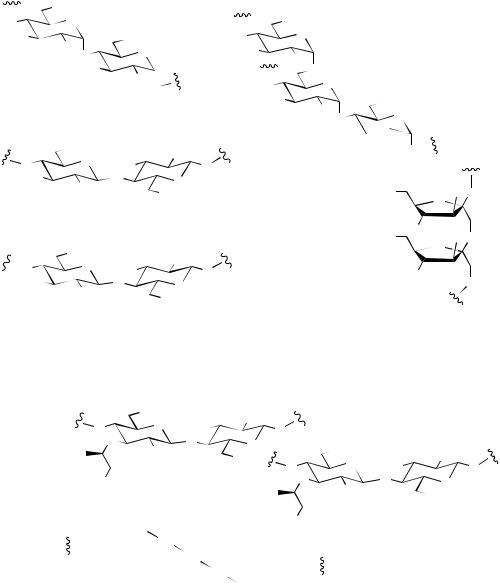
Polysaccharides fulfil two main functions in living organisms, as food reserves, and as structural elements. Plants accumulate starch as their main food reserve, a material that is composed entirely of glucopyranose units but in two types of molecule. Amylose (Figure 8.19) is a linear polymer containing some 1000–2000 glucopyranose units linked α1→4. Amylopectin (Figure 8.19) is a much larger molecule than amylose (the number of glucose residues varies widely but may be as high as 106), and it is a branched-chain molecule. In addition to α1→4 linkages, amylopectin has branches at about every 20 units through α1→6 linkages. These branches continue with α1→4 linkages, but then may have subsidiary branching, giving a treelike structure. The mammalian carbohydrate storage molecule is glycogen, which is analogous to amylopectin in structure, but is larger and contains more frequent branching, about every ten residues. The branching in amylopectin and glycogen is achieved by the enzymic removal of a portion of the α1→4 linked straight chain consisting of several glucose residues, then transferring this short chain to a suitable 6-hydroxyl group. A less common storage polysaccharide found in certain plants of
the Compositae/Asteraceae and Campanulaceae is inulin (Figure 8.19), which is a relatively small polymer of fructofuranose, linked through β2→1 bonds.
Cellulose is reputedly the most abundant organic material on earth, being the main constituent in plant cell walls. It is composed of glucopyranose units linked β1→4 in a linear chain. Alternate residues are ‘rotated’ in the structure (Figure 8.19), allowing hydrogen bonding between adjacent molecules, and construction of the strong fibres characteristic of cellulose, as for example in cotton. The structure of chitin (Figure 8.19) is rather similar to cellulose, though it is composed of β1→4 linked N - acetylglucosamine residues. Chitin is a major constituent in the shells of crustaceans, e.g. crabs and lobsters, and insect skeletons, and its strength again depends on hydrogen bonding between adjacent molecules, producing rigid sheets. Chemical deacetylation of chitin provides chitosan, a valuable industrial material used for water purification because of its chelating properties, and in wound-healing preparations. Bacterial cell walls contain peptidoglycan structures in which carbohydrate chains composed of alternating β1→4 linked N -acetylglucosamine and N -acetylmuramic acid (O-lactyl-N -acetylglucosamine) residues are
474 |
CARBOHYDRATES |
|
|
OH |
α1→4 |
|
|
|
|
|
|||
|
|
O |
|
||
O |
|
||||
|
HO |
1 |
|
OH |
|
|
|
HO |
4 |
|
|
|
|
O |
|
||
|
D-Glc |
O |
|
|
|
|
|
|
|
||
|
HO |
|
|
||
|
|
|
|
||
|
|
|
|
HO |
|
|
|
|
|
O |
|
|
|
amylose |
D-Glc |
|
|
|
|
|
|
||
|
(1000–2000 residues) |
|
|||
|
|
OH |
β1→4 |
OH |
|
|
O |
O |
|
HO |
O |
|
|
1 |
|||
|
HO |
|
O |
O |
|
|
|
|
|||
|
|
HO |
|
4 |
|
|
|
|
OH |
||
|
|
D-Glc |
|
|
|
|
|
cellulose |
D-Glc |
||
|
|
|
|||
|
|
|
|
||
|
|
(~8000 residues) |
|
||
|
|
OH |
β1→4 |
NHAc |
|
|
|
O |
|
|
|
|
O |
|
HO |
O |
|
|
|
1 |
|||
|
HO |
|
O |
O |
|
|
|
|
|||
|
|
AcHN |
|
4 |
|
|
|
|
OH |
||
|
|
|
|
|
|
D-GlcNAc
D-GlcNAc
chitin (500–5000 residues)
|
|
D-Glc |
|
|
|
|
|
|
|
OH |
|
|
|
|
|
|
|
|
|
|
|
|
|
O |
|
|
|
O |
|
|
|
|
||
|
|
|
|
|
||
|
HO |
|
|
1 |
|
|
|
|
|
HO |
α1→6 |
|
|
|
|
|
6 |
O |
|
|
|
|
|
|
|
||
|
|
|
O |
|
|
|
|
|
O |
α1→4 |
|
||
|
|
|
HO |
|
1 |
OH |
|
|
|
|
HO |
4 |
|
|
|
|
|
O |
||
|
|
|
D-Glc |
|
O |
|
|
|
|
|
|
||
HO

HO
D-Glc
HO
D-Fru
HO
amylopectin
(up to 106 residues; branching about every 20 residues)
glycogen
(>106 residues; branching about every 10 residues)
O
HO O
O
2 |
|
1 |
|
HO |
β2→1 |
HO O |
O |
2 |
|
|
D-Fru |
|
HO |
|
O |
GlcN = glucosamine |
inulin |
GlcNAc = N-acetylglucosamine |
(30–35 residues) |
|
|
|
|
|
|
|
|
Figure 8.19 |
|
|
|
|
||||||
N-acetylmuramic acid |
OH β1→4 |
D-GlcNAc |
|
|
|
|
|
|
|
|
|
|||||||
|
|
|
|
|
|
NHAc |
|
|
|
|
|
|
|
|
|
|||
|
|
|
|
|
|
O |
|
|
|
|
|
|
|
|
|
|
||
|
|
O |
HO |
|
|
O |
|
|
|
|
||||||||
|
|
|
|
|
|
|
|
|
||||||||||
|
|
|
O |
|
O |
O |
|
|
|
|
|
|
OH |
|
|
|||
|
|
|
|
|
|
AcHN |
|
|
|
|
|
|
|
|
NHAc |
|||
|
|
|
|
|
|
|
OH |
|
|
|
|
|
|
|
||||
|
|
|
|
|
O |
|
|
|
O |
|
O |
HO |
O |
|||||
|
|
|
|
|
|
|
|
|
|
|
||||||||
|
|
|
|
|
|
|
|
|
|
|
|
|||||||
|
|
|
|
|
|
|
|
|
|
|
||||||||
|
|
Ala |
|
|
|
|
|
|
O |
|
|
O |
O |
|||||
|
|
|
|
|
|
|
|
|
|
|
|
AcHN |
|
|||||
|
|
|
|
|
|
|
|
|
|
|
|
|
|
|
|
|
OH |
|
|
|
D-Glu |
|
|
|
|
|
|
|
|
|
O |
|
|||||
|
|
|
|
|
|
|
|
|
|
|
|
|
||||||
|
|
|
|
|
|
|
|
|
|
|
|
|
||||||
|
|
|
|
|
|
|
|
|
|
|
Ala |
|
|
|||||
|
|
|
|
|
|
|
|
|
|
|
|
|
|
|
||||
|
|
Lys Gly |
|
|
|
|
|
|
|
|||||||||
|
|
|
|
|
|
|
|
|
|
|
|
|
|
|||||
|
|
|
|
|
|
Gly |
|
|
|
|
|
|
|
|
|
|
|
|
|
|
D-Ala |
Gly |
|
|
|
D-Glu |
|
|
|
|
|||||||
|
|
|
|
|
|
|
|
|
|
|
|
|
|
|||||
|
|
|
|
|
|
|
|
|
|
|
|
|
|
|
|
|
|
|
|
|
|
|
|
|
|
|
Gly |
|
Lys |
|
|
|
|
|
|||
|
|
|
|
|
|
|
|
|
|
|
|
|
||||||
|
|
|
|
|
|
|
|
|
|
|
|
|
|
|
|
|
||
|
|
|
|
|
|
|
|
Gly |
|
|
D-Ala |
|
|
|
|
|||
|
|
|
|
|
|
|
|
|
|
|
|
|
|
|||||
|
|
|
|
|
|
|
|
peptidoglycan |
|
|
|
|
||||||
|
|
|
|
|
|
|
(20–130 monosaccharide residues) |
|
|
|||||||||
Figure 8.20
cross-linked via peptide structures. The peptidoglycan of Staphylococcus aureus is illustrated in Figure 8.20, showing the involvement of the lactyl group of the N -acetylmuramic acid to link the peptide with the carbohydrate via an amide/peptide bond. During the cross-linking process, the peptide chains from the N -acetylmuramic acid residues have a terminal –Lys–D-Ala–D-Ala
sequence, and the lysine from one chain is bonded to the penultimate D-alanine of another chain through five glycine residues, at the same time displacing the terminal D-alanine (see Figure 7.36, page 444). The biological activities of the β-lactam antibiotics, e.g. penicillins and cephalosporins (see page 444) and of the last-resort antibiotic vancomycin (see page 426)