

Molecular Fluorescence
.pdf10.3 Fluorescent molecular sensors of cations 295
Fig. 10.13. Polyamine-based PET sensors (PET-8: Huston M. E. et al. (1988) J. Am. Chem. Soc. 110, 4460. PET-9: Fabbrizzi L. et al. (1996) Inorg. Chem. 35, 1733. PET-10: Fabbrizzi L. et al. (1994) Angew. Chem., Int. Ed. Engl. 33, 1975. PET-11: Walkup G. K. et al. (2000) J. Am. Chem. Soc. 122, 5644).
measurable a nity for Ca2þ or Mg2þ. Upon addition of ZnII under physiological conditions, the fluorescence quantum yield increases from 0.39 to 0.87. Concentrations in the nanomolar range can be determined (stability constant: 1:4 109).
Chelators with carboxylic groups are known to e ciently bind divalent hard cations like Ca2þ and Mg2þ. Many selective calcium probes have been designed by Tsien and coworkers for applications in cellular biology, i.e. for probing calcium concentrations in the micromolar range. The so-called BAPTA recognition moiety resembles EDTA, but the nitrogen atoms are linked to a phenyl group in order to avoid pH sensitivity in physiological media. Most of them are not based on the PET principle but on photoinduced charge transfer (see Section 10.3.3.2).
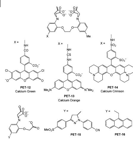
Figure 10.14 shows examples of chelating PET sensors. PET-12 to PET-14 are selective for calcium. In PET sensors, the changes in fluorescence quantum yield are accompanied by proportional changes in excited-state lifetime. Therefore, compounds PET-12 to PET-14 were found to be suitable for fluorescence lifetime imaging of calcium.
The same design principles apply to the magnesium sensors PET-15 and PET-16, in which the recognition moiety has a smaller ‘cavity’.
10.3.2.5 Calixarene-based PET sensors
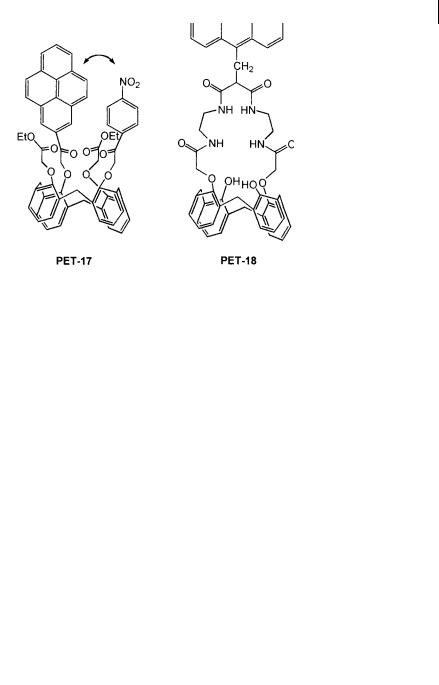
PET-17 has been designed for selective recognition of sodium (Figure 10.15). It contains four carbonyl functions, two of them being linked to pyrene and nitrobenzene at opposite sites on the calixarene lower rim. Complexation with Naþ prevents close approach of pyrene and nitrobenzene and thus reduces the probability of PET. The fluorescence quantum yield increases from 0.0025 to 0.016.
296 10 Fluorescent molecular sensors of ions and molecules
Fig. 10.14. Chelating PET sensors (PET-12, PET-13, PET-14:
Kuhn M. A. (1993), in: Czarnik A. W. (Ed.), Fluorescent
Chemosensors for Ion and Molecule Recognition, ACS Symposium
Series 358. p. 147. PET-15, PET-16: de Silva A. P. et al. (1994) J.
Chem. Soc., Chem. Commun. 1213).
Calixarene containing a dioxotetraaza unit, PET-18, is responsive to transition metal ions like Zn2þ and Ni2þ. Interaction of Zn2þ with the amino groups induces a fluorescence enhancement according to the PET principle. In contrast, some fluorescence quenching is observed in the case of Ni2þ. PET from the fluorophore to the metal ion is a reasonable explanation but energy transfer by electron exchange (Dexter mechanism) cannot be excluded.
10.3.2.6 PET sensors involving excimer formation
PET-19 (Figure 10.16) consists of a diazacrown ether with two pendant pyrene groups. As expected, cation binding results in a large change in the monomer/
298 10 Fluorescent molecular sensors of ions and molecules
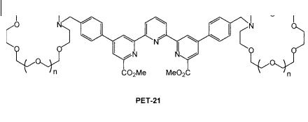
Fig. 10.17. PET sensor involving energy transfer (PET-21: de
Silva A. P. et al. (1997) J. Chem. Soc., Chem. Commun. 1891).
excimer ratio. There is a concomitant increase in the overall fluorescence emission as a result of the reduction in PET from the nitrogen atom to the pyrenyl groups. The monomer/excimer ratio was found to be strongly dependent on the nature of the metal ion. Among the investigated metal ions, the larger stability constants of the complexes were obtained for Kþ and Ba2þ, in accordance with the size of these cations with respect to the crown diameter.
In the same way, the emission spectrum of PET-20, existing as the protonated form in acetonitrile, exhibits an excimer band whose intensity decreases upon binding of Cd2þ and Pb2þ.
10.3.2.7 Examples of PET sensors involving energy transfer
PET-21 (Figure 10.17) contains a terpyridyl diester that can strongly bind EuIII. Excitation energy transfer from this type of ligand to EuIII is known to occur via the triplet state and should result in luminescence from EuIII. However, when the crown is empty, only weak luminescence is detected because of quenching due to PET from the nitrogen atom of the crown. Binding of Kþ causes a very large enhancement of the luminescence quantum yield, as expected from cation-induced reduction of the PET e ciency. PET-21 provides the first example of metaltriggered metal-centered emission. Thanks to the long lifetime of EuIII (hundreds of microseconds), time-delayed detection of the luminescence is possible which enables eradication of the fast parasitic fluorescence of biological samples.
10.3.3
Fluorescent PCT (photoinduced charge transfer) cation sensors
10.3.3.1 Principles
When a fluorophore contains an electron-donating group (often an amino group) conjugated to an electron-withdrawing group, it undergoes intramolecular charge transfer from the donor to the acceptor upon excitation by light. The consequent change in dipole moment results in a Stokes shift that depends on the microenvironment of the fluorophore; polarity probes have been designed on this basis (see Chapter 7). It can thus be anticipated that cations in close interaction with the donor or the acceptor moiety will change the photophysical properties of the fluo-

10.3 Fluorescent molecular sensors of cations 299
rophore because the complexed cation a ects the e ciency of intramolecular charge transfer.
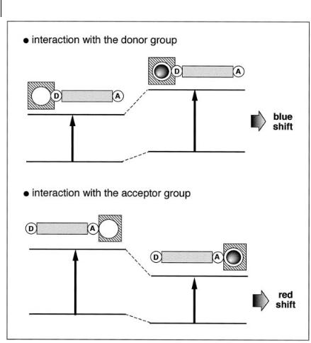
When a group (like an amino group) playing the role of an electron donor within the fluorophore interacts with a cation, the latter reduces the electron-donating character of this group; owing to the resulting reduction in conjugation, a blueshift of the absorption spectrum is expected, together with a decrease in the molar absorption coe cient. Conversely, a cation interacting with the acceptor group enhances the electron-withdrawing character of this group; the absorption spectrum is thus red-shifted and the molar absorption coe cient is increased. The fluorescence spectra are in principle shifted in the same direction as the absorption spectra. In addition to these shifts, changes in quantum yields and lifetimes are often observed. All these photophysical e ects are obviously dependent on the charge and the size of the cation, and selectivity of these e ects is expected.
The photophysical changes on cation binding can also be described in terms of charge dipole interaction. Let us consider only the case where the dipole moment in the excited state is larger than that in the ground state. Then, when the cation interacts with the donor group, the excited state is more strongly destabilized by the cation than the ground state, and a blue-shift of the absorption and emission spectra is expected (however the fluorescence spectrum undergoes only a slight blue-shift in most cases; this important observation will be discussed below). Conversely, when the cation interacts with the acceptor group, the excited state is more stabilized by the cation than the ground state, and this leads to a red-shift of the absorption and emission spectra (Figure 10.18).
10.3.3.2 PCT sensors in which the bound cation interacts with an electron-donating group
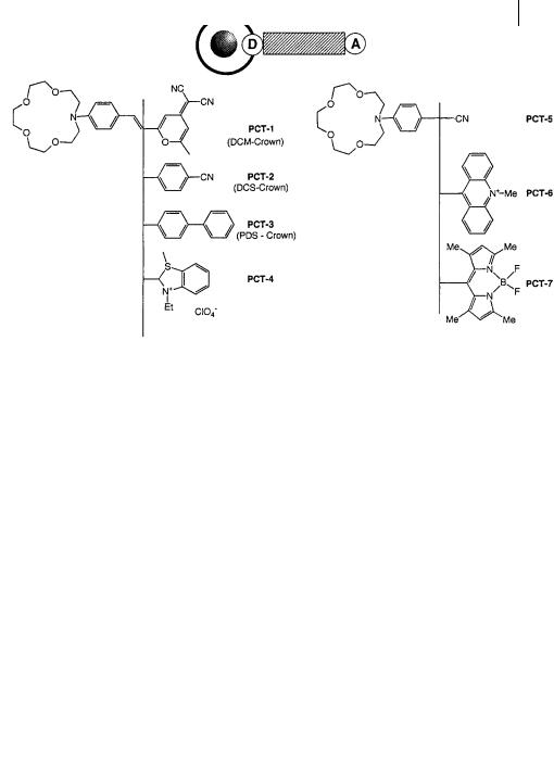
Crown-containing PCT sensors Many fluoroionophores have been designed according to the following principle: the cation receptor is an azacrown containing a nitrogen atom that is conjugated to an electron-withdrawing group (Figure 10.19). Compounds PCT-1 to PCT-4 exhibit a common feature: the blue-shift of the absorption spectrum is much larger than that of the emission spectrum on cation binding. An example is given in Figure 10.20. Such a small shift of the fluorescence spectrum – which at first sight is surprising – can be interpreted as follows. The photoinduced charge transfer reduces the electron density on the nitrogen atom of the crown, and this nitrogen atom becomes a non-coordinating atom because it is positively polarized. Therefore, excitation induces a photodisruption of the interaction between the cation and the nitrogen atom of the crown. The fluorescence spectrum is thus only slightly a ected because most of the fluorescence is emitted from species in which the interaction between the cation and the fluorophore does not exist any more or is much weaker.
This interpretation is supported by a thorough study of the photophysics of PCT-1 and its complexes with Liþ and Ca2þ (Martin et al., 1994). In particular, subpicosecond pump-probe spectroscopy provided compelling evidence for the disruption of the link between the crown nitrogen atom and the cation. A photo-
300 10 Fluorescent molecular sensors of ions and molecules
Fig. 10.18. Spectral displacements of PCT sensors resulting from interaction of a bound cation with an electron-donating or electron-withdrawing group.
disruption was also demonstrated in complexes of PCT-2 and PCT-3. The cationinduced spectral changes in PCT-4 were interpreted in the same way.
Such a photodisruption results in a lower stability of the complexes in the excited state. Therefore, excitation of these complexes by an intense pulse of light is expected to cause some cations to leave the crown and di use away, provided that the time constant for total release of the cation from the crown is shorter than the lifetime t of the excited state (for a discussion on this point, see Valeur et al., 1997).
Intramolecular charge transfer in conjugated donor–acceptor molecules may be accompanied by internal rotation leading to TICT (twisted intramolecular charge transfer) states. A dual fluorescence may be observed as in PCT-5 (Le´tard et al., 1994) (which resembles the well-known DMABN (see section 3.4.4) containing a dimethylamino group instead of the monoaza-15-crown-5): the short-wavelength

302 10 Fluorescent molecular sensors of ions and molecules
band corresponds to the fluorescence from the locally excited state and the longwavelength band arises from a TICT state. The fluorescence intensity of the latter decreases upon cation binding because interaction between a bound cation and the crown nitrogen disfavors the formation of a TICT state, which leads to a concomitant increase in the short-wavelength band.
The formation of a TICT state is often invoked even if no dual fluorescence is observed. For donor–acceptor stilbenes (PCT-2 and PCT-3), the proposed kinetic scheme contains three states: the planar state E reached upon excitation can lead to state P (non-fluorescent) by double-bond twist, and to TICT state A by singlebond twist, the latter being responsible for most of the emission.
The absence of fluorescence of PCT-6 may be due to the formation of a nonfluorescent TICT state, and an acridinium-type fluorescence is recovered upon binding of Hþ and Agþ. The same explanation may hold for the low fluorescence of PCT-7, whose electron-withdrawing group, boron-dipyrromethen, must be twisted due to steric interactions with the phenyl ring: the fluorescence enhancement factor varies from 90 for Liþ to 2250 for Mg2þ. Both PCT-6 and PCT-7 compounds undergo much larger fluorescence enhancements than most PCT molecular sensors (factors of @2–5 are generally observed). They can be considered as limiting cases closely resembling PET sensors but with a virtual spacer.
The crown-containing fluoroionophores described above are of great interest for the understanding of cation–fluorophore interactions. They o er a wide variety of photophysical changes on cation binding that can be used for cation recognition. However, the selectivity of azacrowns towards metal ions is not good enough when stringent discrimination between cations of the same chemical family is required.
Improvement of selectivity can be achieved by the participation of external groups, as shown in Figure 10.21. In PCT-8 (PBFI) and PCT-9 (SBFI), the oxygen atom of the methoxy substituent of the fluorophore can interact with a cation; binding e ciency and selectivity are thus better than those of the crown alone. SBFI has been designed for probing intracellular sodium ions and PBFI for potassium ions. In both compounds, the photophysical changes are likely to be due to the reduction of the electron-donating character of the nitrogen atoms of the diazacrown by the complexed cation. Further improvement of the selectivity towards Kþ with respect to Naþ is desirable. PCT-10 resembles PBFI but it shows greater selectivity for potassium over sodium than PBFI.
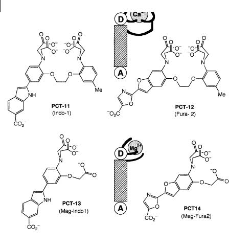
Chelating PCT sensors In light of the preceding considerations on crowned chargetransfer compounds, it is worth examining the photophysical properties of wellknown chelators in the BAPTA series used for the recognition of cytosolic cations and in particular calcium ions (Grynkiewicz et al., 1985). For instance PCT-11 (Indo-1) and PCT-12 (Fura-2) (Figure 10.22) are widely used as calcium indicators. In these compounds, the fluorophore is a donor–acceptor molecule with an amino group as the electron-donating group that participates in the complexation, the ionophore being a chelating group of the BAPTA type.
Upon complexation by Ca2þ in water, the absorption spectrum of Fura-2 is blueshifted, whereas there is almost no shift of the fluorescence spectrum. The same

10.3 Fluorescent molecular sensors of cations 303
Fig. 10.21. Crown-containing bifluorophoric PCT sensors (PCT- 8: Kasner S. E. and Ganz M. B. (1992) Am. J. Physiol. 262, F462. PCT-9: Minta A. and Tsien R. Y. (1989) J. Biol. Chem. 264, 19449. PCT-10: Crossley R. et al. (1994) J. Chem. Soc., Perkin Trans. 2, 513).
interpretation as for the above-described crown-ether-linked compounds can be proposed: the electron density of the nitrogen atom conjugated with the electronwithdrawing group of the fluorophore is reduced on excitation and might even become positively polarized – this causes disruption of the interaction between this nitrogen atom and a bound cation. Consequently, fluorescence emission closely resembles that of the free ligand.
In contrast to Fura-2, the photoinduced charge transfer in Indo-1 may not be su cient to cause nitrogen–Ca2þ bond breaking. This interpretation is consistent with the fact that the fluorescence maximum of free Indo-1 is located at a shorter wavelength than Fura-2 by @30 nm, thus indicating a less polar charge-transfer state.
By keeping the same fluorophores, but reducing the ‘cavity’ size of the ionophore, we obtain PCT-13 (Mag-Indo1) and PCT-14 (Mag-Fura2) (Figure 10.22), which are selective for magnesium. PCT-8, PCT-9 and PCT-11 to PCT-14 are commercially available in the non-fluorescent acetoxymethylester form so that they are cell permeant and they recover their fluorescence upon hydrolysis by enzymes (Molecular Probes, Inc., Eugene, OR).
Cryptand-based PCT sensors The above-described chelators are well suited to the detection of alkaline earth cations but not alkali cations. In contrast, cryptands are very selective towards the latter.
304 10 Fluorescent molecular sensors of ions and molecules
Fig. 10.22. Chelating PCT sensors for calcium and magnesium ions (PCT-11 and PCT-12: Grynkiewicz G. et al. (1985) J. Biol. Chem. 260, 3440. PCT-13 and PCT-14: Haugland R. P.,
Handbook of Fluorescent Probes and Research Chemicals, 6th edn, Molecular Probes, Inc., Eugene, OR).
PCT-15 (FCryp-2) (Figure 10.23) is a nice example of a fluorescent signaling receptor in which the ionophore moiety has been specially designed to determine intracellular free sodium concentration. An indole derivative acts as the fluorophore: on sodium binding, the emission maximum shifts from 460 nm to 395 nm and the fluorescence intensity increases 25-fold. The origin of these photophysical changes has not yet been studied. The large Stokes shift of the free ligand may be accounted for by photoinduced charge transfer with concomitant internal rotation in the excited state leading to a TICT state. The blue-shift of the emission spectrum on sodium binding is likely to be due to the reduction in the electron-donating character of the dye-bound nitrogen atom of the cryptand.