

The Chemistry of Dienes and Polyenes. Volume 1
Edited by Zvi Rappoport
Copyright ∂ 1997 John Wiley & Sons, Ltd.
ISBN: 0-471-96512-X
CHAPTER 10
Analysis of dienes and polyenes and their structure determination
ZEEV AIZENSHTAT
Casali Institute for Applied Chemistry and Department of Organic Chemistry, The Hebrew University of Jerusalem, Jerusalem 91904, Israel
Fax: 972-2-6528-250; e-mail: ZEEV@VMS.HUJI.AC.IL
I. INTRODUCTION . . . . . . . . . . . . . . . . . . . . . . . . . . . . . . . . . . . . . |
481 |
II. SPECTROSCOPIC METHODS . . . . . . . . . . . . . . . . . . . . . . . . . . . . |
482 |
III. SEPARATION AND CHROMATOGRAPHY OF DIENES |
|
AND POLYENES . . . . . . . . . . . . . . . . . . . . . . . . . . . . . . . . . . . . . |
485 |
IV. THERMAL DESORPTION AND ELECTRON ENERGY LOSS |
|
SPECTROSCOPY . . . . . . . . . . . . . . . . . . . . . . . . . . . . . . . . . . . . . |
486 |
V. MASS SPECTROMETRY . . . . . . . . . . . . . . . . . . . . . . . . . . . . . . . |
486 |
VI. CHEMICAL DERIVATIZATIONS . . . . . . . . . . . . . . . . . . . . . . . . . . |
496 |
VII. SELECTED EXAMPLES OF MULTI-PARAMETER ANALYSIS FOR |
|
DIENES AND POLYENES: STRUCTURE DETERMINATION . . . . . . |
499 |
A. Enolic Dienes Derived from Testosterone-17ˇ-acetate . . . . . . . . . . . |
499 |
B. Antiviral and Antifungal Mycoticin (A and B) Partial Structure |
|
Determination . . . . . . . . . . . . . . . . . . . . . . . . . . . . . . . . . . . . . . |
500 |
VIII. ANALYSIS OF CAROTENOIDS AS AN EXAMPLE OF |
|
POLYENE STUDIES . . . . . . . . . . . . . . . . . . . . . . . . . . . . . . . . . . . |
501 |
IX. ACKNOWLEDGEMENTS . . . . . . . . . . . . . . . . . . . . . . . . . . . . . . . |
504 |
X. REFERENCES . . . . . . . . . . . . . . . . . . . . . . . . . . . . . . . . . . . . . . . |
504 |
I. INTRODUCTION
In the last fifteen years most efforts aimed at identification and structure determination of dienes and of polyenes were related to studies of bio-originated compounds. The analysis of dienes and polyenes has not been reviewed, so far. The analysis of double bonds containing molecules utilizes the chemical reactivity of the bonds, and hence conjugated double bonds require different approaches than methods used for non-conjugated double bonds. One example is the use of the Diels-Alder reaction which yields derivatives of conjugated dienes whereas isolated double bonds are not affected. Some of the methods
481
482 |
Zeev Aizenshtat |
reviewed are very basic and have been in use for many years; others are very specific for the family of compounds studied, as exemplified for carotenoids.
The determination of the structure of synthetic dienes and polyenes is somewhat easier than the identification and structure determination of natural products. Obviously, this stems from the need to separate the latter compounds from very complex mixtures.
The theoretical analysis of the spectra (mostly IR and UV-VIS) of polyenes has been reviewed twice in the last 20 years1,2. These reviews concentrate on understanding their biological role and the extension of polyene application. However, both reviews do not cover the structure determination of dienes and polyenes.
One important reason why biologists and biochemists are interested in polyenes is related to the fact that they are light-harvesting antennas and are responsible for triggering the vision signal1,2. Moreover, long-chain polyenes appear in vitamins and carotenoids as well as in many antibiotics. Another reason is that the polymeric form, such as polyacetylene, is a natural photo-conductive matter that upon doping becomes conductive, comparable with copper! These properties have ignited the interest of chemists in the synthesis of polyene polymers for ‘molecular electronics’. These conjugated functional polymers may be designed to serve as tunable electro-light emitting diodes (LED).
II. SPECTROSCOPIC METHODS
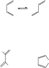
The use of UV-VIS spectra to analyse dienes and polyenes was historically the first method of choice. The spectra of isolated non-conjugated polyenes is actually the superposition of the spectrum of each one of the double bonds. For each double bond the spectrum depends on the various substituents and also on its location in the molecule. It also depends on the stereochemistry, since conjugated double bonds have either E or Z configuration around each -bond but also a cisoid and transoid conformer3 around the single bond marked as s-cis and s-trans4.
s-cis s-trans
Polyenes can undergo rotation easily about the ‘single’ bond at room temperature. The s-trans conformers are generally more stable because steric interactions in them are minimized.
The s-cis conformers tend to be distorted from planarity, and this may influence the UV spectra, as shown by the comparison of butadiene (1) and cyclopentadiene (2).
H |
|
|
CH2 |
H |
|
(1) |
(2) |
λ 220 nm (ε 20900) |
λ 238.5 nm (ε 3400) |
It is evident that the s-cis frozen conformation in the ring of 2 shows a batochromic shift, but a much lower absorption (ε) in comparison with butadiene (1). Woodward and

10. Analysis of dienes and polyenes and their structure determination |
483 |
Fieser and Fieser5 used an empirical correlation, based on a wide range of compounds, between the diene structure and max. This empirical correlation of structure and ultraviolet absorption followed a regular pattern, allowing the calculation of max with reasonable accuracy. The early rules were: parent diene 214 nm; for cisoid add 39 nm, extended conjugated diene 30 nm. In the studies of steroids and other cyclic terpenoids these rules were employed to differentiate between homoannular dienes and exocyclic enes (see also Section VII.A)5. A more theoretical approach is presented for longer conjugated polyenes, i.e. hexatriene, octatetraene, etc., that have geometrical isomers, i.e. cis-polyenes and trans-polyenes. For butadiene, all vibrational frequencies have been observed in the IR or Raman spectra2. The theoretical analysis of the vibrational properties and the frequencies were reviewed in 1991 by Orlandi and coworkers; their review contains 251 references2. However, this review does not offer the analytical tools needed for structural determination. Furthermore, all polyenes examined are conjugated with no branching or ring effects.
In general, it is noteworthy that one can use the lowering of the energies required to either excite (UV-VIS) or stretch (IR, Raman) the CDC bond with the extension of the polyene system. We have mentioned the batochromic effect (Woodward rule)5 and will discuss the CDC stretch frequency which correlates with the length of the polyene (e.g. 1640 cm 1 for butadiene and 1490 cm 1 for the ˇ-carotene homologs, respectively). These correlations can supplement other spectroscopic data to assess the length of the polyene conjugated systems. Of course, the extreme case is cyclic conjugated aromatic systems which are beyond the scope of the present review.
Very powerful tools for the study of dienes and, to some extent, polyenes (in particular annular polyenes) are both 1H and 13C NMR spectroscopies, which will be discussed in a separate section. As previously mentioned 1,3-butadiene is more stable in the s-trans conformation and in the 1H NMR spectrum both butadiene (1) and 2,3,6,7-tetramethyl- 2,4,6-octatriene (3) display the vinyl proton at a low chemical shift value. In these simple examples the υ value can be predicted theoretically. The 1H NMR spectrum of a C25- branched isoprenoid was examined as part of the structural determination for biomarkers and is shown in Figure 16. The other spectral and structure assignments are described later in this review.
|
H δ 5.16 |
|
R |
H |
R |
H |
H δ 5.06 |
H |
|
|
H |
|
|
|
|
||
H |
H |
R |
H |
|
R |
|
δ 6.27 |
|
δ |
6.62 |
R = CH3 |
|
|
|
|||
|
(1) |
|
(3) |
|
|
Figure 1 shows a partial 1H NMR spectrum for H-23, H-5 and H-24 a,b (see the formula of the C25 polyene, 4). These are the hydrogens on the alkenic position and the multiplet between 4.89 5.76 υ (ppm) integrates to four hydrogens, i.e. to a vinyl functionality. Since this is an isoprenoid skeleton it is clear that the position of the double bond must be at C-24. Proton H-23 appears at a lowest field as a heptet due to cis coupling with H-24a, trans coupling with H-24b and an additional coupling with the vicinal H-22. This indicates the presence of a single H-22 allylic proton, thus supporting the assignment. The double bond between C-5 and C-6 shows the influence of the H-4 allylic hydrogens. These assignments and further discussion on decoupling and 13C NMR spectrum appear in Belt and coworkers6.

484 |
Zeev Aizenshtat |
FIGURE 1. The 1H NMR spectrum of the double bonds region (υ 4.8 5.8 ppm) of the high branched C25, compound 4. The signal X is due to an impurity (see Belt and coworkers6)
|
16 |
|
17 |
|
|
18 |
|
19 |
|
|
4 |
6 |
|
8 |
|
12 |
14 |
|
2 |
|
|
10 |
|
|
||
1 |
3 |
|
5 |
7 |
9 |
11 |
13 |
15 |
|
|
|
|
|
|
|
|
|
|
|
|
20 |
|
21 |
|
|
|
|
|
|
|
|
|
|
|
|
|
|
|
23 |
|
22 |
25 |
|
|
|
|
|
H |
|
H |
|
|
|
|
|
|
24a |
|
24b |
|
|
|
|
|
|
|
|
(4) |
|
|
|
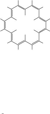
Although we have stated that we will not include aromatic structures, large systems of polyenes give rise to long conjugated systems, with very strong shielding and deshielding effects. The annulene family contains systems having the (4n C 2) -electrons which, according to Huckel,¨ should have a cyclic delocalization but are diatropic. An example is [18] annulene (5), where the 12 outer hydrogens absorb at υ 9.28 ppm and the 6 inner hydrogens absorb at υ 2.99 ppm.
In contrast, if the same polyene were to be ‘open’, no ring current would exist and the NMR spectrum would be very different (see discussion on carotenoids).
Dienes and polyenes show a pronounced molecular ion in the mass spectra and hence the molecular weight of polyenes can be determined by positive ion mass spectra. The easy removal of a -electron from a diene is usually the reason for the distinct MžC . The mass spectral investigation of conjugated polyenes is somewhat similar to that of aromatic structures, due to the high stability of the rearranged ions formed after the
10. Analysis of dienes and polyenes and their structure determination |
485 |
||
H |
|
H |
|
H |
|
H |
|
H |
H |
H |
|
H |
|
H |
|
H |
|
H |
|
H |
H |
H |
|
H |
|
H |
|
H H
(5)
first electron removal. This phenomenon will be discussed in more detail in the section devoted to carotenoids. For a general discussion on the mass spectrum (MS) of dienes, see Budzikiewicz and coworkers7. Since dienes and polyenes are highly sensitive to photodissociation this method was also employed in conjunction with MS8 for the study of branched dienes.
III. SEPARATION AND CHROMATOGRAPHY OF DIENES AND POLYENES
Chemical separation of conjugated dienes and other polyunsaturated hydrocarbons is based on the availability of delocalized electrons. The use of a strong dienophile (e.g. tetracyanoethylene, TCNE) will derivatize only conjugated dienes, thus separating the polyunsaturated compounds into two groups. However, such derivatization is not always reversible since a retro-Diels Alder reaction may require a high temperature. Hence, the retrieved compounds may be the thermostable ones and not those present in the initially analysed mixture.
Even simple dienes and polyenes are difficult to classify in comparison with alkenes. Whereas bromination, oxidation and reaction with tetranitromethane (TNM) can identify the number of double bonds and their location in the molecular structure, conjugated double bonds produce very complex mixtures. Furthermore, many of the tests based on-complexation can also apply for aromatic moieties. An example is the TNM -complex which is yellow with benzene and orange with naphthalene and the tests are therefore non-specific.
Basically, the chromatographic separation of dienes and polyenes is similar to that of alkenes. Both gas chromatography (GC) and liquid chromatography (LC or HPLC) can be employed. For low molecular weight, more volatile diene/GC is usually good enough. If a better separation is needed, this can be enhanced by AgC - -complexation. HPLC is employed either for more polar derivatives of polyenes or for non-volatile high molecular weight compounds (see special discussion on carotenoids). The use of small particle size silver-nitrate-impregnated silica as stationary phase was adapted for HPLC separation of unsaturated hydrocarbons from petroleums and bitumens9,10. The same approach can be used in thin layer chromatography (TLC) to separate the unsaturated components (10% AgNO3/silica gel w/w)10.
We will discuss in some detail examples where various methods of separation, including chemical derivatization, were complemented by spectroscopic identification. However, even the use of the most advanced analytical methods frequently yields only partial

486 |
Zeev Aizenshtat |
structure determination, and for more complex compounds comparison with a model compound is the only solution to total structure determination.
Higher molecular weight dienes and polyenes which are solid and can be crystallized make it possible to study their structure by X-ray diffraction. This, of course, will give information only about the crystalline form (see discussion on steroids and antifungal molecules, Section VII).
IV. THERMAL DESORPTION AND ELECTRON ENERGY LOSS
SPECTROSCOPY
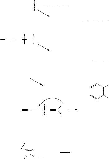
Structure determination of unsaturated compounds can be supplemented by thermal desorption (TD) and electron energy loss (EEL) spectroscopies. The two methods use the chemisorption of cis and trans enes or dienes to the Pt(111) surface over a range of temperatures11. The experimental equipment and procedures described12 show these methods to be employed for dienes such as 1,3-butadiene. At very low temperature the diene is adsorbed on Pt(111) and the thermal desorption is followed by increasing the temperature.
Figure 2 shows a comparison of C4 hydrocarbon desorption spectra of Pt(111) monitoring temperature and m/z of 2, 54, 56, 58 using a mass spectrometer. The TD spectrum for a monolayer of 1,3-butadiene is compared with 1-butene, cis-2-butene and trans-2- butene. Monitoring of the hydrogen thermal desorption shows that for the 1,3-diene no hydrocarbon desorption is recorded, but rather a destructive dehydrogenation of the diene. In the monoenes, up to 300 K one can see the release of m/z 56 (C4H8C ) followed at higher temperature by the release of hydrogen, whereas in the diene (Figure 2c) only H2C (m/z 2) is monitored.
The electron energy loss spectra (EEL) in Figure 3 shows the IR vibrational difference of cis and trans alkenes up to 170 K, arising from different geometry of the two bonds between the metal and the double bond. At 300 K this difference is erased and both form C4H6, by loss of hydrogen11. The bond formed by the diene (1,3-butadiene) is shown to have the same vibrational properties. Hence, the authors conclude that the end product adsorbed is:
H H
C C
C C
Pt(III)
V. MASS SPECTROMETRY
Simple dialkenes of general formula CnH2n 2 produce a fragmentation pattern which depends upon the relative location of the two double bonds (Scheme 1). The nonconjugated dienes fragment corresponding to the respective allylic fission13. The nonconjugated dienes fragmentation pattern is dominated by ˇ-cleavage and the formation of a C3H6C ion if rearrangement is possible (Scheme 1)6. The allylic fission13 is not preferred in conjugated systems, hence the formation of the diene-ion which can be stabilized by cyclization. However, if one examines conjugated dienes and polyenes such as terpenes, the most abundant ion is the [M 1]C base peak formed by loss of a single hydrogen atom. Apart from this ion the other abundant ions are the m/z 53 and m/z 39, formed by

10. Analysis of dienes and polyenes and their structure determination |
487 |
FIGURE 2. Thermal desorption spectra for monoenes and a diene C4 hydrocarbon, chemisorbed on Pt(III). (a) cis-but-2-ene and trans-but-2-ene, (b) but-2-yne and (c) 1,3-butadiene. D 1 (full line) ˇ D 3KS 1 for all. The monitoring of m/z 2, 54, 56, 58 was done by MS (see Avery and Sheppard11 and references cited therein)

488 |
Zeev Aizenshtat |
FIGURE 3. Electron energy loss spectra from 1,3-butadiene, chemisorbed on Pt(III) at (a) 170 K,
(b) 300 K, (c) 385 K and (d) 450 K11
|
|
10. Analysis of dienes and polyenes and their structure determination |
489 |
||||||||||||||||||||||||||||||||||||||||||||||||||||||
[R |
|
|
CH |
|
CH |
|
CH2 |
CH2 |
|
|
|
|
|
CH |
CH R′]•+ |
|
|
|
|
|
|
|
|
|
|
|
|
|
|
|
|
||||||||||||||||||||||||||
|
|
|
|
|
|
|
|
|
|
|
|
|
|
|
|
|
|
|
|
|
|
||||||||||||||||||||||||||||||||||||
|
|
|
|
|
|
|
|
|
|
|
|
|
|
|
|
|
|
|
|
|
|||||||||||||||||||||||||||||||||||||
|
|
|
|
|
|
|
|
|
|
|
|
|
|
|
|
|
|
|
|
|
|
||||||||||||||||||||||||||||||||||||
|
|
|
|
|
|
|
|
[R |
|
|
|
|
|
CH |
|
|
|
|
|
|
CH |
|
|
|
|
|
CH2 ] + |
+ |
|
[R′ |
|
CH |
|
|
|
CH |
CH2 ] • |
|
|||||||||||||||||||
|
|
|
|
|
|
|
|
|
|
|
|
|
|
|
|
|
|
|
|||||||||||||||||||||||||||||||||||||||
|
|
|
|
|
|
|
|
|
|
|
|
|
|
|
|
|
|
||||||||||||||||||||||||||||||||||||||||
|
|
|
|
|
|
|
|
|
|
|
|
|
|
|
|
|
|
||||||||||||||||||||||||||||||||||||||||
|
|
|
|
|
|
|
|
[R′ |
|
|
|
|
|
CH |
|
|
|
|
CH |
|
|
|
|
CH ] + |
+ |
|
[R |
|
|
CH |
|
|
|
CH |
|
|
|
CH ] • |
|
||||||||||||||||||
|
|
|
|
|
|
|
|
|
|
|
|
|
|
|
|
|
|
|
|
|
|
|
|
|
|
||||||||||||||||||||||||||||||||
|
|
|
|
|
|
|
|
|
|
|
|
|
|
|
|
|
|
|
|
|
|
||||||||||||||||||||||||||||||||||||
|
|
|
|
|
|
|
|
|
|
|
|
|
|
|
|
|
|
|
|
|
|
|
|
|
|
|
|
|
|
|
|
|
2 |
|
|
|
|
|
|
|
|
|
|
|
|
|
|
|
|
|
2 |
|
|||||
[R |
|
CH |
CH |
CH2 |
CH |
|
|
|
CH |
|
|
|
R′]•+ |
|
|
] + |
|
+ |
[R′ |
|
|
|
|
|
|
|
CH] • |
|
|||||||||||||||||||||||||||||
|
|
|
|
|
|
|
|
|
|
|
|
|
|
|
|
|
|
||||||||||||||||||||||||||||||||||||||||
|
|
|
|
|
|
|
|
|
|
|
|
|
|
|
|
|
|
[R |
|
|
|
CH |
|
|
|
CH |
|
CH |
|
|
|
CH |
|
|
|
||||||||||||||||||||||
|
|
|
|
|
|
|
|
|
|
|
|
|
|
|
|
|
|
|
|
|
|
|
|
|
|
|
|
|
|
|
|
|
|
||||||||||||||||||||||||
|
|
|
|
|
|
|
|
|
|
|
|
|
|
|
|
|
|
|
|
|
|
|
|
|
|
|
|
||||||||||||||||||||||||||||||
|
|
|
|
|
|
|
|
|
|
|
|
|
|
|
|
|
|
|
|
|
|
|
|
|
|
|
|
|
|
|
|
|
|
||||||||||||||||||||||||
|
|
|
|
|
|
|
|
|
|
|
|
|
|
|
|
|
|
|
|
|
|
|
|
|
|
|
|
|
|
|
|
|
|
|
|
|
|
|
|
2 |
|
|
|
|
|
|
|
|
|
|
|
|
|
|
|
|
|
|
|
|
|
|
|
|
|
|
|
|
|
|
|
|
|
|
|
[R′ |
|
|
|
CH |
|
|
|
|
CH |
|
|
CH2 ] + |
+ |
[R |
|
|
|
CH |
CH] • |
|
|||||||||||||||||||
|
|
|
|
|
|
|
|
|
|
|
|
|
|
|
|
|
|
|
|
|
|
|
|
|
|
|
|
|
|||||||||||||||||||||||||||||
|
|
|
|
|
|
|
|
|
|
|
|
|
|
|
|
|
|
|
|
|
|
|
|
|
|
|
|
||||||||||||||||||||||||||||||
|
|
|
|
|
|
|
|
|
|
|
|
|
|
|
|
|
|
|
|
|
|
|
|
|
|
|
|
|
|||||||||||||||||||||||||||||
[R |
|
|
CH |
|
CH |
|
CH |
|
|
|
CHR′]•+ |
|
|
|
|
|
|
|
|
|
|
|
|
|
|
|
|
|
|
|
|
|
|
|
|
|
|
|
|
|
|
|
|
||||||||||||||
|
|
|
|
|
|
|
|
|
|
|
|
|
|
|
|
|
|
|
|
|
|
|
|
|
|
|
|
|
|
|
|
|
|
|
|
|
|
|
|||||||||||||||||||
|
|
|
|
|
|
|
|
|
|
|
|
|
|
|
|
|
|
|
|
|
|
|
|
|
|
|
|
|
|
|
|
|
|
|
|
|
|
|
|
||||||||||||||||||
|
|
|
|
|
|
|
|
|
|
|
|
−H+ |
|
|
|
|
|
|
|
|
|
|
|
|
|
|
|
|
|
|
|
|
|
|
|
|
|
|
|
|
|
|
|
|
|
|
|
|
|
|
|
|
|||||
|
|
|
|
|
|
|
|
|
|
|
|
|
|
|
|
|
|
|
|
|
|
|
|
|
|
|
|
|
|
|
|
|
|
|
|
|
|
|
|
|
|
|
|
|
|
|
|
|
|
|
|
|
|
|
R′ |
|
|
|
|
|
|
|
|
R |
|
CH |
|
|
CH |
|
|
|
CH |
|
|
|
|
R′ (M−1)+ |
|
|
|
|
|
|
|
+ |
|
|
|
|
R |
|
|||||||||||||||||||||||
|
|
|
|
|
|
|
|
|
|
|
|
|
|
|
|
|
|
|
|
|
|
|
|
|
|
|
|
|
|
||||||||||||||||||||||||||||
|
|
|
|
|
|
|
|
|
|
|
|
|
|
|
|
|
|
|
|
|
|
|
|
|
|
|
|
|
|
||||||||||||||||||||||||||||
|
|
|
|
|
|
|
|
|
|
|
|
|
|
|
|
|
|
|
|
|
|
|
|
|
|
|
|
|
|
|
|
|
|
|
|
|
|
|
|
|
|
|
|
|
|
|
|
|
|
|
|
|
|
|
|
||
|
|
|
|
|
|
|
|
|
|
|
|
|
|
|
|
|
|
|
|
|
|
|
|
|
|
|
|
|
|
|
|
|
|
|
H |
|
|
|
C H + |
|
|
|
|
|
|
|
|
|
|
||||||||
|
|
|
|
H C CH CH CH C |
|
|
|
|
|
|
|
|
|
|
+ C H |
|
|
|
|||||||||||||||||||||||||||||||||||||||
|
|
|
|
2 |
|
|
|
|
|
|
|
|
|
|
|
|
2 |
|
|
|
|
|
|
|
|
|
|
|
|
|
|
|
|
|
|
|
|
|
|
|
|
|
|
3 |
6 |
|
|
2 |
2 |
|
|
|
|
||||
H
SCHEME 1
the cleavage shown in equation 1.
m/z 67 |
|
|
H |
|
|
CH2 |
|
|
C |
C3 H3 + + residue |
(1) |
CH |
CH2 |
|
CH2 |
|
|
m/z 53
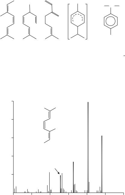
The next conjugated triene of the isoprenoid structure is the allo-ocimene (6). ˇ- Ocimene (7) and myrcene (8) also have a triene moiety but only two of their double bonds are conjugated. Comparison of these three isomeric trienes gives insight into the manner in which the relative positions of the double bonds control the fragmentation (Figure 4). The obvious mass spectral differences show that conjugation yields a higher abundance of the MC (136) and the base peak of compound 6 is derived by the loss of a methyl group (m/z 121). These two ions are much less abundant for 7 and 8, both showing m/z 93 as the most abundent signal whilst 8 shows also m/z 697.
Cyclic dienes of the terpene family are also very interesting. The MS of the C10H16 compounds are discussed extensively in Budzikiewicz and coworkers.7. All of them transform
490 |
Zeev Aizenshtat |
 + CH3 CH CH3
2H
CH3
(6) |
(7) |
(8) |
(9) m/z 136 |
(10) m/z 134 |
into the ion 9, having a structure which is very similar to the ion formed from the aromatic cumene (10) but differing by 2 hydrogens (m/z 136 vs 134), see also compounds 11 14.
The similarity of the MS spectra of isoterpinolene (11), terpinolene (12), ˛-terpinene (13) and the allo-ocimene (7) is striking. Whereas the hydrogen rearrangements suggested to explain this similarity might be speculative, they offer a reasonable explanation for the almost identical MS of the open and closed diene structures with that of the triene (7) spectrum.
The rupture of an allylic bond, followed by an energetically favoured hydrogen migration, leads to the linear, ionized conjugated triene (14). The molecular ion of triene 14 is comparable to the compound 7 molecular ion7.
(a) 100
|
80 |
Abundance |
60 |
|
|
Relative |
40 |
|
|
|
20 |
121 (M-15)
136 (M+)
(6)
91
93
40 |
60 |
80 |
100 |
120 |
140 |
160 |
m/e
FIGURE 4. Mass spectra of (a) allo-ocimene (6), (b) ˇ-ocimene (7) and (c) myrcene (8)7