

Sodium Channels and Neuronal Hyperexcitability
.pdfb SUBUNITS |
143 |
signs. Post-herpetic neuralgia is one of them. If you take skin biopsies from people who are su¡ering from shingles, they have a paucity of nerve endings and reduced innervation in that area, yet they are extraordinarily allodynic. The arguments are vague, but one of them is that if you partially lack the same a¡erent input into the dorsal horn, you get inappropriate sprouting of intact, primary sensory neurons. Perhaps if you have less than normal activity that is present in the nervous system you will get an inappropriate expression of other a¡erent ¢bres there or even have altered descending in£uences. You don’t necessarily need more input or more channel activity, or more channels, in order to get hyperexcitability or positive signs.
Cummins: Herpes can reduce Na + channel density in those neurons. That is one of the e¡ects that has been reported.
One of the problems with the sensory neuronal channels is getting them functionally expressed. There is a feeling that there is some b subunit or other factor that we are missing.
Isom:Whatever itis,itis not homologous to anything we have. Wehave screened a thousand times and nothing else falls out. We are co-immunoprecipitating Na+ channels out of target tissues and then doing Western blotting to identify what we can with the antibodies that we have available, and then we ask what the unidenti¢ed proteins are.
Cummins: Do you see b1, b2 and b3?
Isom: Yes.
References
Aldrich RW, Stevens CF 1987 Voltage-dependent gating of single sodium channels from mammalian neuroblastoma cells. J Neurosci 7:418^431
Bharucha VA, Chen C, Chen Y et al 2000 Mice lacking sodium channel b2 subunits have reduced sodium channel densities and negative shifts in the voltage dependence of channel inactivation. Soc Neurosci Abstr 26:1109
Davis T, Buchberger J, Malhotra JD et al 2001 Association between the a and b1 subunits of neuronal sodium channels and tenascin-R. Soc Neurosci Abstr 27
Kazarinova-Noyes K, Malhotra JD, McEwen DP et al 2001 Contactin associates with Na+ channels and increases their functional expression. J Neurosci 21:7517^7525
Loukas A, Kriegler S, Kazen-Gillespie KA et al 2001 Dose-dependent modulation and suppression of sodium channel currents by mutant b1 subunit associated with GEFS+1 epilepsy. Soc Neurosci Abstr 27
Malhotra JD, Koopman MC, Kazen-Gillespie KA, Hortsch M, Isom LL 2001 Modulation of sodium channel b1 subunit-mediated ankyrin recruitment by an intracellular tyrosine residue. Soc Neurosci Abstr 27
Meadows L, Malhotra JD, Stetzer A, Isom LL, Ragsdale DS 2001 The intracellular segment of the sodium channel b1 subunit is required for e⁄cient association with the sodium channels alpha subunit. J Neurochem 76:1871^1878
Shcherbatko A, Ono F, Mandel G, Brehm P 1999 Voltage-dependent sodium channel function is regulated through membrane mechanics. Biophys J 77:1945^1959
Wallace RH, Wang DW, Singh R et al 1998 Febrile seizures and generalized epilepsy associated with a mutation in the Na + -channel beta 1 subunit gene SCN1B. Nat Genet 19:366^370
Sodium Channels and Neuronal Hyperexcitability.
Novartis 241
Copyright & 2002 JohnWiley & Sons Ltd
Print ISBN 0-471-48530-6 Online ISBN 0-470-84668-2
Modulation of sodium channels in primary a¡erent neurons
Stuart Bevan and Nina Storey
Novartis Institute for Medical Sciences, 5 Gower Place, London WC1E 6BN, UK
Abstract. Electrophysiological studies have revealed that the properties of voltage-gated Na+ channels can be modi¢ed by phosphorylation. Na+ channels have multiple sites for phosphorylation by protein kinases A and C (PKA and PKC). A change in the phosphorylation state of Na+ channels is an important mechanism of neuromodulation for both central and peripheral neurons. In isolated primary a¡erent sensory neurons, application of an in£ammatory mediator, prostaglandin E2 (PGE2), causes an increase in excitability associated with a hyperpolarizing shift in the activation curve of the tetrodotoxin-resistant (TTX-R) Na+ currents. The experimental evidence indicates that the e¡ect of PGE2 is mediated by an elevation in cAMP levels and activation of PKA. This potentiation of TTX-R Na+ channel activity is in marked contrast to the inhibitory e¡ects of PKA and PKC on tetrodotoxin-sensitive (TTX-S) currents in central neurons. Infection of dorsal root ganglion neurons with Herpes simplex virus (HSV) results in an abolition of excitability associated with a selective loss of both TTX-S and TTX-R Na+ currents: voltage-gated Ca2+ and K+ channels are una¡ected by HSV infection. The loss of Na+ current is due to a virally induced internalization process and requires extracellular Na+.
2002 Sodium channels and neuronal hyperexcitability. Wiley, Chichester (Novartis Foundation Symposium 241) p 144^158
Control of sensory neuron
excitability by modulation of Na+ channels
Primary a¡erent neurons transduce and transmit sensory signals (including temperature, pressure, proprioception and pain) from the peripheral tissues and organs to the CNS. Sensory signalling in mammalian a¡erent neurons has been particularly well studied for nociception where a range of in£ammatory mediators, such as prostaglandins, can sensitize the neurons and increase their ability to respond to noxious chemical, mechanical and thermal stimuli. Recent studies have revealed that modi¢cation of voltage-gated tetrodotoxin-resistant (TTX-R) Na+ channel properties can contribute to the sensitization process by increasing the probability that a depolarizing stimulus will evoke action potentials in the sensory neurons. This modi¢cation of TTX-R channel properties involves protein kinases A and C (PKA and PKC) mediated
144
MODULATION OF SODIUM CHANNELS |
145 |
phosphorylation events and is in marked contrast to the e¡ects of phopshorylation of tetrodotoxin-sensitive (TTX-S) channels in CNS neurons which are associated with current inhibition and a consequent reduction in excitability.
Phosphorylation of Na+ channels
There is ample biochemical evidence that the a subunits of brain Na+ channels can be phosphorylated on multiple sites following activation of PKA and PKC (Costa & Catterall 1984a,b). Conversely Na+ channels can be dephoshorylated by phosphatases, notably by calcineurin and phosphatase 2A (Murphy et al 1993, Chen et al 1995), suggesting that Na+ channels are dynamically regulated by the degree of phosphorylation. In general, electrophysiological studies of either native or heterologously expressed cloned TTX-S Na+ channels (notably Nav1.2) have shown that both PKC (Numann et al 1991) and PKA (Li et al 1992, Smith & Goldin 1996) activation lead to a reduction in peak macroscopic Na+ conductance, usually with no shift in the voltage dependences of activation and steady state availability. PKC activation also leads to a slowing of inactivation, which is associated with longer single channel openings and persistent channel activity with prolonged depolarizations (Numann et al 1991). These modi¢cations explain, in part, the ability of agents that activate PKA to reduce the excitability of CNS neurons.
Site-directed mutagenesis studies have identi¢ed the major sites for phosphorylation-mediated alterations in TTX-S Na+ channel activity. The PKAinduced reduction in peak conductance can be ascribed to phosphorylation of serine 571 in the I^II intracellular linker region of the channel (Cantrell et al 1997, Smith & Goldin 1996), while the PKC-induced reduction is due to phosphorylation of serine residues 554 and 573 (Cantrell et al 2000). Mutagenesis studies have also indicated that phosphorylation of serine 1506 is responsible for the PKC-mediated slowing of inactivation (West et al 1992). The PKC and PKA phosphorylation events for TTX-S channels are not independent and PKC phosphorylation is, at least in some experimental conditions, required for the PKA mediated reduction in Na+ conductance (Li et al 1993).
Excitatory e¡ects of PGE2 on DRG neurons are mediated by cAMP and PKA
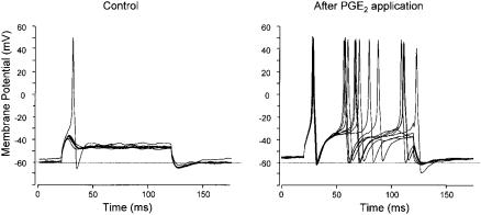
Application of prostaglandin E2 (PGE2) to dorsal root ganglion (DRG) neurons isolated from either adult or neonatal rats leads to an increase in the number of action potentials evoked by depolarizing stimuli (England et al 1996, Fig. 1). This e¡ect can be ascribed, in a large part, to an e¡ect on the TTX-R Na+ channels (England et al 1996, Gold et al 1996) that are a feature of these sensory
146 |
BEVAN & STOREY |
FIG. 1. Responses of DRG neuron to current injection in presence of 500 nM TTX. (Left) 200 pA current pulses evoke no action potentials until 1 M PGE2 added when a single action potential is evoked. (Right) 30 s later same current pulses evoke multiple action potentials. Data from England et al (1996).
neurons, as the facilitatory e¡ects of PGE2 are seen when the TTX-S currents are blocked by high concentrations of TTX. Two types of TTX-R channels have been identi¢ed in DRG neurons (Nav1.8 and Nav1.9); however, it is probable that the reported e¡ects of hyperalgesic agents on TTX-R currents will have re£ected actions on Nav1.8 under the experimental conditions used.
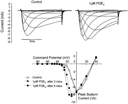
In voltage clamp experiments, application of PGE2 to DRG neurons increased the amplitude of TTX-R currents evoked by depolarizing steps to test potentials of 710 mV. This increase in amplitude occurred over several minutes and was associated with increases in the rates of activation and decay of the currents. Figure 2 shows that the basis of these changes was a *5 mV shift in the voltage dependence of activation to more hyperpolarized potentials (V50 for activation shifted from 74.5 mV to 710.1 mV). A similar hyperpolarizing shift in the steady state inactivation relationship (V50 732.3 to 739.3 mV) was noted after PGE2 treatment (England et al 1996). Although the peak current seen in the current^voltage relationship was increased by about 13% after PGE2 application, this was primarily due to the change in driving force with the hyperpolarizing shift in activation and the reported overall conductance was variable with both increases and reductions reported (England et al 1996, Gold et al 1996).
The PGE2-activated prostanoid receptors on DRG neurons (EP1, EP4, and some splice variants of EP3 receptors . EP3B and EP3C ) can be coupled to the stimulation of adenylate cyclase to raise cAMP levels. Experimental elevation of cAMP with the adenylate cyclase stimulator, forskolin, or application of membrane permeant cAMP analogues, dibutyryl cAMP (dbcAMP) and 8- bromo-cAMP, mimicked the e¡ect of PGE2 treatment by shifting the activation
MODULATION OF SODIUM CHANNELS |
147 |
FIG. 2. (Top) Families of TTX-R currents evoked under control conditions (left) and after exposure to PGE2. (Bottom) Peak current^voltage relationships under control conditions and 3 and 5 minutes after PGE2 addition. Data from England et al (1996).
and steady-state inactivation curves in the hyperpolarizing direction (England et al 1996, Gold et al 1996, Fig. 3). In contrast no such e¡ects were noted with the forskolin analogue, dideoxyforkolin, which does not activate adenylate cyclase. In fact dideoxyforskolin decreased the peak conductance without evoking any shift in the current^voltage relationship for activation (England et al 1996). Prior exposure to dbcAMP also abrogated the e¡ects of subsequent PGE2 applications on TTX-R currents, which argues for a cAMP-mediated pathway for the PGE2 e¡ect. This cAMP pathway appears to involve PKA activation as the e¡ects of PGE2 were inhibited by intracellular application of peptide inhibitors of PKA (England et al 1996, Gold et al 1996).
PKC activity regulates TTX-R currents
PKC regulation of TTX-R currents in DRG neurons was ¢rst shown by application of staurosporine or intracellular administration of the peptide

148 |
BEVAN & STOREY |
FIG. 3. (Left) 1 mM dibutyryl cAMP evokes an increase in TTX-R current evoked by depolarizations to a test potential of 715 mV. (Inset) Currents evoked at times 1,2,3 indicated on graph. (Right) Dideoxyforskolin (10 M) evokes a small reduction in TTX-R current amplitude while a subsequent challenge with forskolin (10 M) increases current amplitude. Data from England et al (1996).
inhibitor PKC19^36 (Gold et al 1998). These treatments decreased peak current without any signi¢cant e¡ect on the voltage dependence of activation.
Conversely, the PKC activators phorbol-12-myristate,13-acetate (PMA) and phorbol 12,13-dibutyrate (PDBu) dose-dependently increased the amplitudes of TTX-R currents in DRG neurons by a mechanism that could be inhibited by the peptide inhibitor PKC19^36. However, although the inhibitory e¡ects of the two PKC stimulators were blocked by this speci¢c inhibitor, there were some di¡erences between the e¡ects of these two agents. The e¡ects of PMA were not associated with any shift in the time course or voltage dependence of activation, whereas PDBu induced changes in the rates of current activation and inactivation without any shift in the voltage dependence of activation. This reason for the di¡erent actions of the two PKC activators is unclear but it has been interpreted to suggest activation of di¡erent PKC isoforms.
PKA and PGE2 modulation of TTX-R currents requires PKC activity
The presence of an interaction between PKC and PKA in modulating TTX-R
currents was demonstrated by the ¢nding that the PKC inhibitors, PKC19^36 and staurosporine, signi¢cantly attenuated the ability of forskolin to modulate TTX-R
currents. In contrast, the PKA inhibitors WIPTIDE and Rp-cAMPs failed to a¡ect the PDBu-mediated changes in increases in peak TTX-R current conductance, although the PDBu e¡ects on kinetics were reduced (Gold et al 1998).
PKC inhibitors were also able to attenuate the e¡ects of PGE2 on TTX-R currents in DRG neurons and prior exposure to PKC activators (PMA and PDBu) occluded the PGE2 e¡ect on TTX-R currents. These data suggest that
MODULATION OF SODIUM CHANNELS |
149 |
PKC activity is necessary to allow PKA-mediated modulation of TTX-R currents by PGE2. This scenario is reminiscent of the permissive role of PKC mediated phosphorylation on PKA induced changes in brain neuron TTX-S currents except that the phopshorylation potentiates TTX-R currents but decreases TTX- S currents.
Phosphorylation of cloned Nav1.8 channels
The likely sites for PKA modulation of this TTX-R Na+ channel have been investigated by site-directed mutagenesis (Fitzgerald et al 1999). The Nav1.8 subunit, like other types of Na+ channel a subunits noted above, has ¢ve consensus PKA phosphorylation sites on the I^II intracellular linker loop. In vitro PKA-induced phosphorylation and tryptic peptide mapping of the channel con¢rmed that these ¢ve serine residues were the major PKA substrates in this region of the channel. Exposure of heterologously expressed wild-type Nav1.8 channels to either forskolin or 8-bromo-cAMP resulted in a hyperpolarizing shift in the current^voltage relationship for activation and an increase in current amplitude evoked by test depolarizations to +5 to +20 mV. In contrast, mutant Nav1.8 channels with the ¢ve serine residues replaced with alanine, were not signi¢cantly a¡ected by either forskolin or 8-bromo-cAMP. These results illustrate the similarity of the response of native and cloned TTX-R currents to agents that raise cAMP levels and indicate that the e¡ect is due to a PKA-induced phosphorylation of one or more of the serine residues on the I^II loop.
TTX-R modulation by other hyperalgesic agents
Two other mediators that are produced in in£ammatory conditions, adenosine and serotonin, can also modulate TTX-R currents in DRG neurons (Gold et al 1996). In both cases, the mediators increased the size of the TTX-R current and shifted the current^voltage relationship for activation in the hyperpolarizing direction. Pharmacological characterization indicated that the serotonin e¡ect was mediated by 5-HT4 receptors (Cardenas et al 1997), which are typically linked to activation of adenylate cyclase.
E¡ects of Herpes simplex virus (HSV) on DRG neuron ion channels
HSV induces a selective loss of sodium currents in DRG neurons
Herpes simplex virus type 1 (HSV-1) is a common neurotropic virus that, in vivo, forms a latent infection in primary a¡erent neurons. HSV infection is associated with abnormal sensations around the site of initial infection including tingling, parasthesia, loss of touch and pain sensations (Andoh et al 1995) which have been
150 |
BEVAN & STOREY |
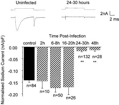
FIG. 4. (Top) Na+ currents evoked by voltage steps from 780 mV to 710 mV in control (left) and HSV-infected DRG neurons (right). (Bottom) Normalized (nA/pF) Na+ current amplitudes with above voltage step in control (¢lled bar) and at various times after HSV (5 plaque forming units/cell) infection show dramatic loss at 24 h.
attributed to alterations in neuronal excitability. HSV infection of DRG neurons in vitro abolishes the excitability of most neurons, and the neurons that remain excitable show action potentials that are smaller in amplitude and longer in duration than normal (Fukuda & Kurata 1981, Mayer et al 1986). Studies of the e¡ects of HSV-1 infection on voltage gated Na+ currents in vitro (Storey et al 1996) have shown that 24 h after HSV-1 infection a voltage step from 780 mV to 10 mV evoked a current in only 28% of neurons and that the amplitude of the current in these neurons was smaller than in uninfected neurons (HSV: 70.017 0.004 nA/ pF; control: 70.14 0.009 nA/pF; n = 84). Although no change in the Na+ current amplitude was noted over the ¢rst 2^20 h after infection, the Na+ current amplitude reduced rapidly and the reduction was maximal after 24 h of infection (Fig. 4). This loss of current was maintained until at least 48 h post-infection. No voltage-gated Na+ currents were unmasked with voltage steps to a wide range of potentials from even more negative holding potentials indicating that the absence of currents was not due to any change in the voltage sensitivity of activation or inactivation. The
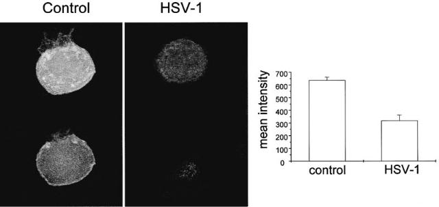
FIG. 5. (Left) Pan Na+ channel antibody staining of control and HSV-infected (24 h) DRG neurons (top, whole cell staining; bottom, cross section image). (Right) Quanti¢ed immuno£uorescence of Na+ channel antibody staining.
152 |
BEVAN & STOREY |
HSV-1 induced loss of Na+ currents was similar for both TTX-S and TTX-R currents. The e¡ects of HSV-1 infection on voltage-gated ion channel function appeared to be restricted to Na+ channels as Ca2+ currents and outwardly rectifying K+ currents were not signi¢cantly changed after HSV-1 infection in vitro.
HSV-induced loss of Na+ currents involves membrane internalization
Exposure of DRG neurons to inhibitors of membrane protein internalization (either ba¢lomycin A or chloroquine) prevented the loss of Na+ conductance normally observed after HSV-1 infection (Storey et al 1998). Furthermore, confocal microscopy of DRG neurons stained with a pan Na+ channel antibody showed that HSV-1 infection resulted in a marked loss of plasma membrane staining and an overall reduction in immuno£uorescence throughout the neurons (Fig. 5). These ¢ndings are consistent with the hypothesis that HSV-1 induces Na+ channel internalization. A possible mechanism of channel loss is suggested by experiments on veratridine-induced loss of surface Na+ channels in neonatal (but not adult) rats CNS neurons where a rapid internalization of Na+ channels (T1/2 *15 min) occurs after a rise in intracellular Na+ concentration (Dargent & Couraud 1990, Dargent et al 1994). HSV-1 induced loss of Na+ currents in adult rat DRG neurons showed a similar dependence on extracellular Na+ and was inhibited when extracellular Na+ was substituted with choline, which raises the possibility that a rise in internal Na+ can mediate the HSV-1-induced internalization.
References
Andoh T, Shiraki K, Kurokawa M, Kuraishi Y 1995 Paresthesia induced by cutaneous infection with herpes simplex virus in rats. Neurosci Lett 190:101^104
Cantrell AR, Smith RD, Goldin AL, Scheuer T, Catterall WA 1997 Dopaminergic modulation of sodium current in hippocampal neurons via cAMP-dependent phosphorylation of speci¢c sites in the sodium channel a subunit. J Neurosci 17:7330^7338
Cantrell AR, Yu F, Sharp EM et al 2000 Molecular mechanisms underlying convergent regulation of brain Na+ channels by protein kinase A and protein kinase C. Soc Neurosc Abstr 26:1908
Cardenas CG, Del Mar LP, Cooper BY, Scroggs RS 1997 5HT4 receptors couple positively to tetrodotoxin-insensitive sodium channels in a subpopulation of capsaicin-sensitive rat sensory neurons. J Neurosci 17:7181^7189
Chen T-C, Law B, Kondratyuk T, Rossie S 1995 Identi¢cation of soluble protein phosphatases that dephosphorylate voltage-sensitive sodium channels in rat brain. J Biol Chem 270:7750^ 7756
Costa MR, Catterall WA 1984a Cyclic AMP-dependent phosphorylation of the alpha subunit of the sodium channel in synaptic nerve ending particles. J Biol Chem 259:8210^8218
Costa MR, Catterall WA 1984b Phosphorylation of the alpha subunit of the sodium channel by protein kinase C. Cell Mol Neurobiol 4:291^297