

Sodium Channels and Neuronal Hyperexcitability
.pdfMOLECULAR BASIS OF Na+ CHANNEL FUNCTION |
33 |
segment, because when you rotate from one side to another there are some residues that are on one side of the electric ¢eld, say intracellular, and then you are rotating them to an extracellular vestibule on the other side. This means that the residues on the back side are moving in the opposite direction. For example, our residues R2 and R3 are on the intracellular side at hyperpolarized voltages. The two neutral residues in between would have to be on the extracellular side. Then they should have to move in opposite directions. It turns out that those two residues are on the same side. This rules out pure rotation, at least for that S4 segment.
Strichartz: You speak about the electric ¢eld like it is an invariant, but it is the gradient of the voltage. You are getting a dielectric relaxation across the gating region when the thing turns, so that the distribution of the driving force is changing during the very process of gating.
Horn: There isn’t a pure hydrophobic slab with charges moving across it. There are some data from Fred Sigworth’s lab showing that these vestibules are not connected to the intracellular surface in terms of electric ¢eld (Islas & Sigworth 2001). The electric ¢eld partly moves through those crevices the way a charged blocker will move through the electric ¢eld to get to a binding site. They are narrow enough that you don’t start out hydrophilic, then hit the electric ¢eld, and then arrive on the other side: it is a gradual movement. You are right: during movement, things can change.
Strichartz: They must change.
References
Featherstone DE, Fujimoto E, Ruben PC 1998 A defect in skeletal muscle sodium channel deactivation exacerbates hyperexcitability in human paramyotonia congenita. J Physiol 506:627^638
Islas LD, Sigworth FJ 2001 Electrostatics and the gating pore of Shaker potassium channels. J Gen Physiol 117:69^89
Isom LL 2002 b subunits: players in neuronal hyperexcitability? In: Sodium channels and neuronal hyperexcitability. Wiley, Chichester (Novartis Found Symp 241) p 124^143
Sodium Channels and Neuronal Hyperexcitability.
Novartis 241
Copyright & 2002 JohnWiley & Sons Ltd
Print ISBN 0-471-48530-6 Online ISBN 0-470-84668-2
Diverse functions and dynamic expression of neuronal sodium channels
Stephen G. Waxman, Theodore R. Cummins, Joel A. Black and Sulayman Dib-Hajj
Department of Neurology and PVA/EPVA Neuroscience Research Center, Yale School of Medicine, New Haven, CT 06510, and Rehabilitation Research Center, VA Connecticut, West Haven CT 06516, USA
Abstract. Nearly a dozen genes encode di¡erent Na+ channels, sharing a common overall motif but with subtly di¡erent amino acid sequences. Physiological signatures have now been established for some Na+ channels and it is clear that, from a functional point of view, Na+ channels are not all the same: di¡erent channels can have di¡erent physiological characteristics, and they can play di¡erent roles in the physiology of excitable cells. Moreover, the expression of Na+ channels within neurons is not a static process. Plasticity of Na+ channel gene expression occurs in the normal nervous system, where it accompanies transitions between di¡erent physiological states (e.g. lowfrequency versus high-frequency ¢ring states) in some types of neurons. Maladaptive changes in Na+ channel gene expression also occur in some pathological neurons. For example, transection of the peripheral axons of spinal sensory neurons triggers downregulation of some Na+ channel genes and up-regulation of others, resulting in changes in Na+ current expression that produce hyperexcitability, thereby contributing to chronic pain. There is also recent evidence for the expression of normally silent Na+ channel genes in Purkinje cells in experimental models of demyelinating diseases and in a human disease, multiple sclerosis; this dysregulation of Na+ channel expression may interfere with neuronal function in these disorders. The diversity and dynamic nature of Na+ channel expression introduce a high degree of complexity into the nervous system and present challenges for neuroscientists. In addition, they may present therapeutic opportunities as selective modulators for various Na+ channel subtypes become available.
2002 Sodium channels and neuronal hyperexcitability. Wiley, Chichester (Novartis Foundation Symposium 241) p 34^60
Elucidation of the function, and underlying molecular structure, of Na+ channels is one of the triumphs of modern neurobiology. The pivotal studies of Hodgkin & Huxley (1952) taught us that, in most neurons, voltage-gated Na+ channels produce the regenerative depolarization that underlies the action potential. Although these pioneering neuroscientists were not able to visualize Na+ channels or directly discern their molecular structure, they were able to infer
34
DIVERSITY AND DYNAMIC EXPRESSION OF Na+ CHANNELS |
35 |
some aspects of the structure of these channels, and to predict, for example, that they possess ‘gates’ which opened as the channels were stimulated, allowing Na+ ions to pass through. Decades later, the molecular revolution permitted the cloning of the ¢rst Na+ channel by Numa and his colleagues (Noda et al 1984).
There were hints, from prior electrophysiological and pharmacological experiments, that there might be more than one type of voltage-gated neuronal Na+ channel. However, the idea that there are multiple neuronal Na+ channels had its formal birth when Numa and his colleagues demonstrated the expression, within the mammalian brain, of three Na+ channel genes, each encoding a di¡erent but related molecule (Noda et al 1986). Following this, other genes encoding additional Na+ channels were discovered. We now know that there are, in fact, nearly a dozen genes, each encoding a Na+ channel with a subtly di¡erent amino acid sequence. Physiological signatures have now been established for some of these Na+ channels, and it is clear that, from a functional point of view, Na+ channels are not all the same: di¡erent channels can have di¡erent physiological characteristics, and they can play di¡erent roles in the physiology of excitable cells.
It has also become clear that the expression of Na+ channels within neurons is not a static process. Although neuronal plasticity has been most extensively studied in terms of synaptic potentiation and depression, sprouting and pruning of neurites, and the recruitment of pre-existing or new neurons into functional circuits, there is now abundant evidence for plasticity of the neuronal electrogenic apparatus: the expression of genes encoding voltage-gated Na+ channels is dynamic.
This chapter will ¢rst review recent progress on the diverse physiological properties and functions of di¡erent Na+ channels. It will then discuss dynamic aspects of Na+ channel expression, both in normal neurons and in diseased neurons.
More than fast threshold devices: Na+ channels can function as ampli¢ers of slow subthreshold depolarizations
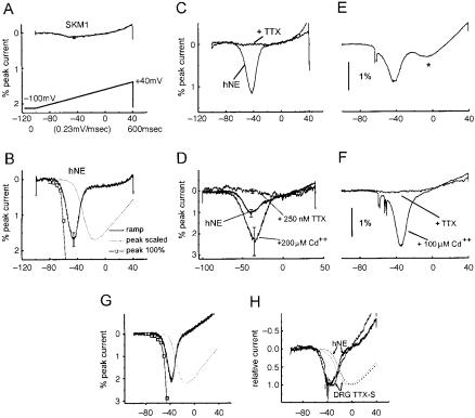
Although the concept of the ‘fast’ transiently-active Na+ channel as a mediator of the depolarizing phase of the action potential is now regarded as classical, it has also become apparent that some Na+ channels can function within a voltage domain that is subthreshold for action potential generation so that they act as boosters of subthreshold depolarizing inputs. An example is provided by the PN1 Na+ channel, which is selectively and prominently expressed within dorsal root ganglion (DRG) neurons (Toledo-Aral et al 1997). To establish the physiological role of PN1, Cummins et al (1998) used patch-clamp techniques to study its human ontology PN1/hNE expressed in a heterologous expression system (HEK293 cells), in a ‘bottom-up’ analysis. The physiological signature of PN1/hNE channels is displayed in Fig. 1 B^H. For comparison, Fig. 1A shows patch-clamp
36 |
WAXMAN ET AL |
FIG. 1. ‘Bottom-up’ and ‘top-down’ analyses reveal similar properties of the PN1/hNE Na+ channel in HEK293 cells and in DRG neurons. (A) SkM1 Na+ channels, transfected into HEK293 cells, do not activate in response to slow ramp-like (0.23 mV/ms) depolarizations.
(B) In contrast, these slow ramp stimuli activate PN1/hNE channels transfected into HEK293 cells, generating distinct inward currents which are evoked close to resting potential. (C) PN1/ hNE currents are blocked by TTX. (D) PN1/hNE currents are enhanced by Cd2+. (E) In a ‘topdown’ analysis, identical ramp stimuli evoke a similar inward current in DRG neurons. (F) Ramp currents in DRG neurons display a similar pharmacologic pro¢le, being blocked by TTX and enhanced by Cd2+. (G) The threshold for activation of ramp currents within intact DRG neurons is similar to that for isolated PN1/hNE currents. (H) PN1/hNE currents in HEK293 cells, and ramp currents in intact DRG neurons, plotted together to facilitate comparison of their voltagedependences. Note that they overlie each other. Modi¢ed from Cummins et al (1998).
recordings from SkM1 (muscle) Na+ channels, transfected into HEK293 cells. Like most traditional Na+ channels, the SkM1 channels require sudden, relatively large depolarizations in order to be activated. In response to slow depolarizations close to resting potential, these channels do not open; when stimulated, for example, by slow ramp-like stimuli (0.23 mV/ms), the SkM1 channels do not generate a response (Fig. 1A). PN1/hNE channels display slow
DIVERSITY AND DYNAMIC EXPRESSION OF Na+ CHANNELS |
37 |
closed-state inactivation and recovery from inactivation. As a result of the slow recovery from inactivation by PN1/hNE channels, cells expressing these channels are not capable of ¢ring at the high frequencies that can be reached by cells expressing SKM1 channels. The presence of PN1/hNE channels does, however, confer a functional advantage. Closed-state inactivation develops slowly in PN1/hNE channels, up to ¢vefold slower than in SkM1 channels (Cummins et al 1998). Thus, in contrast to SkM1, PN1/hNE channels activate and generate a Na+ current in response to slow ramp-like stimuli, even when small depolarizations close to resting potential are used as stimuli (Fig. 1B). The activity of the PN1/hNE channels also exhibits a unique physiological pro¢le, being blocked by tetrodotoxin (Fig. 1C) and enhanced by cadmium (Fig. 1D).
Having established the functional signature for PN1/hNE channels with this bottom-up approach, Cummins et al (1998) next used this information to design a top-down approach, to determine whether these channels play a similar role in their native environment within DRG neurons. Because ramp-like depolarizations constitute an e¡ective stimulus for the PN1/hNE Na+ channel in HEK293 cells, depolarizing ramps were used in the stimulus protocol for intact DRG neurons, and were found to evoke depolarizing responses within these cells (Fig. 1E), similar to those seen in the HEK expression system (Fig. 1B). The Na+ current elicited by these ramp stimuli within intact DRG neurons displayed responses to TTX and cadmium (Fig. 1F) that are identical to those of PN1/hNE channels studied in the HEK293 expression system. As seen in Fig. 1H, moreover, the voltage-dependence of the TTX-sensitive ramp current in DRG neurons is nearly identical to that of the PN1/hNE current in HEK cells. The bottom-up and top-down analyses thus converge and demonstrate that, in intact DRG neurons, the PN1/hNE channel responds to small, slow depolarizations close to resting potential, activating so as to produce inward (depolarizing) currents. These functional properties suggest that PN1/hNE channels respond to small depolarizing inputs, acting as boosters that amplify them. PN1/hNE channels are localized at the distal ends of neurites of spinal sensory neurons in culture (ToledoAral et al 1997); if these channels were similarly deployed in vivo, their localization would place them close to the sites of generator potential activity in nerve terminals.
Na+ channels can contribute to resting potential and modulate neuronal excitability
Dib-Hajj et al (1998a) cloned and sequenced another channel, NaN (also termed SNS 2 by Tate et al 1998), which is preferentially expressed in small DRG neurons. The presence of a serine at position 355 in NaN suggests that it is TTX-resistant (Dib-Hajj et al 1998a). The gene for NaN is localized, together with genes
38 |
WAXMAN ET AL |
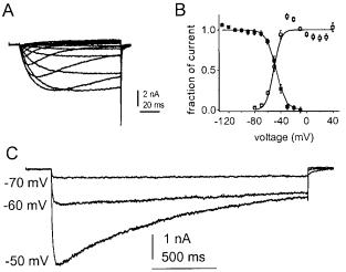
FIG. 2. Persistent TTX-resistant Na+ currents are produced by NaN channels in small DRG neurons. (A) Representative TTXresistant Na+ currents recorded from a DRG neuron from a SNS-null mouse with 100 ms test pulses. (B) Activation (un¢lled squares) and steady-state inactivation (¢lled squares) curves for the NaN current show signi¢cant overlap. Steady-state inactivation was measured in SNS-null neurons with 500 ms prepulses. (C) NaN currents from an SNS-null neuron, elicited with 2 s step depolarizations to the voltages indicated. Recordings
were made with 250 nM TTX, 100 mM cadmium (to block Ca2+ currents) and Vhold = 7120 mV in an SNS-null neuron. Modi¢ed from Cummins et al (1999).
encoding two other TTX-resistant Na+ channels, SNS and hSkM2, within a conserved linkage group at 3p21-3p24 within human chromosome 3 (Dib-Hajj et al 1999a). NaN and SNS are the only TTX-resistant Na+ channels that are present within DRG neurons. Thus, SNS-knockout mutant mice (Akopian et al 1999) provide a model system in which NaN can be studied in isolation. Cummins et al (1999) observed a TTX-resistant (Ki = 39 mM) persistent Na+ current (Fig. 2A, C) attributable to NaN in SNS-knockout DRG neurons. A similar TTX-resistant persistent current, which appears to be produced by NaN, can be recorded from wild-type mouse and rat DRG neurons (Cummins et al 1999) and human DRG neurons (Dib-Hajj et al 1999b). The persistent current is unique in exhibiting a hyperpolarized dependence of activation (threshold 770 mV; midpoint of activation = 741 mV in mouse DRG neurons) and steady-state inactivation (midpoint = 744 mV), with a substantial overlap between activation and steadystate inactivation curves (Fig. 2B) which extends from 770 mV to 730 mV (Cummins et al 1999). Since the resting potential of small DRG neurons is close to 755 mV (Ca¡rey et al 1992), these results suggest that some NaN channels should be active, generating a ‘window current’, near resting membrane potential.
DIVERSITY AND DYNAMIC EXPRESSION OF Na+ CHANNELS |
39 |
Moreover, the low threshold for activation of NaN channels suggests that they should open in response to small subthreshold depolarizations close to membrane potential, thus contributing to subthreshold electrogenesis and regulating the excitability of DRG neurons. Computer simulations suggest that NaN channels contribute a 10 mV to 15 mV depolarizing in£uence to resting potential, and amplify small depolarizing inputs by more than 50%, in small DRG neurons (Herzog et al 2001).
Na+ channel gene expression changes as neurons pass from one functional state to another
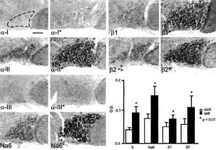
It is of course well known that, as neurons pass from a quiescent state to a highfrequency ¢ring state, they use their (pre-existing) Na+ channels di¡erently, i.e. they activate these channels repetitively. But do neurons produce a di¡erent set of channels when they make these state transitions? To determine whether Na+ channel gene expression changes as neurons pass from a lowto a high-frequency ¢ring state, Tanaka et al (1999) used magnocellular neurosecretory cells within the supraoptic nucleus as a model. These specialized neurons send their axons to the neurohypophysis where they release vasopressin after becoming active in response to increases in plasma osmolality. In their basal state these cells are relatively quiescent, ¢ring irregularly at low frequencies (53 impulses/s) but, in response to changes in the osmotic milieu, these cells respond by generating highfrequency bursts of action potentials which trigger the release of vasopressin (Li & Hatton 1996). The hypothesis, that the transition from the quiescent to the bursting state is accompanied by a change in Na+ channel gene expression, was tested by studying magnocellular neurons under normal conditions and following salt-loading, which triggers a transition to a bursting state. In situ hybridization was used to measure the transcription of mRNA for the various Na+ channels. Notably, these studies revealed that salt loading is accompanied by a distinct up-regulation of mRNAs for two Na+ channels, a-II and Na6, in these neurons (Fig. 3).
Because the expression of ion channels within the cell membrane is controlled at both the transcriptional and translational levels, increased mRNA levels are not necessarily accompanied by increased synthesis of channel protein. As a next step, Tanaka et al (1999) thus studied control and salt-loaded magnocellular neurons using immunocytochemical and immunoblotting methods with an antibody directed against a conserved region of Na+ channels. An increase in the level of Na+ channel protein within the magnocellular neurons of salt-loaded rats, showed that the changes in Na+ channel gene transcription were paralleled by increases in Na+ channel protein.
40 |
WAXMAN ET AL |
FIG. 3. a-II and Na6 Na+ channel mRNAs are up-regulated, together with Na+ channel b1 and b2 mRNA, in supraoptic magnocellular neurons following salt loading. The micrographs, from control (left column) and salt-loaded (right column) rats, were digitally enhanced to show in situ hybridization with subtype-speci¢c riboprobes for Na+ channel subunits a-I, a-II, a-III and Na6. a-I and a-III mRNA are not detectable, and low levels of a-II and Na6 mRNA are present in the control supraoptic nucleus (no asterisks). Expression of the a-II and Na6 transcripts is upregulated following salt-loading (asterisks). Optical densities from unenhanced micrographs (graph, lower right) provide a quantitative measure of mRNA levels and show a signi¢cant increase in a-II and Na6 mRNA following salt loading. *P50.01. Bar = 100 mm. Modi¢ed from Tanaka et al (1999).
Having demonstrated changes in Na+ channel mRNA and protein expression as magnocellular neurons made the transition from one functional state to another, the next question was whether these molecular changes were associated with functional changes in these neurons. To answer this question, Tanaka et al (1999) used patch-clamp recording to study these cells. The presence of two di¡erent Na+ channels (a-II and Na6) in the magnocellular neurons suggested that these cells should produce several distinct Na+ currents. It was known, from studies on other neuronal cell types such as Purkinje cells, that the Na6 Na+ channel can produce a slow or persistent Na+ current (Vega-Saenz de Miera et al 1997, Raman et al 1997). The a-II channel, in contrast, has been shown to produce a fast transient current (Noda et al 1986, Auld et al 1988). Consistent with the expression of these two types of Na+ channels, patch-clamp recordings
DIVERSITY AND DYNAMIC EXPRESSION OF Na+ CHANNELS |
41 |
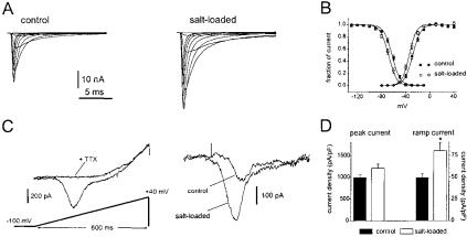
FIG. 4. Two distinct Na+ currents in supraoptic neurons show di¡erential increases following salt loading. (A) Recordings from representative supraoptic neurons acutely isolated from control (left panel) or salt-loaded (right panel) rats, showing the fast transient Na+ current. The currents were elicited by 40 ms test pulses to various potentials from 760 to 30 mV. Cells were held at 7100 mV. (B) Normalized activation (circles) and steady-state inactivation (squares) curves show only small di¡erences between control (¢lled symbols) and salt-loaded (open symbols) neurons. Error bars indicate SEM. (C) Ramp currents are elicited by slow ramp-like depolarizations (extending from 7100 to +40 mV over 600 ms) in supraoptic neurons. The left panel shows that TTX (250 nM) blocks the ramp current in salt-loaded supraoptic neurons, demonstrating that this current is produced by Na+ channels. The right panel shows the TTX-sensitive ramp currents in representative control and salt-loaded supraoptic neurons. Note the larger amplitude in salt-loaded neurons. (D) Fast transient and ramp current densities (estimated by dividing the maximum currents by the cell capacitance) are both increased following salt-loading; however, the increase is proportionately greater for the ramp currents. Error bars indicate SEM, *indicates P50.005. From Tanaka et al (1999).
demonstrated two distinct Na+ currents in control magnocellular neurons: ¢rst, a fast transient Na+ current which would be expected to contribute to the rapid depolarizing upstroke of the action potential (Fig. 4A); and second, a persistent ‘threshold’ Na+ current which activates closer to resting potential (Fig. 4C). The patch-clamp experiments showed that there was an increase of 20% in the density of the fast transient Na+ current in salt-loaded rats. In contrast, the density of the persistent threshold current was increased by approximately 60% (Fig. 4D). The two Na+ currents were increased to signi¢cantly di¡erent degrees. The disproportional increases in the two currents encoded by these two channels would be expected to lower the threshold for action potential generation.
Magnocellular neurons represent an example of molecular and functional remodelling of the neuronal electrogenic apparatus in the normal nervous
42 |
WAXMAN ET AL |
system. As these cells move from a quiescent to a bursting state, they alter their pattern of Na+ channel gene activation, a change that alters the threshold of these neurons.
Expression of some Na+ channel genes is down-regulated in injured neurons
The striking plasticity in Na+ channel expression that can occur in normal neurons raises the question of whether there are alterations in Na+ channel expression in injured neurons. Dorsal root ganglion (DRG) neurons have been especially well studied in this respect. As illustrated in Fig. 5 (middle and bottom rows), following transection of the peripheral axons of DRG neurons (by ligation of the sciatic nerve), expression of mRNA for the SNS (Dib-Hajj et al 1996) and NaN (DibHajj et al 1998a) Na+ channels is down-regulated. The reductions in SNS and NaN transcript expression are paralleled by reduced levels of SNS and NaN Na+ channel protein (Sleeper et al 2000).
While transection of the peripheral axons of DRG neurons triggers a downregulation of SNS and NaN, transection of the centrally-direct branches of DRG neurons (dorsal rhizotomy) does not alter the expression of these channels (Sleeper et al 2000). These results suggest that down-regulation of SNS and NaN might be due to loss of access to trophic factors produced by peripheral targets. Consistent with this hypothesis, nerve growth factor (NGF) upregulates SNS expression (Black et al 1997), while glial derived neurotrophic factor (GDNF) up-regulates NaN and SNS expression (Fjell et al 1999) in DRG neurons in vitro. Similarly, delivery of NGF (Dib-Hajj et al 1998b) and GDNF (Cummins et al 2000) to the injured nerve stump in vivo can rescue the expression of SNS and NaN, respectively.
In parallel with the down-regulation of SNS and NaN mRNA and protein, the slowly-inactivating and persistent TTX-resistant Na+ currents produced by these channels are reduced in axotomized neurons (Cummins & Waxman 1997, Sleeper et al 2000) (Fig. 6A, B). These physiological changes persist for at least 60 days post-axotomy (Cummins & Waxman 1997). Loss of NaN (persistent TTXresistant) channels would be expected to shift resting potential in a hyperpolarizing direction, reducing resting inactivation of fast Na+ channels, and thereby producing DRG neuron hyperexcitability which can contribute to pain and parasthesia (Cummins & Waxman 1997). Consistent with a contribution of these changes to the pathogenesis of neuropathic pain, Dib-Hajj et al (1999c) observed a similar (although quantitatively smaller) down-regulation in SNS and NaN channels and their currents in DRG neurons in the chronic constriction injury model of neuropathic pain.