

Primer on molecular genetics (Human Genome Project, DOE)(44s)
.pdfPrimer on Molecular Genetics
1

DOE Human Genome Program
Primer on Molecular Genetics
Date Published: June 1992
U.S. Department of Energy
Office of Energy Research
Office of Health and Environmental Research
Washington, DC 20585
The "Primer on Molecular Genetics" is taken from the June 1992 DOE Human Genome 1991-92 Program Report. The primer is intended to be an introduction to basic principles of molecular genetics pertaining to the genome project.
Human Genome Management Information System
Oak Ridge National Laboratory
1060 Commerce Park
Oak Ridge, TN 37830
Voice: 865/576-6669
Fax: 865/574-9888
E-mail: bkq@ornl.gov
2

Contents
Primer on
Molecular
Genetics
Revised and expanded by Denise Casey (HGMIS) from the primer contributed by Charles Cantor and Sylvia Spengler (Lawrence Berkeley Laboratory) and published in the
Human Genome 1989– 90 Program Report.
Introduction ............................................................................................................. |
5 |
DNA............................................................................................................................... |
6 |
Genes............................................................................................................................ |
7 |
Chromosomes ............................................................................................................... |
8 |
Mapping and Sequencing the Human Genome ...................................... |
10 |
Mapping Strategies ..................................................................................................... |
11 |
Genetic Linkage Maps ............................................................................................ |
11 |
Physical Maps......................................................................................................... |
13 |
Low-Resolution Physical Mapping...................................................................... |
14 |
Chromosomal map ......................................................................................... |
14 |
cDNA map ...................................................................................................... |
14 |
High-Resolution Physical Mapping ..................................................................... |
14 |
Macrorestriction maps: Top-down mapping ................................................... |
16 |
Contig maps: Bottom-up mapping.................................................................. |
17 |
Sequencing Technologies ........................................................................................... |
18 |
Current Sequencing Technologies ......................................................................... |
23 |
Sequencing Technologies Under Development ..................................................... |
24 |
Partial Sequencing to Facilitate Mapping, Gene Identification ............................... |
24 |
End Games: Completing Maps and Sequences; Finding Specific Genes .................. |
25 |
Model Organism Research.............................................................................. |
27 |
Informatics: Data Collection and Interpretation ..................................... |
27 |
Collecting and Storing Data ........................................................................................ |
27 |
Interpreting Data ......................................................................................................... |
28 |
Mapping Databases .................................................................................................... |
29 |
Sequence Databases .................................................................................................. |
29 |
Nucleic Acids (DNA and RNA)................................................................................ |
29 |
Proteins .................................................................................................................. |
30 |
Impact of the Human Genome Project ....................................................... |
30 |
Glossary... .............................................................................................................. |
32 |
3

4
Introduction
The complete set of instructions for making an organism is called its genome. It contains the master blueprint for all cellular structures and activities for the lifetime of
the cell or organism. Found in every nucleus of a person’s many trillions of cells, the human genome consists of tightly coiled threads of deoxyribonucleic acid (DNA) and associated protein molecules, organized into structures called chromosomes (Fig. 1).
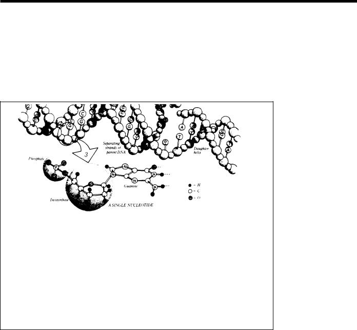
Fig. 1. The Human Genome at Four Levels of Detail. Apart from reproductive cells (gametes) and mature red blood cells, every cell in the human body contains 23 pairs of chromosomes, each a packet of compressed and entwined DNA (1, 2). Each strand of DNA consists of repeating nucleotide units composed of a phosphate group, a sugar (deoxyribose), and a base (guanine, cytosine, thymine, or adenine) (3). Ordinarily, DNA takes the form of a highly regular doublestranded helix, the strands of which are linked by hydrogen bonds between guanine and cytosine and between thymine and adenine. Each such linkage is a base pair (bp); some 3 billion bp constitute the human genome. The specificity of these base-pair linkages underlies the mechanism of DNA replication illustrated here. Each strand of the double helix serves as a template for the synthesis of a new strand; the nucleotide sequence (i.e., linear order of bases) of each strand is strictly determined. Each new double helix is a twin, an exact replica, of its parent. (Figure and caption text provided by the LBL Human Genome Center.)
5

Primer on Molecular Genetics
If unwound and tied together, the strands of DNA would stretch more than 5 feet but would be only 50 trillionths of an inch wide. For each organism, the components of these slender threads encode all the information necessary for building and maintaining life, from simple bacteria to remarkably complex human beings. Understanding how DNA performs this function requires some knowledge of its structure and organization.
Fig. 2. DNA Structure.
The four nitrogenous bases of DNA are arranged along the sugarphosphate backbone in a particular order (the DNA sequence), encoding all genetic instructions for an organism. Adenine (A) pairs with thymine (T), while cytosine (C) pairs with guanine (G). The two DNA strands are held together by weak bonds between the bases.
A gene is a segment of a DNA molecule (ranging from fewer than
1 thousand bases to several million), located in a particular position on a specific chromosome, whose base sequence contains the information necessary for protein synthesis.
DNA
In humans, as in other higher organisms, a DNA molecule consists of two strands that wrap around each other to resemble a twisted ladder whose sides, made of sugar and phosphate molecules, are connected by “rungs” of nitrogen-containing chemicals called bases. Each strand is a linear arrangement of repeating similar units called nucleotides, which are each composed of one sugar, one phosphate, and a nitrogenous base (Fig. 2). Four different bases are present in DNA—adenine (A), thymine (T), cytosine (C), and guanine (G). The particular order of the bases arranged along the sugar-phosphate backbone is called the DNA sequence; the sequence specifies the exact genetic instructions required to create a particular organism with its own unique traits.
Phosphate Molecule |
||
Deoxyribose |
||
Sugar Molecule |
||
|
Nitrogenous |
|
|
|
Bases |
|
A |
T |
|
|
|
C |
|
G |
|
|
|
|
G |
C |
|
|
|
T |
|
A |
|
|
|
|
|
Weak Bonds |
|
|
Between |
|
|
Bases |
Sugar-Phosphate |
||
|
Backbone |
|
The two DNA strands are held together by weak bonds between the bases on each strand, forming base pairs (bp). Genome size is usually stated as the total number of base pairs; the human genome contains roughly 3 billion bp (Fig. 3).
Each time a cell divides into two daughter cells, its full genome is duplicated; for humans and other complex organisms, this duplication occurs in the nucleus. During cell division the DNA molecule unwinds and the weak bonds between the base pairs break, allowing the strands to separate. Each strand directs the synthesis of a complementary new strand, with free nucleotides matching up with their complementary bases on each of the separated strands. Strict basepairing rules are adhered to—adenine will pair only with thymine (an A-T pair) and cytosine with guanine (a C-G pair). Each daughter cell receives one old and one new DNA strand (Figs. 1 and 4). The cell’s adherence to these base-pairing rules ensures that the new strand is an exact copy of the old one. This minimizes the incidence of errors (mutations) that may greatly affect the resulting organism or its offspring.
6

Genes
Each DNA molecule contains many genes—the basic physical and functional units of heredity. A gene is a specific sequence of nucleotide bases, whose sequences carry the information required for constructing proteins, which provide the structural components of cells and tissues as well as enzymes for essential biochemical reactions. The human genome is estimated to comprise at least 100,000 genes.
Human genes vary widely in length, often extending over thousands of bases, but only about 10% of the genome is known to include the protein-coding sequences (exons) of genes. Interspersed within many genes are intron sequences, which have no coding function. The balance of the genome is thought to consist of other noncoding regions (such as control sequences and intergenic regions), whose functions are obscure. All living organisms are composed largely of proteins; humans can synthesize at least 100,000 different kinds. Proteins are large, complex molecules made up of long chains of subunits called amino acids. Twenty different kinds of amino acids are usually found in proteins. Within the gene, each specific sequence of three DNA bases (codons) directs the cell’s protein-synthesizing machinery to add specific amino acids. For example, the base sequence ATG codes for the amino acid methionine. Since 3 bases code for
1 amino acid, the protein coded by an average-sized gene (3000 bp) will contain 1000 amino acids. The genetic code is thus a series of codons that specify which amino acids are required to make up specific proteins.
The protein-coding instructions from the genes are transmitted indirectly through messenger ribonucleic acid (mRNA), a transient intermediary molecule similar to a single strand of DNA. For the information within a gene to be expressed, a complementary RNA strand is produced (a process called transcription) from the DNA template in the nucleus. This
Comparative Sequence Sizes |
Bases |
|
Largest known continuous DNA sequence |
350 |
Thousand |
(yeast chromosome 3) |
|
|
Escherichia coli (bacterium) genome |
4.6 |
Million |
Largest yeast chromosome now mapped |
5.8 |
Million |
Entire yeast genome |
15 |
Million |
Smallest human chromosome (Y) |
50 |
Million |
Largest human chromosome (1) |
250 |
Million |
Entire human genome |
3 |
Billion |
|
|
|
Fig. 3. Comparison of Largest Known DNA Sequence with Approximate Chromosome and Genome Sizes of Model Organisms and Humans. A major focus of the Human Genome Project is the development of sequencing schemes that are faster and more economical.
7
Primer on Molecular Genetics
mRNA is moved from the nucleus to the cellular cytoplasm, where it serves as the template for protein synthesis. The cell’s protein-synthesizing machinery then translates the codons into a string of amino acids that will constitute the protein molecule for which it codes (Fig. 5). In the laboratory, the mRNA molecule can be isolated and used as a template to synthesize a complementary DNA (cDNA) strand, which can then be used to locate the corresponding genes on a chromosome map. The utility of this strategy is described in the section on physical mapping.
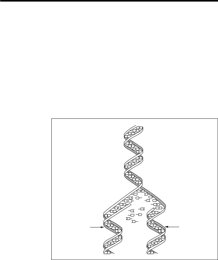
Fig. 4. DNA Replication.
During replication the DNA molecule unwinds, with each single strand becoming a template for synthesis of a new, complementary strand. Each daughter molecule, consisting of one old and one new DNA strand, is an exact copy of the parent molecule. [Source: adapted from Mapping Our Genes—The Genome Projects: How Big, How Fast? U.S. Congress, Office of Technology Assessment, OTA-BA-373 (Washington, D.C.: U.S. Government Printing Office, 1988).]
Chromosomes
The 3 billion bp in the human genome are organized into 24 distinct, physically separate microscopic units called chromosomes. All genes are arranged linearly along the chromosomes. The nucleus of most human cells contains 2 sets of chromosomes, 1 set given by each parent. Each set has 23 single chromosomes—22 autosomes and an X or Y sex chromosome. (A normal female will have a pair of X chromosomes; a male will have an X
ORNL-DWG 91M-17361
DNA Replication
A T
C G
T A
G C
A T
C G
T A |
Parent |
|
|
||
G C |
Strands |
|
A T |
||
|
||
C G |
|
T A
G C
A T
C G
TA G C
|
|
|
A |
T |
|
|
|
|
C |
|
|
G |
|
|
|
T A |
|
A |
|
|
|
|
|
G C |
|
||
|
G |
C |
|
|
|
|
|
A T |
|
|
|
A T |
|
|
C G |
|
|
T |
C G |
|
|
T A |
G |
|
|
|
|
|
|
|
T A |
|
||
|
|
|
A |
|
|
|
|
G C |
C |
|
G C |
|
|
|
|
|
|
|||
Complementary |
A T |
T |
|
|
A T |
|
G |
|
|
|
|||
|
|
|
|
|
|
|
New Strand |
C G |
|
|
|
C G |
Complementary |
T A |
|
|
|
T A |
||
|
G C |
|
|
|
G C |
New Strand |
|
|
|
|
|
|
|
|
A T |
|
|
|
A T |
|
|
C G |
|
|
|
C G |
|
|
T A |
|
|
|
T A |
|
|
G C |
|
|
|
G C |
|
|
A T |
|
|
|
A T |
|
8
and Y pair.) Chromosomes contain roughly equal parts of protein and DNA; chromosomal DNA contains an average of 150 million bases. DNA molecules are among the largest molecules now known.
Chromosomes can be seen under a light microscope and, when stained with certain dyes, reveal a pattern of light and dark bands reflecting regional variations in the amounts of A and T vs G and C. Differences in size and banding pattern allow the 24 chromosomes to be distinguished from each other, an analysis called a karyotype. A few types of major chromosomal abnormalities, including missing or extra copies of a chromosome or gross breaks and rejoinings (translocations), can be detected by microscopic examination; Down’s syndrome, in which an individual's cells contain a third copy of chromosome 21, is diagnosed by karyotype analysis (Fig. 6). Most changes in DNA, however, are too subtle to be detected by this technique and require molecular analysis. These subtle DNA abnormalities (mutations) are responsible for many inherited diseases such as cystic fibrosis and sickle cell anemia or may predispose an individual to cancer, major psychiatric illnesses, and other complex diseases.
ORNL-DWG 91M-17360
NUCLEUS |
Free Amino Acids |
|
Gene |
mRNA |
tRNA Bringing |
|
Amino Acid to |
|||
DNA |
Copying |
Ribosome |
|
DNA in |
|||
|
|
||
|
Nucleus |
|
mRNA
mRNA
CYTOPLASM
Growing
Protein Chain
RIBOSOME incorporating amino acids into the growing protein chain
 Amino
Amino
Acids
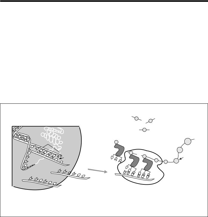
Fig. 5. Gene Expression. When genes are expressed, the genetic information (base sequence) on DNA is first transcribed (copied) to a molecule of messenger RNA in a process similar to DNA replication. The mRNA molecules then leave the cell nucleus and enter the cytoplasm, where triplets of bases (codons) forming the genetic code specify the particular amino acids that make up an individual protein. This process, called translation, is accomplished by ribosomes (cellular components composed of proteins and another class of RNA) that read the genetic code from the mRNA, and transfer RNAs (tRNAs) that transport amino acids to the ribosomes for attachment to the growing protein. (Source: see Fig. 4.)
9
Primer on
Molecular
Genetics
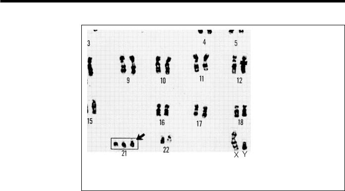
Fig. 6. Karyotype. Microscopic examination of chromosome size and banding patterns allows medical laboratories to identify and arrange each of the 24 different chromosomes (22 pairs of autosomes and one pair of sex chromosomes) into a karyotype, which then serves as a tool in the diagnosis of genetic diseases. The extra copy of chromosome 21 in this karyotype identifies this individual as having Down’s syndrome.
Mapping and Sequencing the Human Genome
A primary goal of the Human Genome Project is to make a series of descriptive dia- grams—maps—of each human chromosome at increasingly finer resolutions. Mapping involves (1) dividing the chromosomes into smaller fragments that can be propagated and char-acterized and (2) ordering (mapping) them to correspond to their respective locations on the chromosomes. After mapping is completed, the next step is to determine the base sequence of each of the ordered DNA fragments. The ultimate goal of genome research is to find all the genes in the DNA sequence and to develop tools for using this information in the study of human biology and medicine. Improving the instrumentation and techniques required for mapping and sequencing—a major focus of the genome project—will increase efficiency and cost-effectiveness. Goals include automating methods and optimizing techniques to extract the maximum useful information from maps and sequences.
A genome map describes the order of genes or other markers and the spacing between them on each chromosome. Human genome maps are constructed on several different scales or levels of resolution. At the coarsest resolution are genetic linkage maps, which depict the relative chromosomal locations of DNA markers (genes and other identifiable DNA sequences) by their patterns of inheritance. Physical maps describe the chemical characteristics of the DNA molecule itself.
10