

Патфиз1 -13 / 25
.pdfCurrent Pharmaceutical Design, 2008, 14, 3549-3564 |
3549 |
Inflammation as Therapeutic Objective in Stroke
Joaquín Jordán1,*, Tomás Segura2, David Brea3, Maria F. Galindo4 and José Castillo3
1Grupo de Neurofarmacología. Departamento de Ciencias Médicas. Facultad de Medicina. Universidad Castilla-La Mancha. Centro Regional de Investigaciones Biomédicas. Spain; 2Servicio de Neurología. Complejo Hospitalario Universitario de Albacete. Albacete. Spain; 3Department of Neurology, Clinical Neuroscience Research Laboratory, Hospital Clínico Universitario de Santiago de Compostela, University of Santiago de Compostela. Spain and 4Unidad de Neuropsicofarmacología Translacional. Complejo Hospitalario Universitario de Albacete. Albacete. Spain
Abstract: Ischemic stroke is the most frequent cause of persistent neurologic disability in modern Western societies. Albeit it is still not clear whether inflammation is merely an epiphenomenon or rather has a disease-promoting function, accumulating evidence implicates inflammation in many forms of acute neurodegenerative disorders including ischemia. The immune cell influx during a neuropathological event is thought to be elicited by glial cells, especially microglia. This article reviews the cellular and molecular pathways involved in stroke-induced inflammatory response in the CNS. We focused on how CNS innate immune cells including microglia and macrophages play integral roles in receiving and propagating inflammatory signals, and how activated microglia secrete a wide range of factors. We present the relevance of the expression of adhesion molecules after ischemia including selectin, immunoglobulin superfamily, integrins, and the role of inflammatory mediators such as cytokines, chemokines and matrix metalloproteinases. Further, we explore the role of transcription factors in inflammation, and the function of immunomodulation and innate and adaptive immunity in brain ischemia, focusing on immunosupression therapies for acute stroke. Although several approaches for anti-inflammatory treatment have proven effective in animal models, clinical trials of immune system modulation therapy after stroke have not yet proved successful. There is still much to be done in order to translate interesting findings into therapies, but undoubtedly studying the cellular and molecular pathways may not only improve our understanding of inflammatory mechanism but also serve as a basis for designing effective therapies.
1. INTRODUCTION
Stroke is the second leading cause of death and the burden of disease in high income countries [1]. Stroke occurs due to a loss of blood supply to part of the brain, initiating the ischemic cascade. As oxygen or glucose becomes depleted in ischemic brain tissue, the production of high energy phosphate compounds such as adenosine triphosphate (ATP) fails leading to failure of energy dependent processes (such as ion pumping) necessary for tissue cell survival. This sets off a series of interrelated events that result in cellular injury and death by necrosis. Dead cells by necrosis produce the release of all cytoplasmatic content into the extracellular space activating the corresponding inflammatory response.
The central nervous system (CNS) has for long been regarded as an immune privileged organ, with the blood–brain barrier (BBB) tightly regulating the influx of immune cells and mediators from the vascular compartment to the brain parenchyma [2]. Inflammation is generally a beneficial response of an organism to infection but, when prolonged or inappropriate, it can be detrimental. Neuronal loss in acute (e.g. stroke and head injury) and chronic [e.g. multiple sclerosis and Alzheimer's disease] CNS diseases has been associated with inflammatory processes systemically and in the
*Address correspondence to this author at the Grupo de Neurofarmacologia, Departamento de Ciencias Médicas, Facultad de Medicina, Universidad Castilla-La Mancha. Centro Regional de Investigaciones Biomédicas, Avda Almansa, 14, 02006-Albacete, Spain; Tel: 34-967599200; Fax: 34-967- 599327; E-mail: Joaquin.jordan@uclm.es
brain. Brain inflammation is characterized by activation of microglia and astrocytes, expression of key inflammatory mediators, but limited invasion of circulating immune cells. Inflammation induces rapid expression of key inflammatory mediators -cytokines, chemokines and prostaglandinswhich in turn up-regulate adhesion molecules, increase permeability of the BBB, facilitating invasion of peripheral immune cells, induce release of potentially toxic molecules and compromise brain cells. Because the BBB is disrupted after stroke, the immune system comes into contact with CNS antigens, in both the brain and periphery [3].
In recent years, major advances in the study of the role of the immune system and inflammation in brain ischemia have been done. There are many evidences that inflammation and immune response play an important role in the outcome of ischemic stroke patients, and they have been associated to larger brain damage. However, on the other hand, these mechanisms might be necessary for the resolution of dead cells and the initiation of repairing mechanisms. In this work we review the inflammatory response to brain ischemia, the role of innate immunity, and the potential role of endogenous anti-inflammation and immune-modulation processes.
2. CELLULAR INFLAMMATORY RESPONSE
The CNS innate immune cells including microglia and macrophages play integral roles in receiving and propagating inflammatory signals. Microglia are a highly responsive population of cells with a well established role in regulating the immune surveillance of the nervous system [4,5]. Found
1381-6128/08 $55.00+.00 © 2008 Bentham Science Publishers Ltd.
3550 Current Pharmaceutical Design, 2008, Vol. 14, No. 33
scattered throughout the CNS, microglia constitute 5–15% of the total brain cell population and form a meshed network able to detect and react to modifications of the local environment [6]. In the mature brain, microglia typically exist in a resting state characterized by ramified morphology, and monitor the brain environment. The CNS endogenous microglia share many properties with macrophages, having developed from a common precursor cell [7-9]. Early in fetal development, monocyte-like cells infiltrate the CNS and develop into parenchymal microglia. In the adult, parenchymal microglia in contrast to perivascular “microglia” are not frequently repopulated by fresh monocytes [10].
Microglia are primarily involved in immune surveillance [6, 11], but when activated have macrophage-like capabilities including phagocytosis, inflammatory cytokine production, and antigen presentation [12]. Normally these neuroinflammatory changes are transient with microglia returning to a resting state as the immune stimulus is resolved. Aging or neurological disease, however, may provide a brain environment where microglia are more “reactive or primed” to a peripheral immune challenge [13]. In response to certain cues such as brain injury or immunological stimuli, however, microglia are readily activated. Activated microglia secrete a wide range of factors, some of which can actively trigger apoptosis in neuronal cell cultures. At the same time, microglia are also reported to increase neuronal survival through the release of trophic and anti-inflammatory factors.
Features common to microglia and systemic macrophages include the expression of innate immune receptors and the ability to phagocyte pathogens, cells or cellular debris [7, 14]. Microglia is activated within minutes of ischemia onset and produces a plethora of inflammatory mediators, which exacerbate tissue damage [15]. Furthermore, microglia responses are segregated not only respect to time, but some extend also spatially. After focal cortical ischemia, there is a second involvement of the ipsilateral thalamus attributable to retrograde degeneration of thalamo-cortical projection fibres [16]. The PET tracer 11C-PK11195 binds to the peripheral benzodiazepine receptor, which is abundant on brain-derived activated microglia, and that has been employed in experimental and clinical studies addressing ischaemia-related inflammation, has shown that microglial activation becomes significant after a few days of stroke and persists for over 30 days, with a peak around 2 weeks for stroke onset [17]. Notably, the spatial distribution of 11C- PK11195 binding evolves to include not only the core but also the rescued penumbra where inflammation may be secondary to selective neuronal damage [18].
Recently it has been shown that patients with acute stroke had significantly better outcome with minocycline treatment compared with placebo [19], by involving minocycline antiinflammatory properties, especially its ability to suppress activation of microglia, are likely to contribute to cytoprotection in the CNS [20].
3. EXPRESSION OF ADHESION MOLECULES AFTER ISCHEMIA
Inflammation after stroke involves leukocytes infiltration in brain parenchyma, specially neutrophils, that contribute to cerebral damage after ischemia [21] through reperfusion or
Jordán et al.
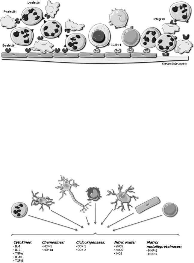
secondary injury mechanisms. Infiltration through the endothelium involves rolling, adhesion and transendothelial migration of leukocytes. Therefore, adhesion molecules in leukocytes and endothelial cells are key molecules that contribute to cerebral damage. Adhesion molecules that participate in this process are classified in three types; selectins, the superfamily of immunoglobulin and integrins.
3.1. Selectins
L-selectin, E-selectin and P-selectin are glycoproteins involved in the initial interaction between leucocytes and endothelial cells in the periphery of the infarct. They interact with P-selectin glycoprotein ligand (PSGL1) and other glycosylated ligand [22], to induce a transitory and reversible action that leads to other secondary cellular interactions mediated by a different group of adhesins. As a consequence of the activation of adhesins, there is a recruitment of leucocytes, taking place their aggregation and adhesion to the vascular wall in a later stage. Such changes are responsible for the obstruction of the microvascularization and for the phenomenon of ‘no-reflux’. The conversion of the endothelium into a prothrombotic state, the production of free radicals and the rise in vascular permeability are other factors responsible for cellular damage mediated by inflammation.
At the individual level, P-selectin is found in platelets [23, 24] and endothelial cells, being its counter receptor, which contains the oligosaccharide sialyl Lewis X, present in leukocytes [25]. The existence of stores of P-selectin in the cytoplasm of endothelial cell [26] allows its mobilization to the cell surface within minutes from the activation of endothelial cells by thrombin, complement, and histamine [27].
Knowledge about the role of P-selectin has been provided from both experimental models and clinical studies. From clinical studies it has been stated that surface expression of P-selectin on platelets is related to clinical worsening after acute ischemic stroke, and a positive correlation between P-selectin expression and NIHSS scores has been observed [28]. Studies on experimental models of cerebral ischemia have revealed that the lack of P-selectin or its blockage with monoclonal antibodies [29] or inhibitors [30, 31] was associated with improved neurological outcome and reduced cerebral infarct volumes.
The role of E-selectin and L-selectin in cerebral ischemia is not so clear. E-selectin can be found in endothelial cells and leukocytes, playing an important task in the development of inflammation in vivo [32]. E-selectin is also upregulated in microvessels of animal models of ischemia, 2 hours after reperfusion [32,33]. L-selectin is found in endothelial cells and leukocytes mediating on leukocyte rolling, but its role in brain ischemia is less clear.
3.2. Anti-Selectin Treatment Possibilities
Studies in P-selectin knockout mice, and alternatively functional blockade of P-selectin with a monoclonal antibody in wild-type mice, have shown decreased infarct volumes after transient permanent middle cerebral artery occlusion (MCAO) respect to controls. Furthermore, administration of sCRsLex, an inhibitor of selectin-mediated plateletleukocyte interactions, reduces infarct volumes in experimental models. However results varied from focal to global
Inflammation as a Therapy for Stroke
experimental models of ischemia, since P-selectin blockade or deficiency was associated to better outcome and smaller infarcts in focal ischemia, while animals treated with antibodies against P-selectin showed reduced survival periods in models of gobal ischemia [34].
Knowledge about E-selectin and L-selectin is not such clear, therefore treatments against these selectins are less studied. Although it has been shown that E-selectin can induce immune tolerance and reduce injury upon intranasal administration to animals [35]. By the other hand, L-selectin treatments with L-selectin antibody has been shown ineffectiveness in animal models of stroke [36].
3.3. Immunoglobulin Superfamily
The immunoglobulin superfamily includes 5 members: intercellular adhesion molecule-1 (ICAM-1), intercellular adhesion molecule-2 (ICAM-2), vascular adhesion molecule- 1 (VCAM-1), platelet–endothelial cell adhesion molecule-1 (PECAM-1), and the mucosal vascular addressing cell adhesion molecule 1 (MAdCAM-1). All of them are expressed on activated endothelial cells, mediating in the adhesion of leukocytes to endothelia, and creating stronger attachments than selectins [37].
ICAM1 is constitutively expressed, although its expression in cerebral microvascular endothelial cells is increased by IL-1 , TNF- , lipopolysaccharide (LPS) [38], as observed in in vitro models of ischemia-like insults [39]. There are some other evidences that indicate its role in cerebral ischemia. Thus, increased expression of ICAM-1 has been found in areas of cerebral ischemia in rats submitted to MCAO [40]. Knock-out mice for ICAM-1 gene presented less cerebral damage than wild-type mice subjected to cerebral ischemia [23]. The implication of ICAM-1 in cerebral ischemia has also been demonstrated in clinical studies, and a rise in serum concentrations of soluble adhesion molecules has been found after cerebral ischemia. Soluble ICAM-1 concentrations are elevated [41] and peak in patients within 24 hours of acute ischemic stroke, being these levels correlated with the infiltration of polimorphonuclear leukocytes [42]. Furthermore, ICAM-1 expression in brain microvessels is significantly increased in the cerebral infarcts of patients who die from ischemic stroke [43].
Localized ICAM-1 expression has been found in histological studies of normal human carotid bifurcation [44], a region of high-risk for the development of atherosclerotic plaque, while endothelial ICAM-1 expression is increased in symptomatic versus asymptomatic carotid plaque [45]. Patients with carotid atherosclerosis who may be described as “prestroke stage” have raised concentrations of soluble (s)ICAM-1 [46]. This is in concordance with elevated soluble ICAM-1 levels that were found in patients with stroke risk factors [41].
Little is known about other intercellular adhesion molecules such as ICAM-2, VCAM-1, or PECAM-1. ICAM-2 may serve to stimulate the ligand binding of ICAM-1 to CD11b/CD18 [29]. Significantly, ICAM-2 is expressed not only by endothelial cells but also by resting and activated platelets [29]. In contrast to the other 2 integrin ligands, ICAM-1 and ICAM-3 (CD50), which are not found on plate-
Current Pharmaceutical Design, 2008, Vol. 14, No. 33 3551
lets, ICAM-2 may be involved in the mediation of leukocyteplatelet interactions in the microvessels during inflammation and thrombosis. The role of VCAM-1 in stroke is controversial. While some authors have described an increase in VCAM-1 mRNA after cerebral ischemia [29], others have failed to observe significant changes [47].
PECAM-1 is expressed at constant levels and plays additional roles in attaching endothelial cells among them, and in negotiating with leukocytes [37]. These data support the role of cell adhesion molecules in the tight adhesion of leukocytes after cerebral ischemia, including the unique role of ICAM-2 in leukocyte-platelet interactions.
3.4. Immunoglobulin Superfamily Treatment Possibilities
A few studies have evaluated the possible treatment for ischemic stroke blocking receptors of the immunoglobulin superfamily.
Anti-ICAM1 antibody treatment has been shown to diminish cerebral damage [32], neutrophil accumulation, apoptosis and neurological deficits [49] in animal models of cerebral ischemia. Furthermore, experimental studies performed on cultured endothelial cells have demonstrated an increase in the expression of ICAM-1 in cells subjected to ischemia [38].
Although ICAM-1 involvement in cerebral ischemia has been demonstrated in vivo, in vitro and in clinical studies, clinical trials with anti-ICAM-1 antibody have showed the existence of side effects and no improvement in outcome at 90 days, compared to control patients. The causes of these negative results could be that the antibody was obtained from mouse, and that included patients were not recanalized.
In a study of global cerebral ischemia in rats, leukotriene receptor antagonist improved neurological deficits and reduced neuron death by inhibiting the ischemia-induced upregulation of VCAM-1 in the hippocampus of ischemic rats [50]. However, another study showed that treatment with VCAM-1 antibodies did not have any effect on stroke outcome suggesting that VCAM-1 may not play a significant role in ischemic brain injury [51].
3.5. Integrins
Integrins are another family of adhesion molecules that consist of heterodimeric membrane glycoproteins, with a common subunit and a variable subunit, that play a role in cell-cell and in cell-extracellular matrix interactions. There are three subfamilies of subunits, denoted 1–3. Members of the 1 subfamily bind collagen, laminin and fibronectin and are involved in the structure of the extracellular matrix. 2 integrins (CD18) are involved in leukocyte cell adhesion, and 3 integrins, also known as the cytoadhesins, include the platelet glycoprotein IIb/IIIa ( IIb/ 3) and the vitronectin receptor ( v/ 3), which are factors involved in clot generation and stabilization. At the level of the basal lamina, integrins link endothelial cells to components of the extracellular matrix, such as laminin and collagen while in the brain, integrins join the endothelial cells, astrocytes, and basal lamina that comprise the blood-brain barrier, being crucial for the maintenance of the integrity of the cerebral microvasculature [52]. Leukocyte integrins are activated
3552 Current Pharmaceutical Design, 2008, Vol. 14, No. 33
by chemokines, cytokines, and other chemoattractants. In order to bind leukocytes to activated endothelium, integrins must be expressed on the cell surface, to be able to recognize endothelial cell adhesion molecules. Although leukocyte rolling is believed to be mediated primarily by P-selectin and E-selectin [27], the resulting firm cohesion to the vascular endothelium requires the expression of ICAM-1 in endothelial cells, and the interaction with the leukocyte integrin CD11b/CD18 [53]. In vitro studies have revealed that hypoxia causes an increase of neutrophil CD11b expression compared to normoxia, and that this injury was protected by aprotinin because of the reduction of the upregulation of neutrophil CD11b [54].
3.6. Anti-Integrin Therapy as Treatment to Reduce Ischemic Stroke Damage
Furthermore, in in vivo studies conducted on rats subjected to MCAO, administration of anti-CD11b or anti-CD18 monoclonal antibodies reduced infarct volumes and apoptosis, and was associated to decreased accumulation of neutrophils [55,56].
Despite the positive experimental data, when this approach was performed in clinical studies, anti-integrin therapies with antibodies against CD11/CD18 in acute stroke patients resulted negative, being these studies terminated prematurely due to a lack of effect on predetermined endpoints [57, 58].
4. INFLAMMATORY MEDIATORS 4.1. Cytokines
Most inflammatory reactions are mediated by cytokines which may potentiate ischemic brain injury. Cytokines are a group of small glycoproteins that play a significant role as activators of adhesion molecules. In the brain there are different cell types capable to secrete cytokines such as; microglia, astrocytes, endothelial cells and neurons. In addition, it has been shown that peripherally derived cytokines are involved in brain inflammation. Thus, peripherally derived mononuclear phagocytes, T-lymphocytes, natural killer (NK) cells and PMN’s, produce and secrete cytokines and might contribute to inflammation of the CNS. Cytokines are upregulated in the brain in response of a variety of stimulus including ischemia, being IL-1, interleukin-6 (IL-6), TNF- , interleukin-10 (IL-10) and TGF- , the most studied cytokines related to inflammation in stroke.
Interleukine-1 (IL-1)
Members of the family of IL-1 family are expressed at low level or undetectable levels in healthy brain but their expression is rapidly up-regulated by ischemia. IL-1 has two isoforms, one called IL-1 and the second one named IL-1 . These two isoforms and its endogenous inhibitor, IL-1 receptor antagonist (IL-1ra) are the most studied ones in experimental stroke. IL-1 acts through two different receptor types (type I and II) [50, 60]. Type I receptor can be found on a variety of cell types and it binds to both IL-1 forms. On the contrary, type II receptor can be found on the cell surface of neutrophils, type B lymphocytes and macrophages and binds IL-1 with higher affinity [61].
Jordán et al.
Both IL-1 and ILare synthesized as precursor proteins that lack a leader sequence. As a precursor IL-1 is fully active, whereas IL-1 is inactive and needs to be cleaved to mature IL-1 by the cysteine-aspartate protease caspase 1 [62].
IL-1 is produced in the CNS by various cell types such as; microglia, astrocytes, neurons and endothelium [63]. It has been shown that IL-1 mRNA is expressed following several brain injury types including kainite excitotoxicity [64] and LPS [65]. Furthermore, rising of mRNA within 15– 30 minutes after ischemia has been demonstrated [66] which leads to an increase in protein a few hours later [67]. Twenty minutes after transient global cerebral ischemia in rats, IL1beta mRNA and protein expression were increased not only during early reperfusion (1 h), but also at later times (6–24 h) indicating a biphasic expression [68].
There are studies that correlate an increase on the levels of IL-1 after ischemia and a worsening of the infarct severity. However, evidences that IL-1 is neurotoxic are also considerable though controversial. IL-1 injected into a healthy brain does not cause any overt damage. Likewise, IL-1 added directly to pure neurons in culture does not cause death. Most IL-1 effects have been described in astrocytes. IL-1 promotes astrocytes proliferation and activation, which leads to astrogliosis [69].
IL-1 in the Treatment of Stroke
IL-1 is a potent pyrogen and increased body temperature exacerbates after experimental injury and worsen prognosis in acute stroke patients [70]. All studies have demonstrated an association between IL-1 upregulation and the infarct size increment, or a bad outcome, however they only show the existence of such association yet they did not comment any therapeutic possibilities using IL-1 as therapeutic target. In this context other studies have introduced some controversy to this issue pointing to certain neuroprotective role of IL-1 [71, 72]. Thus, pretreatment of cultures of mouse primary cortical neurons with IL-1 or IL-1ß showed an attenuation of neurotoxicity induced by NMDA. Such neuroprotection mediated by IL-1 resulted inhibited when a neutralizing antibody to neuronal growth factor (NGF) was used [71]. Additionally, treatment of rat primary cortical neuron cultures with IL-1ß attenuated neuronal death induced by exposure to excytotoxic amino acids (glutamate, NMDA, AMPA, and kainate) [72]. The neuroprotective effects attributed to IL-1ß seem to be partially mediated by induction of NGF. In that study [72], IL-1ß was added to culture media both before (pretreatment) and after (post-treatment) exposure to excitatory amino acids. It was not determined whether pretreatment alone would have conferred greater neuroprotection nor if post-treatment alone would have exacerbated neurotoxicity. The common factor in both studies appears to be that the neuroprotection afforded by IL-1 depends on exposure of cultures to IL-1 prior to injury (pretreatment). Other studies have demonstrated that intraventricular injection of recombinant IL-1B after MCAO increases the formation of brain edema, the volume of the size and the influx of neutrophils [73]. Those authors demonstrate that these phenomena happen when IL-1 is administered to rats [73], and observed that IL-1 deficient mice presented smaller infarcts in comparison with wild type mice.
Inflammation as a Therapy for Stroke
Furthermore, overexpression or treatment with IL-1ra reduced the size of the infarcts and the severity of neurologic deficits [74, 75] while IL-1ra deficient mice exhibited a dramatic increase in ischemic damage [76].
Interleukin 6 (IL-6)
IL-6 is a plethoric cytokine with several detrimental effects which may contribute to early inflammatory injury in the brain. IL-6 is involved in the regulation of neuronal apoptosis [77]. IL-6 is up-regulated following cerebral ischemia [78]. Different studies suggest that IL-6 has detrimental effects in cerebral ischemia. Thus, raised plasma concentrations of IL-6 are a powerful predictor for early neurological deterioration [79] and are associated with greater infarct volumes [80] and bad outcome [81]. Furthermore, as demonstrated by our group, the association between IL-6 and early neurological worsening, prevails without regard to the initial size, topography, or mechanism of the ischemic infarction [81].
Tumor Necrosis Factor- (TNF- )
In the CNS, the pro-inflammatory cytokine TNFis considered the principal mediator of neuroinflamattion that elicits a cascade of cellular events culminating in neuronal death. TNForchestrates a diverse array of functions involved in immune surveillance and defense, cellular homeostasis, and protection against certain neurological insults [82]. TNFis upregulated in the brain after ischemia. Induction of TNFmRNA has been proved to happen in ischemic cortex after both permanent [83, 84] and transient MCAO [78] in rats. Barone et al., [85] have reported that following MCAO, the induction of TNFwas associated with exacerbation of neurological deficits and infarct size. Analysis of the temporal profile of mRNA expression of cytokines in ischemic rats have revealed that the up-regulation of TNFmRNA is proportional to IL-1 [86] and IL-6 [87] up-regula- tion. Initial increases are seen 1-3 h after ischemia onset [83], and have a two-phase pattern of expression with a second peak at 24-36 h [88, 89]. In clinical studies it has been shown that TNFis upregulated in the brain tissue of patients with acute cerebral infarction [90], and appears sequentially in the infarction core and peri-infarct areas before it is expressed in the contralateral hemisphere and other remote brain areas [91]. Concentration of TNFin cerebrospinal fluid (CSF) are increased in patients with acute ischemic stroke [79], including those with pronounced white matter lesions [92]. Serum concentrations of TNFare also increased in most studies with acute ischemic stroke patients [79, 93] and raised TNFconcentrations in plasma of patients suffering from lacunar infarctions are associated with early neurologic deterioration and poor functional outcome [94].
TNFin the Treatment of Stroke
There are a considerable number of studies about the neurotoxicity of TNFbut its role is still controversial [95]. Some of these studies support the deleterious effects of TNF-in experimental stroke studies. Inhibition of TNFreduces ischemic brain injury [96], while administration of recombinant TNFprotein after stroke onset worsens ischemic brain damage [85]. TNFbinding protein, an endogenously produced inhibitor of TNFsignaling, is a soluble protein produced by cleavage of the TNFextracellular binding do-
Current Pharmaceutical Design, 2008, Vol. 14, No. 33 3553
main of its membrane-bound receptor [97]. The action of TNFcan be blocked using specific TNFneutralizing antibody (TNF- ab) or TNFbinding protein, that binds TNFand prevents it from interacting with its receptors. Administration of TNF- ab or TNFbinding protein [98] after cerebral ischemia has demonstrated to be beneficial [99]. TNFdeficient mice showed a clear reduction of the infarction, compared to wild-type mice, while infusion of TNFworsen infarct volume in focal cerebral ischemia [85]. However, TNFmay also protect the brain under certain circumstances. TNFappears to be involved in the ischemic tolerance process [100], since TNF-receptor deficient mice present larger infarcts than wild-type ones [101].
The biological actions of TNFare mediated through two distinct cell surface receptors, receptor 1 (TNFR1, p55) and TNF receptor 2 (TNFR2, p75), to which it exhibits fairly equal affinity. Most effects induced by TNFare mediated by TNFR1, which contains a death domain (DD) that interacts directly with TNFR1 and may act as a bifurcation point for signaling related to cell death or cell survival. The differential patterns of localization of TNF receptors on neuronal or glial cells, their expression profile and activational state on these cells, and the down-stream effectors that they activate, are thought to play a critical role in determining if TNFwill have protective or cytotoxic role [102].
Ischemic preconditioning causes up-regulation of neuronal TNFR1, and intracerebral administration of TNFR1 antisense oligodeoxynucleotide, which causes a reduction in TNFR1 expression, inhibits the ischemic preconditioninginduced protective effect, suggesting that TNFR1 upregulation is implicated in the phenomenon of ischemic tolerance [101]. Finally preconditioning with TNFalso appears to be neuroprotective in ischemic cerebral injury. Intracisternal administration of TNFsignificantly reduced infarct size and decreased microglial activation in a MCAO model of cerebral ischemia [103].
Interleukin 10 (IL-10)
Interleukin-10 is an anti-inflammatory cytokine, mainly secreted by lymphocytes and monocytes/macrophages, which acts by inhibiting IL-1 and TNF- , and by suppressing cytokine receptor expression and receptor activation as well. IL-10 is synthesized in the CNS and is up-regulated in experimental stroke [104]. In acute ischemic stroke, elevated concentrations of IL-10 in CSF have also been found [105]. Clinical data have shown that subjects with reduced production levels of IL-10 have an increased risk of stroke, supporting a protective role for this cytokine [93]. In addition, we have demonstrated that low plasma concentrations of IL-10 (<6 pg/ml) are associated with clinical worsening, independently of hyperthermia, hyperglycemia or neurological condition on admission [106].
IL-10 could be a Potential Anti-Inflammatory Therapy for Stroke
Since IL-10 has been shown as an anti-inflammatory cytokine, exogenous administration of this cytokine could be a possible therapeutic strategy to reduce brain damage after stroke. This strategy was showed to be a good approach in animal models. IL-10–deficient mice have an increased stroke lesion size after MCAO [107], and administration
3554 Current Pharmaceutical Design, 2008, Vol. 14, No. 33
[108] and gene transfer of IL10 [109] in models of cerebral ischemia seems to have beneficial effects independently of stroke subtype [110].
Transforming Growth Factor- (TGF- )
Increased expression of TGFmRNA has been shown in ischemic tissues 1-6 hours after ischemic insult in rodent models [111], remaining elevated up to 15 days [112]. Such expression may coincide with the influx of monocytes and macrophages and with microglial proliferation in injured tissues [113]. In this context, some experimental studies have tried to elucidate the potential neuroprotective or neurotoxic role of TGFin ischemic stroke.
TGFcould be a Neuroprotectant Drug in Stroke
Overexpression of TGFusing an adenoviral vector resulted in mouse brain protection from ischemic stroke and in a reduction of the accompanying inflammatory response [114]. Another study has shown that TGFprotect cultured neurons from ischemia-like insults [115]. In addition, TGFhas demonstrated its neuroprotective role when administered before ischemic insult [116]. Other authors have reported both beneficial and insignificant effects when TGFis administered after an ischemic insult [117]. It has been pointed out that results could be dependent on the TGFadministration site since one study showed that TGFreduced infarct volume when administered into the penumbra area of rats, 1 hour after MCAO, while its neuroprotective effect was absent when injected in the core area of the infarct [118].
It has been proposed that TGFcould be neuroprotectant by blocking apoptosis, or that participates in the recovery of ischemic stroke because its effect is visible in the penumbra area and it is present in the recovery phase of some CNS diseases [119].
4.2. Chemokines
Chemokines are a class of cytokines, generally small ones, which incite neutrophils and macrophages to migrate toward the source of the chemokine. They play important roles in cellular communication and inflammatory cell recruitment. Expression of chemokines after cerebral ischemia is thought to be deleterious by increasing leukocyte infiltration [120]. In this context, levels of a variety of chemokines such as monocyte chemoattractant protein-1 (MCP-1), IL-8 and macrophage inflammatory protein-1 (MIP-1 ), have been found to increase in animal models of ischemia, and its inhibition or deficiency has been associated with reduced injury [121]. MCP-1 is a potent chemoattractant of monocytes and its expression induces an increment of monocytes infiltration in cerebral parenchyma after ischemia. A significant increase of MCP-1 levels in CSF was found in patients with acute ischemic stroke [122].
In addition to their chemotactic properties, it has been found that chemokines affect the permeability of the blood brain barrier (BBB) and thus, addition of MCP-1 enhanced 17-fold the permeability of the barrier in an in vitro model, suggesting their implication in the opening of the BBB in cerebral ischemia [123]. Finally it has been proposed that chemokines could have an important role in homing stem cells to injured regions and could also be involved in marrow derived stromal cell migration into ischemic brain [124].
Jordán et al.
On the other hand, chemokines are also signaling molecules that down regulate microglia activity. On such molecule is fractalkine (CX3CL1), which is primarily expressed by neurons and which has been shown before to inhibit secretion of pro-inflammatory cytokines by activated microglia [125]. After transient focal cerebral ischemia, fractalkinedeficient mice had a reduction in infarction size and lower mortality rate, when compared to wild-type littermates [126]. Fractalkine acts through its G-protein-coupled receptor CX3CR1 and may participate in the activation and chemoattraction of microglia into the infarcted tissue. Fractalkine contributes to the control of leukocyte trafficking from blood vessels into the injured area. After ischemia, fractalkine immunoreactivity was strongly increased in morphologically intact cortical neurons of the ischemic penumbra and its synthesis was also induced in endothelial cells of the infarcted area after ischemia. CX3CR1 expression was detected in the activated microglial cells of the ischemic tissue after ischemia, and became strongly up-regulated in macrophages/ phagocytic microglia inside the infarted tissue after ischemia [127]. Indeed, Lavergne et al., proposed that the extra adhesion of monocytes observed in individuals carrying rare alleles of CX3CR1 may favour mechanisms leading to stroke [128].
4.3. Cyclooxygenase (COX) and their Possible role in Stroke Therapy
There are two isoforms of COX. COX-1 is constitutively expressed in many cells types, including microglia and leukocytes during brain injury [129]. COX-2 is constitutively expressed in excitatory neurons, whereas in many organs its expression is regulated by a variety of stimuli, such as inflammatory mediators or mitogens [130].
It has been shown that COX-1 deficient mice have increased vulnerability to focal brain ischemia [131], although COX-1 inhibition increased the number of healthy neurons in hippocampus in transient global ischemia [132]. These discrepancies could be due to differences in the focal versus the global ischemic models.
COX-2, the rate-limiting enzyme of prostanoid synthesis, is associated with the production of free radicals and toxic prostanoids and is induced during inflammation and cerebral ischemia. COX-2 is up-regulated 12-24 hours after ischemia [133] and it is expressed in neurons and vascular cells in the border of the ischemic territory [134] and in other cerebral zones including remote regions from the infarct [135]. It has been proposed that COX-2 metabolites are deleterious in cerebral ischemia. Furthermore, COX-2 inhibitors treatment has been demonstrated to improve neurological outcome after cerebral ischemia [134,136], and COX-2 deficient mice have reduced injury after NMDA exposure [131], whereas COX-2 overexpression exacerbates brain injury [137].
4.4. Nitric Oxide (NO) and Nitric Oxide Synthase (NOS) and their Potential as Therapeutic Targets
NO is an important signaling molecule involved in physiological processes such as neuronal communication, host defense, and regulation of the vascular tone [138]. This relatively stable gas readily diffuses into cells and cell membranes where it reacts with molecular targets. NO is synthe-
Inflammation as a Therapy for Stroke
sized by nitric oxide synthetase (NOS). There are three known isoforms of NOS; 1) neuronal NOS (nNOS, NOS I), localized in particular groups of neurons; 2) inducible NOS (iNOS, NOS II), that is induced during pathological states, associated to inflammation, and 3) endothelial NOS (eNOS, NOS III), mainly found in endothelial cells, [139]. eNOS and nNOS are constitutively expressed and are regulated by intracellular calcium while iNOS is inducible, and it is not regulated by intracellular calcium.
NO may cause DNA damage in cerebral ischemia trough the formation of peroxynitrite [140], but its presence at normal levels is also important. The beneficial or detrimental effects of this molecule will depend on where and when is expressed [141]. After induction of ischemia, vasodilator effect of NO produced by eNOS, is beneficial because induces vasodilatation and limits blood flow reduction [142]. However, when ischemia has been developed, NO produced by iNOS contribute to brain injury [141].
iNOS is expressed in the postischemic brain, reaching its peak level in infiltrating cells 48 hours from the onset. It has been demonstrated that its expression is detrimental and therefore its inhibition has yield in reduced infarct volumes and reduced neurological deficits [143, 144]. Furthermore, knock-out mice for iNOS gene have smaller infarcts than wild type mice when they are submitted to MCAO [143]. All these studies demonstrate the deleterious role of iNOS in cerebral ischemia, providing evidence that iNOS could be a therapeutic target for ischemic stroke.
4.5. Matrix Metalloproteinases (MMPs)
These proteases are a family of over 20 endopeptidases and during the development they play a central role in the modulation of extracellular matrix allowing neurite outgrowth and cellular migration. MMPs are responsible for remodeling the extracellular matrix and that can degrade all its constituents. MMPs are secreted as pro-enzymes that need to be activated. Tissues contain inhibitors to their action, such as 2-macroglobulin and tissue inhibitors of metalloproteinase.
MMP-2 (gelatinase A) and MMP-9 (gelatinase B) have been implicated in cerebral ischemia [145]. Elevated MMP-9 levels were found in brain tissue and in serum from patients with acute ischemic stroke [146], and had been claimed to be responsible for the rupture of the BBB, leading to the development of vasogenic edema and facilitation of hemorrhagic transformation of infarctions [147, 148].
The relationship between inflammatory response and expression of the MMPs is being revealed nowadays. Both IL-6 and TNFare cytokines capable of expressing MMP- 9. The gene promoter region of MMP-9 contains a union place for the activated protein 1 (AP-1) and for the NF- B that respond to a large number of inflammatory stimulants. The immediate response genes (c-fos and c-jun) form the heterodimer AP-1 that activates the MMP-9 gene [149].
MMPs as Possible Therapeutic Agents or Targets
Inhibition of MMPs in experimental models of ischemia have been shown to reduce infarct size and brain edema [150]. MMP-9 deficient mice had smaller infarcts than wildtype controls subjected to ischemia [151]. Since transplanted
Current Pharmaceutical Design, 2008, Vol. 14, No. 33 3555
mice with bone marrow obtained from MMP-9 deficient mice had smaller injury than mice transplanted with cells containing MMP-9, it is considered that such deleterious effects are due to MMP-9, derived from peripheral inflammatory cells.
However despite its deleterious effects, it is thought that MMP has potential beneficial effects in ischemic stroke, since its elevation in later phases of cerebral ischemia seems to be related to plasticity and recovery. Since, although an early increase in MMPs has been associated with the BBB breakdown and aggravation of ischemic injury, delayed expression MMPs in the peri-infarct cortex has been associated with neurovascular remodeling and stroke recovery [152]. MMP-9 is associated with several growth factors such as vascular endothelial growth factor (VEGF), which is involved in angiogenesis. Furthermore, increased ischemic injury was observed at 14 days after MCAO, in rats treated with MMP inhibitors [152].
5. ROLE OF TRANSCRIPTION FACTORS IN INFLAMATION
Largely, inflammation promotes an orchestral response involving the rapid upregulation and activation of a variety of genes. Cerebral ischemia induces massive changes in gene expression. Recent studies have shown that transcription factors like P53, peroxisome proliferator-activated receptors (PPARs), interferon regulatory factor (IRF)-1, signal transducer and activator of transcription (STAT)-3 and nuclear factor (NF)-kappaB promote inflammatory gene expression and thus precipitates severe neuronal damage.
The p53 tumor suppressor gene is a sequence-specific transcription factor that activates the expression of genes engaged in promoting growth arrest or cell death in response to multiple forms of cellular stress [153]. In the mature nervous system, numerous studies indicate that p53 plays a key role in neuronal death following certain number of insults including ischemia. PPARs are ligand-activated transcription factors of the nuclear hormone receptor superfamily. The 3 PPAR isoforms (alpha, delta/beta and gamma) are known to control many physiological functions including glucose absorption, lipid balance, and cell growth and differentiation. Of interest, PPAR-gamma activation was recently shown to mitigate the inflammation associated with chronic and acute neurological insults. After focal ischemia, PPAR-gamma expression was observed to be increased in the brain, especially in the peri-infarct area. Nuclear factor kappa-B (NFkB) is one of the most important transcription factors playing a pivotal role in mediating inflammatory responses to a variety of signals, including inflammatory cytokines, bacterial and viral products, oxidative stress, hypoxia-reoxygenation and irradiation.
As the transcription of inflammatory genes is the first step of any inflammatory cascade, therapies that target the proinflammatory transcriptional events will potentially curtail inflammation at the very beginning of the signalling process.
5.1. Transcription Factors as Neuroprotectant Targets in Stroke
Transcription factors are being studied as molecular targets for therapeutic repair, since they intricately regulate a
3556 Current Pharmaceutical Design, 2008, Vol. 14, No. 33
variety of genes that modulate cellular functions. Transcriptional activation can be viewed as a double-edged sword since individual transcription factors can induce either neuroprotective or neurotoxic genes. p53-deficient mice show reduced neuronal death after ischemia [154] and the antisense knockdown of p53 resulted in a significant increase in neuronal survival after ischemia [155]. Moreover, inhibition of NF B signaling pathway has been associated with alteration in activity-dependent synaptic plasticity, suggesting that that the NF B signaling pathways are actively involved in modulation of important neuropsychological functions such as synaptic remodeling and plasticity [156].
PPAR-alpha and the PPAR-gamma agonists protect against stroke and that this beneficial outcome is associated with improved endothelial relaxation, reduced oxidative stress and decreased VCAM1 and ICAM1 expression. In adult rodents, pre-treatment with rosiglitazone or pioglitazone one day prior to ischemia resulted in decreased microglial activation and macrophage infiltration, as well as decreased expression of proinflammatory COX2, iNOS and IL- 1 mRNA in the ischemic hemisphere [157]. Rosiglitazone pre-treatment in both rats and mice also significantly decreased the infarct volume following focal ischemia and this effect was completely reversed when a specific PPARgamma antagonist, GW9662 was administered prior to thiazolidinediones treatment [157]. Finally, mild hypothermia, a small decreases in brain temperature to 30-34 degrees, which inhibits inflammation after experimental stroke and brain inflammation, and results in reduced inflammatory cell infiltrate [158].
6. IMMUNOMODULATION IN STROKE
Since inflammation has an important role at all stages of the disease process and it is produced by the immune system, immune system could be a therapeutic target in the treatment of cerebrovascular disease. Secondary brain damage originated by inflammation is influenced by innate and adaptive immune responses. Innate immunity represents a fast but relatively blunt inflammatory and toxic response to invading microorganisms but also interacts with several modified endogenous antigens. Adaptive immunity is much more specific than innate immunity but may take several days or even weeks to be fully mobilized. It involves a stochastic rearrangement process in immunoblasts, leading to generation of a large number of T and B cell receptors and immunoglobulins, which can recognize foreign antigens.
After stroke organism suffers a series of changes some of them produce inflammation and other of them try to contain the inflammatory response. This response is known as immunomodulation. Evidences that immunomodulation takes part in stroke is the high rate of infections in stroke patients. This process should be studied and its knowledge could help to establish new immunomodulatory therapies. In this part we review the role of the immune system and the immunomodulation process in stroke.
7. INNATE INMUNITY IN STROKE
Innate immunity represents the first line of defense against pathogens and does not require prior exposure to foreign antigens to be activated. Macrophages, neutrophils,
Jordán et al.
dendritic cells, NK cells, are the cell types that constitute the innate immune system, which in the CNS includes microglia and perivascular macrophages. On the other hand, the induction of adaptive immunity requires some signals provided by the innate immune system to facilitate the expansion of antigen specific T and B lymphocytes, which are important for the production of antibodies and for the formation of longliving memory cells. Unlike adaptive immunity, in which a huge number of potential antigens can be recognized by T and B cells, in the innate immune system, cells must recognize their cognate antigens by using a predetermined subset of germline encoded receptors. Because of their limited expression of receptors, cells of the innate immune system may not be able to recognize every possible antigen. Thus, these cells focus on a few highly conserved structures, expressed by large groups of microorganisms. The conserved structural patterns are called pathogen-associated molecular patterns (PAMPs), calling pattern recognition receptors (PRRs) to the receptors of the innate immune system that recognize these structures.
The family of the Toll-like receptors (TLR) is the best characterized kind of PRRs in mammalian species. Although exact gene numbers may differ from one specie to another, it is likely that most mammals have 10 to 15 TLRs. TLRs detect multiple PAMPS [159]. TLR4 detects lipopolysaccharide (LPS), TLR2 detects bacterial lipoproteins and lipoteichoic acids, TLR5 detects flagellin, TLR9 detects unmethylated CpG DNA of bacteria and viruses, TLR3 detects double stranded RNA, and TLR7 detects single-stranded viral RNA [160].
TLRs also detect endogenous ligands that might signal other dangerous conditions, such as the presence of degradation products from macromolecules, products from proteolytic cascades, intracellular components of broken cells and products from genes that are activated by inflammation, such as hyaluronan, fibrinogen, fibronectin, HSP60, HSP70, and others [159-164].
TLR signalling pathway has been studied above all in TLR2 and TLR4 and it has been demonstrated that is very similar to IL-1 receptor pathway. In Fig. (2) is showed a schematic representation of TLR pathway and the principal effects of its activation. TLR pathway is activated when endogenous or exogenous ligands interact with their receptor. This receptor has an adaptor protein named as MyD88. MyD88 interact with the kinase IRAK-1 (IL-1R associated kinase) and IRAK-2 that activates another adaptor protein, TRAF6 (TNF Receptor Activated Factor-6). This adaptor protein activates NF- B because it allows I B degradation. Therefore, NF- B is translocated to the nucleus and activates the transcription of several genes such as IL-1, TNF- , IL-6, iNOS, COX-2, ICAM-1, VCAM-1… This process starts inflammatory response [165].
TLRs have been detected in CNS, such as TLR2, TLR3, TLR4 and TLR9. All of them are expressed by microglia, astrocytes express TLR2, TLR3 and TLR4 and neurons express TLR2 and TLR4 [166]. Furthermore other cells from immune system express TLRs in the CNS, such as monocytes, neutrophils, basophils, eosinophils, NK cells… Monocytes are important cells in the immune system that express TLRs and contribute to generate adaptive immune response.
Inflammation as a Therapy for Stroke |
Current Pharmaceutical Design, 2008, Vol. 14, No. 33 3557 |
Fig. (1). Adhesion molecules expression and function in blood vessels when stroke occurs.
Monocytes can differentiate to dendritic cells (DC) and these cells promote T cells differentiation to Th1 or Th2 [160] (Fig. 3).
So far there are only few studies that examine the role of TLRs in cerebral ischemia, and all of them suggest that TLRs are involved in the enhancement of cell damage following ischemia and that their absence is related to lower infarction volumes in experimental models of ischemia [166168]. Furthermore psychological stress causes an inflammatory response in the brain and is able to exacerbate brain damage caused by experimental stroke, and this process has been showed to be TLR4 dependent [169].
As TLR has been involved in cerebral brain damage in stroke, TLR inhibition or blocking could be a potential therapy to reduce ischemic brain damage. Furthermore, TLRs are a family of receptors whose activation occurs at the starting of inflammatory response and so their blocking could eliminate all inflammatory response.
In addition, apart from being involved in the cellular damage, it seems that TLRs could also be involved in me-
chanisms of tissue regeneration. This point has been demonstrated in studies of liver regeneration after hepatectomy, epithelium regeneration, and in spinal cord cell proliferation [170]. In this context, TLR activation promotes TNFex- pression. TNFcan activate two different receptors; TNFR- 1 (Tumor Necrosis Factor Receptor-1) that is related with neurotoxic mechanisms and TNFR-2 (Tumor Necrosis Factor Receptor-2), that is expressed by neuroblasts and whose activation promote progenitor cells to proliferate. So, it is possible that TLR activation can be involved in CNS repairing after cerebral ischemia. Furthermore, TLR activation promotes IL-1 expression, too. IL-1 can activate IL-1R in astrocytes and these could respond expressing neutrophins and growth factors that stimulate neuroblasts proliferation (Fig. 2). In summary, TLR activation can trigger neuroblasts proliferation in different pathways and so TLR activation could be involved in neurogenesis process.
8. ADAPTIVE INMUNITY IN STROKE
The inflammatory response of CNS after stroke is followed by at least one or two phases of T cell infiltration.
Fig. (2). Schematic representation of the inflammatory mediators that participates in stroke and the cells that express them.
3558 Current Pharmaceutical Design, 2008, Vol. 14, No. 33
Surprisingly, whether T cells play a beneficial or detrimental role in these processes is still controversial. T-cells are differentiated in two different subsets, T-helper1 (Th1) and T- helper2 (Th2). Th1 cells secrete proinflammatory cytokines, including interleukin-2 (IL-2), IL-12, interferon-gamma, and tumor necrosis factor-alpha, that may play a key role in the pathogenesis of stroke, whereas CD4+ Th2 cells may play a protective role through anti-inflammatory cytokines such as IL-4, IL-5, IL-10, and IL-13 (Fig. 3). Therefore, Th1 cells promote inflammation and the following secondary brain damage and Th2 cells promote anti-inflammatory responses reducing secondary brain injury. However, inflammation could be necessary to remove cell debris from necrotic tissue that dies after ischemia. Therefore, Th1-Th2 balance could be the clue of a regulated inflammatory response. It has been demonstrated that stroke induces a Th1/Th2 shift in mice. This phenomenon could be a neuroprotective response of the brain to reduce inflammatory response [171].
T-cells should be considered as therapeutic targets for ischemic stroke. However, because infection is a leading cause of mortality in the postacute phase of ischemic stroke, and considering the anti-inflammatory role of Th2 cells, treatment targeting T-cells should be carefully designed to reduce deleterious and enhance protective actions of T-cells [172].
9. STROKE INDUCES IMMUNODEPRESSION
As it has been already exposed, innate and adaptive immunity play an important role in the outcome following cerebral ischemia. Regarding to this, we must consider the existence of an important balance between inflammation and immunodepression. The unbalance of this system involves an increase of secondary damage to the tissue caused by inflammatory damage or by infection.
Eighty-five percent of all stroke patients have relevant complications, being infection the most frequent one, to such an extent that infection is the leading cause of death in patients suffering from stroke. To support this, it has been shown that after cerebral ischemia, mice develop spontaneous pneumonia and septicemia due to apoptotic loss of lymphocytes, shift of Th cells to Th2 cytokine production, and lost of monocytes [173]. There are some evidences, such as infection rate, showing that stroke induces immunodepression, however the mechanisms and the signaling that down-regulate immune responses after ischemia remains unclear. It has been proposed that pro-inflammatory cytokines produced by damaged brain tissue can directly lead to hypothalamic-pituitary axis and CNS activation, resulting in immune dysfunction [174].
Although immunodepression has been related with secondary brain damage, it could also be a neuroprotective mechanism to reduce brain damage. Therefore, depletion of circulating T-cell populations and suppression of IFN , mechanisms of immunodepression in stroke, might counteract the inflammatory brain after stroke. Indeed, stroke suppress autoaggressive Th1 responses. Furthermore, brain epitopes that usually are inaccessible to immune system are exposed after the disruption of the BBB, in stroke. That may induce to the immune system to attack the CNS. Therefore,
Jordán et al.
immunodepression could be a neuroprotective mechanism to avoid autoimmunity [174].
10. IMMUNE SYSTEM MODULATION
There are two major ways to manipulate immune system: immunosuppression with different drugs and immunization. Immunization can either be active, in which immune response is induced through exposure to an antigen, or passive, in which preformed antibodies are administered directly.
10.1 Immunosupression Therapies for Acute Stroke
Since inflammation is deleterious in acute stroke, immunosupression could be beneficial for acute ischemic stroke patients. The immunosuppressive drugs available today include; anti-inflammatory corticosteroids, cytotoxic drugs such as azothioprine and cyclosphosphamide, fungal and bacterial derivates inhibiting T-cell activation such as cyclosporine A and rapamycin and non-conventional immunosuppressive drugs whose immunosupressor role has been recently known. In this context statins and Granulocyte Colony Stimulating Factor (G-CSF) could be promising immunomodulators therapies.
G-CSF as Immunomodulator
There is compelling evidence that G-CSF exerts immunoregulatory effects in adaptive immunity. G-CSF enhances the total lymphocyte count in both bone marrow and peripheral blood and increases CD3+, CD4+ and CD8+ cells as well as CD3-CD16+CD56+ NK cells, while the increase in CD4+ and CD8+ T cells results from CD45RO+ memory T cells and from cells expressing the CD38 activation marker 2. G-CSF not only alters T cell numbers, but also T cell functions through the shift of T cell subsets in both bone marrow and peripheral blood. G-CSF polarizes T cell differentiation from Th1 to Th2 cells and induces Th2 responses with the production of IL-4 and IL-10 [175-177] accompanied by a decrease in production of IFNand IL-2 [178], thereby suppressing T cell proliferative responses to allogeneic stimulation [177]. In addition, G-CSF increased the production of TGF- [179], and decreased the production of TNF- . G-CSF treatment elevates a CD4+CD25+ T cell subset that constitute functional regulatory T cells [180]. Since these cells act secreting anti-inflammatory cytokines they have a role as immunomodulators.
Furthermore, G-CSF treatment induces tolerogenic dendritic cells in the peripheral blood. These cells are poor stimulators of Th1 cells but they are good stimulators of Th2 cells which are the T cells that express anti-inflammatory mediators [181].
Statins could have a Role as Immunomodulators
There are some evidences indicating that statins have some beneficial effects independent of cholesterol reduction. Statins inhibit recruitment and activation of immunecompetent cells, such as macrophages, and they inhibit the IFN- -induced expression of class II major histocompatibility complexes (MHCII) on antigen-presenting cells. MHCII are required for antigen presentation and T-cell activation via the T-cell receptor (TCR). TCR activation may trigger both proliferation and differentiation of T cells and influences