

Патфиз1 -13 / Stroke-2013-Mould-STROKEAHA.111.000411
.pdfMinimally Invasive Surgery Plus Recombinant Tissue-type Plasminogen Activator for Intracerebral Hemorrhage Evacuation Decreases Perihematomal Edema
W. Andrew Mould, J. Ricardo Carhuapoma, John Muschelli, Karen Lane, Timothy C. Morgan, Nichol A. McBee, Amanda J. Bistran-Hall, Natalie L. Ullman, Paul Vespa, Neil A. Martin, Issam Awad, Mario Zuccarello, Daniel F. Hanley and for the MISTIE Investigators
Stroke. published online February 7, 2013;
Stroke is published by the American Heart Association, 7272 Greenville Avenue, Dallas, TX 75231 Copyright © 2013 American Heart Association, Inc. All rights reserved.
Print ISSN: 0039-2499. Online ISSN: 1524-4628
The online version of this article, along with updated information and services, is located on the World Wide Web at:
http://stroke.ahajournals.org/content/early/2013/02/07/STROKEAHA.111.000411
Permissions: Requests for permissions to reproduce figures, tables, or portions of articles originally published in Stroke can be obtained via RightsLink, a service of the Copyright Clearance Center, not the Editorial Office. Once the online version of the published article for which permission is being requested is located, click Request Permissions in the middle column of the Web page under Services. Further information about this process is available in the Permissions and Rights Question and Answer document.
Reprints: Information about reprints can be found online at:
http://www.lww.com/reprints
Subscriptions: Information about subscribing to Stroke is online at:
http://stroke.ahajournals.org//subscriptions/
Downloaded from http://stroke.ahajournals.org/ by guest on November 25, 2013
Original Contribution
Minimally Invasive Surgery Plus Recombinant Tissuetype Plasminogen Activator for Intracerebral Hemorrhage Evacuation Decreases Perihematomal Edema
W. Andrew Mould, BA*; J. Ricardo Carhuapoma, MD*; John Muschelli, ScM; Karen Lane, CCRP; Timothy C. Morgan, MPH; Nichol A. McBee, MPH; Amanda J. Bistran-Hall, BS;
Natalie L. Ullman, BS; Paul Vespa, MD; Neil A. Martin, MD; Issam Awad, MD; Mario Zuccarello, MD; Daniel F. Hanley, MD; for the MISTIE Investigators
Background and Purpose—Perihematomal edema (PHE) can worsen outcomes after intracerebral hemorrhage (ICH). Reports suggest that blood degradation products lead to PHE. We hypothesized that hematoma evacuation will reduce PHE volume and that treatment with recombinant tissue-type plasminogen activator (rt-PA) will not exacerbate it.
Methods—Minimally invasive surgery and rt-PA in ICH evacuation (MISTIE) phase II tested safety and efficacy of hematoma evacuation after ICH. We conducted a semiautomated, computerized volumetric analysis on computed tomography to assess impact of hematoma removal on PHE and effects of rt-PA on PHE. Volumetric analyses were performed on baseline stability and end of treatment scans.
Results—Seventy-nine surgical and 39 medical patients from minimally invasive surgery and rt-PA in ICH evacuation phase II (MISTIE II) were analyzed. Mean hematoma volume at end of treatment was 19.6±14.5 cm3 for the surgical cohort and 40.7±13.9 cm3 for the medical cohort (P<0.001). Edema volume at end of treatment was lower for the surgical cohort: 27.7±13.3 cm3 than medical cohort: 41.7±14.6 cm3 (P<0.001). Graded effect of clot removal on PHE was observed when patients with >65%, 20% to 65%, and <20% ICH removed were analyzed (P<0.001). Positive correlation between PHE reduction and percent of ICH removed was identified (ρ=0.658; P<0.001). In the surgical cohort, 69 patients underwent surgical aspiration and rt-PA, whereas 10 underwent surgical aspiration only. Both cohorts achieved similar clot reduction: surgical aspiration and rt-PA, 18.9±14.5 cm3; and surgical aspiration only, 24.5±14.0 cm3 (P=0.26). Edema at end of treatment in surgical aspiration and rt-PA was 28.1±13.8 cm3 and 24.4±8.6 cm3 in surgical aspiration only (P=0.41).
Conclusions—Hematoma evacuation is associated with significant reduction in PHE. Furthermore, PHE does not seem to be exacerbated by rt-PA, making such neurotoxic effects unlikely when the drug is delivered to intracranial clot. (Stroke.
2013;44:XX-XX.)
Key Words: brain edema ■ clot aspiration ■ intracerebral hemorrhage ■ minimally invasive surgery ■ MISTIE ■ rt-PA ■ thrombolysis
ntracerebral hemorrhage (ICH) remains a devastating form |
and refinement of minimally invasive surgery (MIS) in recent |
Iof stroke. The initial injury induced by the mechanical |
years has allowed testing of new modalities of clot evacuation. |
effect of the hematoma on surrounding brain tissue as well as |
Concomitant use of direct aspiration, endoscopic removal, or |
the subsequent cascade of processes, such as perihematomal |
ultrasound-enhanced thrombolysis of intraparenchymal clots |
edema (PHE), account for the high 30-day mortality and poor |
have been reported, suggesting positive results regarding |
neurological outcome in the surviving victims. Attempts at |
the safety and efficacy of such techniques.4,5 In particular, |
clot removal via craniotomy and hematoma evacuation with |
the administration of recombinant tissue-type plasminogen |
operative hemostasis have failed to provide an effective |
activator (rt-PA) using MIS has been reported by several |
treatment alternative for most ICH patients.1–3 The advent |
groups in the last decade showing important results favoring |
Received December 10, 2012; first revision received December 24, 2012; accepted December 27, 2012.
From the Department of Neurology, Division of Brain Injury Outcomes (W.A.M., K.L., T.C.M., N.A.M., A.J.B-H., N.L.U., D.F.H.), Departments of Neurology, Neurosurgery and Anesthesiology/Critical Care Medicine (J.R.C.), Johns Hopkins Medical Institutions, Baltimore, MD; Department of Biostatistics, Johns Hopkins Bloomberg School of Public Health, Baltimore, MD (J.M.); Departments of Neurology and Neurosurgery, UCLA School of Medicine, Los Angeles, CA (P.V., N.A.M.); Department of Neurosurgery, University of Chicago Medicine and Biological Sciences, Chicago, IL (I.A.); and Department of Neurosurgery, University of Cincinnati, Cincinnati, OH (M.Z.).
*Mr Mould and Dr Carhuapoma contributed equally to this work.
The online-only Data Supplement is available with this article at http://stroke.ahajournals.org/lookup/suppl/doi:10.1161/STROKEAHA. 111.000411/-/DC1.
Correspondence to J. Ricardo Carhuapoma, MD, The Johns Hopkins Hospital, 600 N Wolfe St, Meyer 8–140, Baltimore, MD 21287. E-mail jcarhua1@ jhmi.edu
© 2013 American Heart Association, Inc.
Stroke is available at http://stroke.ahajournals.org |
DOI: 10.1161/STROKEAHA.111.000411 |
Downloaded from http://stroke.ahajournals1 .org/ by guest on November 25, 2013

2 Stroke March 2013
accelerated clot thrombolysis with an acceptable safety profile.6–8 As a result of this, the minimally invasive surgery and rt-PA in ICH evacuation phase II (MISTIE II) was designed and conducted in 2 stages, dose finding and safety, from 2005 to 2012 to determine the safety and efficacy of using MIS combined with rt-PA administration.9
Interest in the physiopathology of PHE has gained significant momentum in recent years. The role of inflammation and blood–brain barrier breakdown in the genesis of this form of edema has been known for some time. Thrombin, iron, microglia, neutrophils, matrix metalloproteinases, and cytokines have been identified as playing key roles in the process of edema formation.10–12 Experimental studies have shown promise in ameliorating the cascade of secondary neuronal injury leading to PHE by modifying the process of inflammation involved in this response.13 In humans, knowledge of the natural history of this form of edema and its independent impact on neurological outcome is still incomplete. Early clinical studies seem to suggest delayed worsening of mass effect owing to cerebral edema.14 Wijman et al15,16 have better defined the natural history of PHE using magnetic resonance imaging techniques. Fainardi et al17 have also identified the longitudinal changes of apparent diffusion coefficient in the perihematoma regions that evolve from elevated apparent diffusion coefficient (vasogenic edema) to reduced apparent diffusion coefficient (cytotoxic edema). Staykov et al18 using computerized tomographic (CT) studies reported that PHE can double the original hematoma volume from 7 to 11 days after the ictus. Unlike cerebral edema after ischemic stroke, however, the relation of this form of edema to treatment, tissue injury, and neurological outcome after ICH remains poorly understood.19,20 Therefore, improved knowledge of PHE in patients with ICH is necessary.
MISTIE II enrollment was completed in 2012. We tested the hypothesis that hematoma removal in patients treated with MIS and rt-PA would lead to concomitant reduction of edema volume at the end of treatment (EOT) as compared with ICH patients treated with medical management. As part of this analysis, we also tested the hypothesis that MISTIE II patients treated with intraclot rt-PA do not develop PHE exacerbation in the process of thrombolysis as compared with patients treated with the clot aspiration only.
Subjects and Methods
Subjects
MISTIE II (R01NS046309) was a multicenter, randomized, prospective trial testing image-guided catheter-based removal of blood clot in subjects with hypertensive ICH. Patients were recruited by 27 sites. This 2-stage trial included a dose finding and a safety phase. Eightyone patients were assigned to MIS and 42 patients to standard medical care (either as pilot or randomized subjects). Five patients were excluded, 3 medical, and 2 surgical, because of previous craniotomy or poor image quality. In the surgical arm, 69 patients received surgical aspiration and rt-PA (S + rt-PA) (Alteplase, Genentech, Inc, South San Francisco, CA), , whereas 10 patients received surgical aspiration only (SO). A list of inclusion/exclusion criteria as well as an outline of the surgical technique is provided in the online-only Data Supplement.
Thrombolysis Protocol
After the postoperative CT scan, intraclot rt-PA administration followed by a sterile flush was initiated. After each assigned dose, the
system was closed for 1 hour to allow drug clot interaction. After 1 hour, the system was opened for gravitational drainage. Subsequent doses of 0.3 mL (18 patients) or 1.0 mL (51 patients) were given every 8 hours, up to 9 doses, or until an end point was reached. Clinical endpoints included reduction of clot to 20% of original size, or clot size is reduced to ≤10 cm3. Additional end points include any clinically significant rebleeding event or any new hemorrhage (treatment failures). CT scans were obtained every 24 hours to evaluate drainage or as clinically indicated.
Medical Treatment Protocol
The medical management of these patients followed the MISTIE II protocol, which followed the American Heart Recommendations for the treatment of Spontaneous ICH.21
Volumetric Analysis of Hematoma and PHE
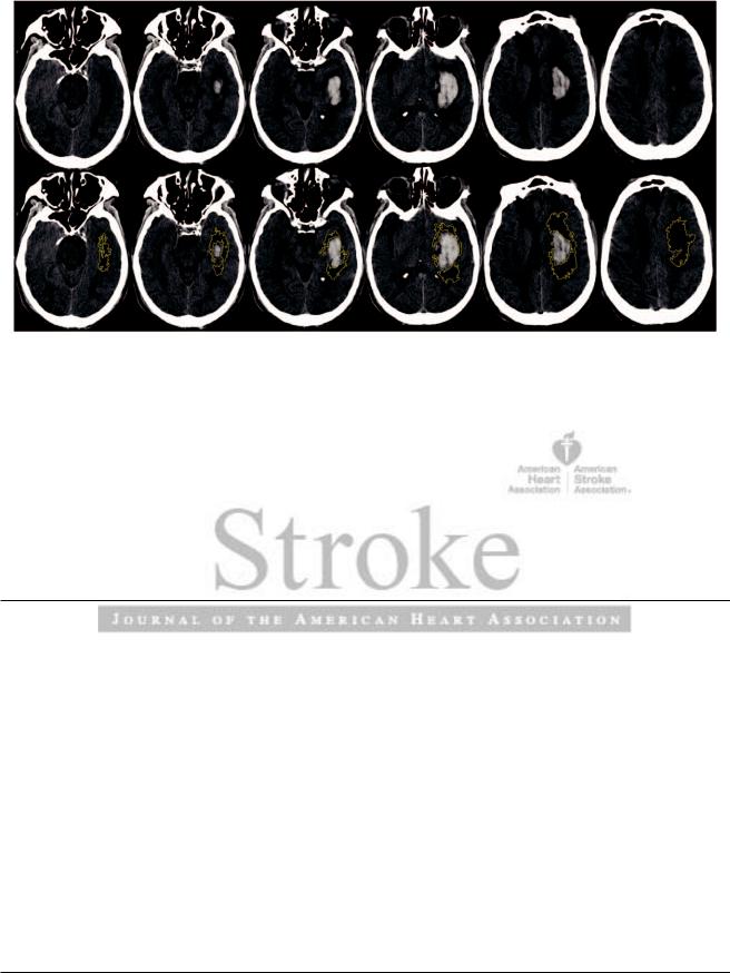
Independent and adjudicated volumetric measurements of all intraparenchymal clot and edema volumes were performed by W.A.M. and J.R.C. using an open source DICOM viewer software program for MAC (Osirix v. 4.1, Pixmeo; Geneva, Switzerland). Generous regions were drawn by hand to include areas of ICH and PHE susceptible to the computerized analysis, as determined by the reader. A semiautomated threshold-based approach using a Hounsfield unit range of 5 to 33 HU was then used to identify regions of PHE, as previously reported by Volbers et al.22 Using such range, a fixed lower value of 5 HU was set. The upper limit and absolute maximum of 33 HU was adjusted to obtain the best delineation of edema and avoid artifact introduced by leukoaraiosis. Once these HU limits were determined, Osirix created edema regions and produced a volume in cubic centimeters (cm3) by computing region of interest and slice thickness. Volumes were calculated using a similar threshold-based segmentation on well-definable boundaries of blood on CT (Figure 1).
For the purpose of this study, we identified the baseline stability (BLS) scan as the closest gradable scan before randomization. An EOT scan was defined as the scan performed 24 hours (±12) post last dose for S+rt-PA or post operative treatment for SO. A homologous time window was then ascribed to the medical cohort to perform the statistical analysis (closest scan to 3.9 days post onset).
Statistical Analysis
T tests were done to test differences in means for continuous variables. ANOVA was used to determine differences across groups. Wilcoxon rank-sum tests and a Kruskal–Wallis test also determined that inferences were not different at a probability value of 0.05 using these nonparametric tests. Fisher exact tests were done to determine differences in the distributions of categorical variables across groups. locally weighted scatterplot smoothing smoothing was used to determine the relationship between variables.23 Spearman ρ was used to determine the association between variables when the relationship seemed monotonic but not necessarily linear. We choose direct comparisons of preand post treatment edema volume as the most specific primary analysis of data to support or reject our hypothesis. We performed multivariate linear models to assess factors with the possibility to affect edema reduction.
Results
MISTIE II was composed of 2 stages: (1) dose finding (2005– 2009) and (2) safety (2009–2012). One hundred and twentythree patients were prospectively enrolled into 1 of 2 treatment groups, MIS plus rt-PA (surgery) or best medical management (medical), as shown in Table 1. Eighty-one patients were randomized to receive MIS, whereas 42 were randomized to medical management. Imaging of 5 patients (3 medical, 2 surgical) was not graded owing to instance of prior craniotomy creating image artifact and therefore poor image quality. The data of 118 patients are reported in this communication.
Downloaded from http://stroke.ahajournals.org/ by guest on November 25, 2013
Mould et al Perihematomal Edema Following MISTIE 3
Figure 1. Computed tomography scan at EOT for a medical patient with (right) and without (left) the semiautomated threshold-based segmentation of perihematomal edema. EOT indicates end of treatment scan.
Demographic and Clinical Data |
Neuroradiologic Features |
The surgical and medical cohorts were similar in age, sex, race, hematoma location, and admission Glasgow Coma Scale (Table 1). It is important to note that instance of symptomatic hemorrhage and central nervous system infection, within the analysis window (BLS-EOT), was low. Three symptomatic hemorrhages occurred in the surgical cohort (P=0.30), and 2 instances of central nervous system infection compared with 1 in the medical cohort (P=1.00).
Data on 118 patients were analyzed; time from ictus to BLS, ictus to EOT, and number of patients with intraventricular involvement were similar for the surgical and medical cohorts (Table 2).
ICH and edema volumes at BLS for surgical patients were similar compared with the medical cohort: surgical ICH, 43.8±17.2 cm3, PHE, 33.3±19.5 cm3; medical ICH 42.2±14.8 cm3, PHE, 30.3±12.0 cm3. Neither of these
Table 1. Demographic and Clinical Characteristics of the Study Patients
|
|
Surgical (n=79) |
Medical (n=39) |
P |
SO (n=10) |
S+rt-PA (n=69) |
P |
||||
|
|
|
|
|
|
|
|
|
|
||
Symptom onset age, y |
60.6 |
(11.5) |
61.0 |
(12.4) |
0.87 |
68.9 (9.2) |
59.4 |
(11.4) |
0.01 |
||
Enrollment GCS |
10.1 |
(2.9) |
10.4 |
(3.8) |
0.67 |
11.5 (2.8) |
9.8 |
(2.9) |
0.09 |
||
% Male |
53 |
(67.1%) |
26 |
(66.7%) |
1.00 |
6 |
(60.0%) |
47 |
(68.1%) |
0.72 |
|
Race |
|
|
|
|
0.60 |
|
|
|
|
0.81 |
|
|
White |
44 |
(55.7%) |
21 |
(53.8%) |
|
7 |
(70.0%) |
37 |
(53.6%) |
|
|
Black |
25 |
(31.6%) |
10 |
(25.6%) |
|
2 |
(20.0%) |
23 |
(33.3%) |
|
|
Hispanic |
9 |
(11.4%) |
5 |
(12.8%) |
|
1 |
(10.0%) |
8 |
(11.6%) |
|
Asian or Pacific Islander |
1 |
(1.3%) |
2 |
(5.1%) |
|
0 |
(0.0%) |
1 |
(1.4%) |
|
|
|
Unknown |
0 |
(0.0%) |
1 |
(2.6%) |
|
0 |
(0.0%) |
0 |
(0.0%) |
|
Clot location |
|
|
|
|
0.22 |
|
|
|
|
0.10 |
|
|
Thalamus |
4 |
(5.1%) |
1 |
(2.6%) |
|
0 |
(0.0%) |
4 |
(5.8%) |
|
|
Putamen |
46 |
(58.2%) |
24 |
(61.5%) |
|
3 |
(30.0%) |
43 |
(62.3%) |
|
|
Lobar |
22 |
(27.8%) |
14 |
(35.9%) |
|
6 |
(60.0%) |
16 |
(23.2%) |
|
Globus pallidus |
7 |
(8.9%) |
0 |
(0.0%) |
|
1 |
(10.0%) |
6 |
(8.7%) |
|
|
CNS infection* |
2 |
(2.5%) |
1 |
(2.6%) |
1.00 |
0 |
(0.0%) |
2 |
(2.9%) |
1.00 |
|
Symptomatic bleed* |
3 |
(3.8%) |
0 |
(0.0%) |
0.55 |
0 |
(0.0%) |
3 |
(4.3%) |
1.00 |
|
Emergent ICP therapy† |
14 |
(17.7%) |
10 |
(25.6%) |
0.34 |
1 |
(10.0%) |
13 |
(18.8%) |
0.68 |
|
Emergent osmotherapy |
10 |
(12.7%) |
9 |
(23.1%) |
0.18 |
1 |
(10.0%) |
9 |
(13.0%) |
1 |
|
CNS indicates central nervous system; GCS, Glasgow Coma Scale; ICP, intracranial pressure; SO, surgery only; and S+tPA, surgery + rt-PA. *Within analysis window of BLS to EOT; † ICP therapy included osmotherapy, aggressive hyperventilation, and surgical decompression.
Downloaded from http://stroke.ahajournals.org/ by guest on November 25, 2013

4 Stroke March 2013
Table 2. Radiological and Volumetric Data of the Study Patients
|
Surgical (n=79) |
Medical (n=39) |
P |
SO (n=10) |
S+rt-PA (n=69) |
P |
||||
|
|
|
|
|
|
|
|
|
|
|
Symptom onset to EOT scan, days |
3.9 |
(1.1) |
3.7 |
(0.5) |
0.23 |
3.6 |
(0.7) |
4.0 |
(1.1) |
0.27 |
Patients w/ IVH extension |
48 |
(60.8%) |
25 |
(64.1%) |
0.84 |
5 |
(50.0%) |
43 |
(62.3%) |
0.50 |
BLS ICH volume, mL |
43.8 |
(17.2) |
42.2 |
(14.8) |
0.61 |
40.1 |
(15.7) |
44.4 |
(17.4) |
0.46 |
EOT ICH volume, mL |
19.6 |
(14.5) |
40.7 |
(13.9) |
<0.001 |
24.5 |
(14.0) |
18.9 |
(14.5) |
0.26 |
BLS edema volume, mL |
33.3 |
(19.5) |
30.3 |
(12.0) |
0.37 |
6.4 |
(12.9) |
34.3 |
(20.1) |
0.24 |
EOT edema volume, mL |
27.7 |
(13.3) |
41.7 |
(14.6) |
<0.001 |
24.4 |
(8.6) |
28.1 |
(13.8) |
0.41 |
Reduction in edema (BLS-EOT) |
5.6 |
(15.1) |
−11.4 (9.6) |
<0.001 |
2.1 |
(10.6) |
6.2 |
(15.7) |
0.43 |
|
Relative PHE stability |
0.8 |
(0.3) |
0.7 |
(0.3) |
0.66 |
0.7 |
(0.2) |
0.8 |
(0.4) |
0.24 |
Reduction in edema/stability ICH* |
0.1 |
(0.3) |
−0.3 (0.3) |
<0.001 |
-0.0 |
(0.3) |
0.1 |
(0.3) |
0.31 |
|
BLS indicates baseline stability scan; EOT, end of treatment scan; ICH, intracerebral hemorrhage; IVH, intraventricular hemorrhage; PHE, perihematomal edema; SO, surgery only; and S + rt-PA, surgery + rt-PA.
*Relative PHE difference, edema reduction divided by BLS ICH.
comparisons was statistically significant (Table 2). Surgical patients had lower EOT ICH volume, 19.6±14.5 cm3, as compared with their medical counterparts, 40.7±13.9 cm3 (P<0.001). EOT edema volume was lower in surgical patients, 27.7±13.3 cm3, when compared with medical patients, 41.7±14.6 cm3 (P<0.001) as shown in Figure 2.
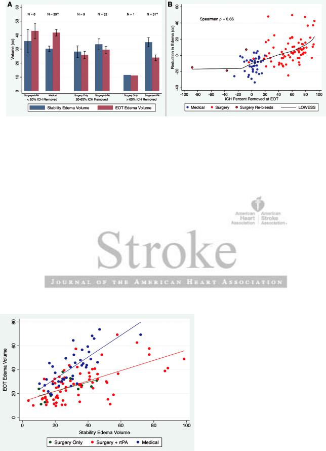
When patients were subdivided into roughly equally sized tertiles respecting the trial goal of clot removal of >65% (n=32), 20% to 65% (n=39), and <20% (n=8) clot removal from BLS to EOT, surgical patients with >65% clot removal demonstrated PHE reduction of 10.7±13.9 cm3, whereas medical patients, all with <20% resolution by EOT, showed an increase in PHE of 11.4±9.6 cm3 (P<0.001), as depicted in Figure 3. A significant graded effect of clot removal on PHE was observed overall (ANOVA P<0.001). Furthermore, a positive correlation between PHE reduction and percent of ICH removed was identified (Spearman ρ=0.66; P<0.001) as represented in Figure 3. In multivariate analyses, this relation was unaltered by dose of rt-PA, osmotherapy, or intracranial pressure therapy.
In the surgical arm, 69 patients received S+rt-PA, whereas 10 patients received SO. Both treatment subgroups were comparable for enrollment Glasgow Coma Scale, intraventricular involvement, time from symptom onset to BLS or EOT, baseline ICH volume, and baseline PHE volume but differed for age: SO, 68.9±9.2 years old and S+rt-PA, 59.4±11.4 years old (P=0.01). Both treatment cohorts achieved similar blood clot reduction: S+rt-PA, 18.9±14.5 cm3 and SO, 24.5±14.0 cm3 (P=0.26). Mean edema at EOT in patients treated with S+rt-PA was 28.1±13.8 cm3, while in patients treated with SO was 24.4±8.6 cm3 (P=0.41). Edema levels for both arms of the surgical cohort, S+rt-PA and SO, at BLS and EOT are shown in Figure 4.
Conclusions
We report on the effect of hematoma removal using MIS and rt-PA on PHE formation in ICH patients. We identified a significant PHE reduction in patients who underwent successful clot evacuation after the MISTIE procedure. Furthermore, administration of rt-PA for clot lysis in addition to initial
Figure 2. BLS and EOT edema volumes for the surgical (S+rt-PA and SO) and medical cohorts. BLS indicates baseline stability scan; EOT, end of treatment scan; SO, surgery only; and S + rt-PA, surgery + rt-PA.
Downloaded from http://stroke.ahajournals.org/ by guest on November 25, 2013
Mould et al Perihematomal Edema Following MISTIE 5
Figure 3. A, BLS and EOT edema volumes for patients separated by treatment group (medical, surgical aspiration only, and surgery plus rt-PA) and trichotimized by order of percent intracerebral hemorrhage (ICH) removed. BLS indicates baseline stability scan; EOT, end of treatment scan. B, Percent of ICH removed as calculated by ([BLS ICH volume−EOT ICH volume]/BLS volume) in a continuous fashion vs reduction in edema (BLS edema volume−EOT edema volume) for patients receiving medical management (blue) and MIS (red). S + rt-PA indicates surgery plus rt-PA; and SO, surgical aspiration only. *denotes statistical significance.
aspiration did not enhance edema formation in relation to patients treated with clot aspiration only.
PHE is an almost universal occurrence after ICH. Early interpretations of perihematomal events included cerebral ischemia, which found some support in animal experiments.24–31 Subsequent human studies using surrogates of cerebral ischemia, such as single photon emission CT and perfusion weighted magnetic resonance imaging, corroborated the presence of hypoperfused tissue surrounding parenchymal clots.32–34 Only when studies measuring cerebral metabolism were performed did it become clear that hypoperfusion was likely the result of hypometabolism.35 This metabolic state of hibernation is hypothesized to be associated to vasogenic cerebral edema. Attempts to indirectly quantify blood– brain barrier disruption using diffusion weighted magnetic resonance imaging have suggested a cause-effect or doseresponse association between the volume of ICH, intensity of apparent diffusion coefficient elevation PHE volume.36–38
The clinical significance of PHE remains unclear. Volumetric analyses of PHE using CT and magnetic resonance imaging studies have repeatedly demonstrated edema volumes reaching 2- to 3-fold the original hematoma volume.38 Delayed neurological deterioration as late as 2 to 3 weeks after the ictus, likely the result of PHE, has also been described. However, the independent impact of these events on long-term neurological outcome remains unknown. Gebel et al39,40 reported on the paradoxical improved functional outcome predicted by relative PHE in the initial 24 hours. Using data from the Intensive Blood Pressure Reduction in Acute Cerebral Hemorrhage (INTERACT) trial, Arima et al20 reported differently. These investigators found PHE to be significantly associated to the underlying hematoma volume but lacking independent effect on the outcome of ICH patients. These studies and ours are limited by difficulty completely blinding the edema analysis of surgical subjects and our still limited knowledge of factors that provoke and mitigate edema.
Figure 4. Relationship between BLS and EOT edema volumes for patients separated by treatment group (medical, surgical aspiration only, and surgery plus rt-PA). BLS indicates baseline stability scan; EOT, end of treatment scan; and rtPA, recombinant tissue-type plasminogen activator.
Downloaded from http://stroke.ahajournals.org/ by guest on November 25, 2013
6 Stroke March 2013
Targeted therapies for this form of edema for ICH are lacking; thus the differential impact of PHE modification on neurological outcome is largely unknown. Therapeutic trials for ICH have concentrated primarily on clot evacuation.2,3 Several early clinical trials comparing best medical therapy alone versus best medical therapy and surgical evacuation of the hematoma have been completed. Minimally invasive neurosurgical procedures seem to minimize trauma to viable brain tissue. Studies using MIS and clot aspiration, thrombolysis, and endoscopic evacuation have reported on their safety and potential for efficacy when used in selected ICH patients. These studies not only suggest that hematoma evacuation is safe but that a parallel response between hematoma volume reduction and PHE volume exists.41–43 Our results confirm such observations in this prospectively recruited cohort of patients. The combined effect of this form of hematoma evacuation and edema volume attenuation on neurological recovery after ICH awaits testing in a properly powered prospective clinical trial.
rt-PA has been used in several paradigms of brain injury, more conspicuously as the thrombolytic agent for acute recanalization during acute ischemic stroke. The safety and efficacy profiles of rt-PA in this setting have been evaluated in several studies before its recommendation as thrombolytic agent for the treatment of acute cerebral ischemia using the intravenous administration route. The experimental use of rt-PA in the treatment of intraventricular and ICH, however, has opened 2 new modalities of drug delivery that have not been previously tested. After completing proof of concept and dose escalation studies, Clot Lysis: Evaluating Accelerated Resolution of Hemorrhage with rt-PA Intraventricular Hemorrhage (CLEAR IVH) and MISTIE II have reported on the efficient and safe clot evacuation from the intraventricular and intraparenchymal compartments using rt-PA. Concerns of toxicity produced from the direct exposure of the drug to neuronal tissue have, however, been raised by some investigators.44,45 Early reports of retinal toxicity when tissue plasminogen activator and L-arginine when used in the treatment of vitreal hemorrhage exist.46–48 Furthermore, first in a pig model and more recently in a clinical study, rt-PA has been postulated to worsen vasogenic edema when used in the treatment of intraventricular hemorrhage and ICH.49,50 Nonetheless, no evidence for this toxicity was noted when histological assessments in large animal intracranial and retinal hemorrhage models were performed.51–55 Additionally, recent mouse tissue plasminogen activator knockout studies suggest amelioration of blood-clot-related neuronal and glial tissue injury by rt-PA.56 Finally, no signs of human neuronal rt-PA toxicity have been noted in the current treatment of ischemic stroke, despite administration under conditions of blood–brain barrier disruption.57
Our study is the first a priori investigation that uses a semiautomated volumetric analysis for prospectively obtained group of patients treated with clot aspiration alone versus clot aspiration and thrombolytic therapy with rt-PA. Both groups achieved similar clot volume reduction without experiencing differences in PHE volumes, confirming the overall positive impact of hematoma removal using rt-PA on PHE volumes, reported by our group as well as others.
Our analysis of 118 patients enrolled in the MISTIE II trial is consistent with the hypothesis that successful hematoma evacuation leads to significant edema volume reduction. In
2008, we did report on such association after the retrospective analysis of a convenience cohort of ICH patients treated using a similar approach with MIS and thrombolysis. This is the first time such an observation is confirmed in a prospectively obtained cohort of ICH patients. Hematoma evacuation and its impact on vasogenic edema formation leading to improved neurological outcomes after ICH remains under investigation. MISTIE III offers to test for such association. In the meantime, our results demonstrate that efficient hematoma evacuation using a combined approach of MIS and aspiration with or without rt-PA leads to a significant reduction in PHE.
Acknowledgments
We thank the patients and families who volunteered for this study, Genentech Inc for the donation of study drug (Alteplase), and the following people for their support in assisting data collection, Lucas First, Saman Nekoovaght-Tak, Johanna Block.
List of PIs and Surgeons for MISTIE II
Allegheny General Hospital, Pittsburgh, PA, Khaled Aziz, MD, PI; Bronson Methodist Hospital, Kalamazoo, MI, Jeffrey Fletcher, MD, PI, Bratislav Velimirovic, MD, Coinvestigator, Daryl Warder, MD, Coinvestigator; Duke University Medical Center, Durham, NC, Gavin Britz, MBBCh, MPH, Coinvestigator, Carmelo Graffagnino, MD, PI; Hartford Hospital, Hartford, CT, Inam Kureshi, MD, PI; Johns Hopkins Medical Institutions, Baltimore, MD, Judy Huang, MD, PI; Medical University of South Carolina, Charleston, NC, Byron Bailey, MD, PI, Dilantha Ellegala, MD, PI, Angela Hays, MD, PI, Marc LaPointe, PharmD, PI; Montreal Neurological Institute at McGill University, Montreal, QC, Canada, David Sinclair, MD, PI; Mount Sinai Medical Center, New York, NY, Joshua B Bederson, MD, PI, Henry Moyle, MD, PI; Newcastle University, Newcastle on Tyne, United Kingdom, Professor A David Mendelow, PI, Prokopios Panaretos, Coinvestigator; New Jersey Neuroscience Institute at JFK Medical Center, Edison, NJ, Martin Gizzi, MD, PhD, PI, Thomas Steineke, MD, PhD, Coinvestigator; Rush University, Chicago, IL, Lorenzo Munoz, MD, Coinvestigator, Shaun T O’Leary, MD, Coinvestigator, Richard E Temes, MD, PI; Stanford University School of Medicine, Palo Alto, CA, Robert Dodd, MD, Coinvestigator, Cristanne Wijman, MD, PhD, PI; St. Luke’s Hospital, Kansas City, MO, Paul Camarata, MD, PI; Temple University, Philadelphia, PA, Jack Jallo, MD, PhD, PI, Christopher Loftus, MD, PI, Michael Weaver, MD, Coinvestigator; University of Alabama at Birmingham, Birmingham, AL, Mark Harrigan, MD, PI; University of California, Los Angeles, Los Angeles, CA, Neil Martin, MD, PI, Paul Vespa, MD, PI; University of California, San Diego, San Diego, CA, Bob Carter, MD, PhD, PI; University of Chicago, Chicago, IL, Issam Awad, MD, PI, Fernando Goldenberg, MD, PI; University of Cincinnati, Cincinnati, OH, Andrew Ringer, MD, Coinvestigator, Mario Zuccarello, MD, PI; University of Maryland, Baltimore, MD, E. Francois Aldrich, MD, PI; University of Texas, Houston, Houston, TX, William Ashley, MD,Coinvestigator,PengRocChen,MD,Coinvestigator,GeorgeLopez, MD, PI; University of Texas, San Antonio, San Antonio, TX, Jean-Louis Caron, MD, PI; Universitätsklinikum Heidelberg, Heidelberg, Germany, Dr. med. Daniel Haux, Coinvestigator, Berk Orakcioglu, Coinvestigator, Dr. med. Sven Poli, PI, Thorsten Steiner, MD, PhD, PI; Virginia Commonwealth University, Richmond, VA, William C Broaddus, MD, PhD, PI, R. Scott Graham, MD, Coinvestigator.
Sources of Funding
National Institute of Health/National Institute of Neurological Disorders and Stroke supported this research with grants number R01Ns046309 and 5U01NS062851.
Disclosures
Dr Daniel F. Hanley was awarded significant research support of grants number R01Ns046309 and 5U01NS062851. Johns Hopkins University holds a use patent for intraventricular tissue plasminogen activator.
Downloaded from http://stroke.ahajournals.org/ by guest on November 25, 2013

Mould et al Perihematomal Edema Following MISTIE 7
References
1.Mendelow AD, Gregson BA, Fernandes HM, Murray GD, Teasdale GM, Hope DT, et al; STICH Investigators. Early surgery versus initial conservative treatment in patients with spontaneous supratentorial intracerebral haematomas in the International Surgical Trial in Intracerebral Haemorrhage (STICH): a randomised trial. Lancet. 2005;365:387–397.
2.Mendelow AD, Unterberg A. Surgical treatment of intracerebral haemorrhage. Curr Opin Crit Care. 2007;13:169–174.
3.Prasad K, Mendelow AD, Gregson B. Surgery for primary supratentorial intracerebral haemorrhage. Cochrane Database Syst Rev. 2008;(4):CD000200.
4. Newell DW, Shah MM, Wilcox R, Hansmann DR, Melnychuk E, Muschelli J, et al. Minimally invasive evacuation of spontaneous intracerebral hemorrhage using sonothrombolysis. J Neurosurg. 2011;115:592–601.
5.Wu G, Li C, Wang L, Mao Y, Hong Z. Minimally invasive procedures for evacuation of intracerebral hemorrhage reduces perihematomal glutamate content, blood-brain barrier permeability and brain edema in rabbits. Neurocrit Care. 2011;14:118–126.
6.Zhou H, Zhang Y, Liu L, Huang Y, Tang Y, Su J, et al. Minimally invasive stereotactic puncture and thrombolysis therapy improves long-term outcome after acute intracerebral hemorrhage. J Neurol. 2011;258:661–669.
7.Carhuapoma JR, Barrett RJ, Keyl PM, Hanley DF, Johnson RR. Stereotactic aspiration-thrombolysis of intracerebral hemorrhage and its impact on perihematoma brain edema. Neurocrit Care. 2008;8:322–329.
8.Miller CM, Vespa PM, McArthur DL, Hirt D, Etchepare M. Frameless stereotactic aspiration and thrombolysis of deep intracerebral hemorrhage is associated with reduced levels of extracellular cerebral glutamate and unchanged lactate pyruvate ratios. Neurocrit Care. 2007;6:22–29.
9.Morgan T, Zuccarello M, Narayan R, Keyl P, Lane K, Hanley D. Preliminary findings of the minimally-invasive surgery plus rtPA for intracerebral hemorrhage evacuation (MISTIE) clinical trial. Acta Neurochir Suppl. 2008;105:147–151.
10.Masada T, HuaY, Xi G,Yang GY, Hoff JT, Keep RF. Attenuation of intracerebral hemorrhage and thrombin-induced brain edema by overexpression of interleukin-1 receptor antagonist. J Neurosurg. 2001;95:680–686.
11.Peeling J, Yan HJ, Corbett D, Xue M, Del Bigio MR. Effect of FK-506 on inflammation and behavioral outcome following intracerebral hemorrhage in rat. Exp Neurol. 2001;167:341–347.
12.Gong Y, Xi GH, Keep RF, Hoff JT, Hua Y. Complement inhibition attenuates brain edema and neurological deficits induced by thrombin. Acta Neurochir Suppl. 2005;95:389–392.
13.Ziai WC. Proceedings of the Princeton Conference: Hematology and Inflammatory Signaling of Intracerebral Hemorrhage. Stroke. In Press
14.Zazulia AR, Diringer MN, Derdeyn CP, Powers WJ. Progression of mass effect after intracerebral hemorrhage. Stroke. 1999;30:1167–1173.
15.Olivot JM, Mlynash M, Kleinman JT, Straka M, Venkatasubramanian C, Bammer R, et al; DASH Investigators. MRI profile of the perihematomal region in acute intracerebral hemorrhage. Stroke. 2010;41:2681–2683.
16.Venkatasubramanian C, Mlynash M, Finley-Caulfield A, Eyngorn I, Kalimuthu R, Snider RW, et al. Natural history of perihematomal edema after intracerebral hemorrhage measured by serial magnetic resonance imaging. Stroke. 2011;42:73–80.
17.Fainardi E, Borrelli M, Saletti A, Sarubbo S, Roversi G, Bernardoni A, et al. Temporal changes in perihematomal apparent diffusion coefficient values during the transition from acute to subacute phases in patients with spontaneous intracerebral hemorrhage. Neuroradiology. September 2012. doi: 10.1007/s00234-012-1093-x. Accessed January 16, 2013.
18.Staykov D, Wagner I, Volbers B, Hauer EM, Doerfler A, Schwab S, et al. Natural course of perihemorrhagic edema after intracerebral hemorrhage. Stroke. 2011;42:2625–2629.
19.Gebel JM Jr, Jauch EC, Brott TG, Khoury J, Sauerbeck L, Salisbury S, et al. Natural history of perihematomal edema in patients with hyperacute spontaneous intracerebral hemorrhage. Stroke. 2002;33:2631–2635.
20.Arima H, Wang JG, Huang Y, Heeley E, Skulina C, Parsons MW, et al.; INTERACT Investigators. Significance of perihematomal edema in acute intracerebral hemorrhage: the INTERACT trial. Neurology. 2009;73:1963–1968.
21.Morgenstern LB, Hemphill JC III, Anderson C, Becker K, Broderick JP, Connolly ES Jr, et al; American Heart Association Stroke Council and Council on Cardiovascular Nursing. Guidelines for the management of spontaneous intracerebral hemorrhage: a guideline for healthcare professionals from the American Heart Association/American Stroke Association. Stroke. 2010;41:2108–2129.
22.Volbers B, Staykov D, Wagner I, Dörfler A, Saake M, Schwab S, et al. Semi-automatic volumetric assessment of perihemorrhagic edema with computed tomography. Eur J Neurol. 2011;18:1323–1328.
23.Cleveland WS; Taylor & Francis Group. Robust locally weighted regression and smoothing scatterplots. J Am Stat Assoc. 1979;74:829–36.
24.Ropper AH, Zervas NT. Cerebral blood flow after experimental basal ganglia hemorrhage. Ann Neurol. 1982;11:266–271.
25.Nath FP, Jenkins A, Mendelow AD, Graham DI, Teasdale GM. Early hemodynamic changes in experimental intracerebral hemorrhage. J Neurosurg. 1986;65:697–703.
26.Sinar EJ, Mendelow AD, Graham DI, Teasdale GM. Experimental intracerebral hemorrhage: effects of a temporary mass lesion. J Neurosurg. 1987;66:568–576.
27.Kingman TA, Mendelow AD, Graham DI, Teasdale GM. Experimental intracerebral mass: description of model, intracranial pressure changes and neuropathology. J Neuropathol Exp Neurol. 1988;47:128–137.
28.Bullock R, Brock-Utne J, van Dellen J, Blake G. Intracerebral hemorrhage in a primate model: effect on regional cerebral blood flow. Surg Neurol. 1988;29:101–107.
29.Nehls DG, Mendelow DA, Graham DI, Teasdale GM. Experimental intracerebral hemorrhage: early removal of a spontaneous mass lesion improves late outcome. Neurosurgery. 1990;27:674–682; discussion 682.
30.Nehls DG, Mendelow AD, Graham DI, Sinar EJ, Teasdale GM. Experimental intracerebral hemorrhage: progression of hemodynamic changes after production of a spontaneous mass lesion. Neurosurgery. 1988;23:439–444.
31.Yang GY, Betz AL, Chenevert TL, Brunberg JA, Hoff JT. Experimental intracerebral hemorrhage: relationship between brain edema, blood flow, and blood-brain barrier permeability in rats. J Neurosurg. 1994;81:93–102.
32.Mayer SA, Lignelli A, Fink ME, Kessler DB, Thomas CE, Swarup R, et al. Perilesional blood flow and edema formation in acute intracerebral hemorrhage: a SPECT study. Stroke. 1998;29:1791–1798.
33.Kidwell CS, Saver JL, Mattiello J, Warach S, Liebeskind DS, Starkman S, et al. Diffusion-perfusion MR evaluation of perihematomal injury in hyperacute intracerebral hemorrhage. Neurology. 2001;57:1611–1617.
34.Carhuapoma JR, Wang P, Beauchamp NJ, Hanley DF, Barker PB.
Diffusion-perfusion MR evaluation and spectroscopy before and after surgical therapy for intracerebral hemorrhage. Neurocrit Care. 2005;2:23–27.
35.Zazulia AR, Diringer MN, Videen TO, Adams RE, Yundt K, Aiyagari V, et al. Hypoperfusion without ischemia surrounding acute intracerebral hemorrhage. J Cereb Blood Flow Metab. 2001;21:804–810.
36.Carhuapoma JR, Wang PY, Beauchamp NJ, Keyl PM, Hanley DF, Barker PB. Diffusion-weighted MRI and proton MR spectroscopic imaging in the study of secondary neuronal injury after intracerebral hemorrhage. Stroke. 2000;31:726–732.
37.Carhuapoma JR, Barker PB, Hanley DF, Wang P, Beauchamp NJ.
Human brain hemorrhage: quantification of perihematoma edema by use of diffusion-weighted MR imaging. AJNR Am J Neuroradiol. 2002;23:1322–1326.
38.Carhuapoma JR, Hanley DF, Banerjee M, Beauchamp NJ. Brain edema after human cerebral hemorrhage: a magnetic resonance imaging volumetric analysis. J Neurosurg Anesthesiol. 2003;15:230–233.
39.Gebel JM, Brott TG, Sila CA, Tomsick TA, Jauch E, Salisbury S, et al. Decreased perihematomal edema in thrombolysis-related intracerebral hemorrhage compared with spontaneous intracerebral hemorrhage. Stroke. 2000;31:596–600.
40.Gebel JM Jr, Jauch EC, Brott TG, Khoury J, Sauerbeck L, Salisbury S, et al. Relative edema volume is a predictor of outcome in patients with hyperacute spontaneous intracerebral hemorrhage. Stroke. 2002;33:2636–2641.
41.Barrett RJ, Hussain R, Coplin WM, Berry S, Keyl PM, Hanley DF, et al. Frameless stereotactic aspiration and thrombolysis of spontaneous intracerebral hemorrhage. Neurocrit Care. 2005;3:237–245.
42.Zhou H, Zhang Y, Liu L, Han X, Tao Y, Tang Y, et al. A prospective controlled study: minimally invasive stereotactic puncture therapy versus conventional craniotomy in the treatment of acute intracerebral hemorrhage. BMC Neurol. 2011;11:76.
43.Vespa P, McArthur D, Miller C, O’Phelan K, Frazee J, Kidwell C, et al. Frameless stereotactic aspiration and thrombolysis of deep intracerebral hemorrhage is associated with reduction of hemorrhage volume and neurological improvement. Neurocrit Care. 2005;2:274–281.
44.Tsirka SE, Rogove AD, Strickland S. Neuronal cell death and tPA. Nature. 1996;384:123–124.
Downloaded from http://stroke.ahajournals.org/ by guest on November 25, 2013

8 Stroke March 2013
45.Wang YF, Tsirka SE, Strickland S, Stieg PE, Soriano SG, Lipton SA. Tissue plasminogen activator (tPA) increases neuronal damage after focal cerebral ischemia in wild-type and tPA-deficient mice. Nat Med. 1998;4:228–231.
46.Chen SN, Yang TC, Ho CL, Kuo YH, Yip Y, Chao AN. Retinal toxicity of intravitreal tissue plasminogen activator: case report and literature review. Ophthalmology. 2003;110:704–708.
47.Mali RS, Cheng M, Chintala SK. Plasminogen activators promote exci- totoxicity-induced retinal damage. FASEB J. 2005;19:1280–1289.
48.Oh HS, Kwon OW, Chung I, Lee SC, Koh HJ, Lee SH, et al. Retinal toxicity of commercial tissue plasminogen activator is mediated by the induction of nitric oxide in the mouse retinal primary cells. Curr Eye Res. 2005;30:291–297.
49.Ducruet AF, Hickman ZL, Zacharia BE, Grobelny BT, Narula R, Guo KH, et al. Exacerbation of perihematomal edema and sterile meningitis with intraventricular administration of tissue plasminogen activator in patients with intracerebral hemorrhage. Neurosurgery. 2010;66: 648–655.
50.Thiex R, Küker W, Müller HD, Rohde I, Schröder JM, Gilsbach JM, et al. The long-term effect of recombinant tissue-plasminogen-activator (rt-PA) on edema formation in a large-animal model of intracerebral hemorrhage. Neurol Res. 2003;25:254–262.
51.Pang D, Sclabassi RJ, Horton JA. Lysis of intraventricular blood clot with urokinase in a canine model: part 1. Canine intraventricular blood cast model. Neurosurgery. 1986;19:540–546.
52.Pang D, Sclabassi RJ, Horton JA. Lysis of intraventricular blood clot with urokinase in a canine model: part 2. In vivo safety study of intraventricular urokinase. Neurosurgery. 1986;19:547–552.
53.Pang D, Sclabassi RJ, Horton JA. Lysis of intraventricular blood clot with urokinase in a canine model: part 3. Effects of intraventricular urokinase on clot lysis and posthemorrhagic hydrocephalus. Neurosurgery. 1986;19:553–572.
54.Wagner KR, Xi G, HuaY, Zuccarello M, de Courten-Myers GM, Broderick JP, et al. Ultra-early clot aspiration after lysis with tissue plasminogen activator in a porcine model of intracerebral hemorrhage: edema reduction and blood-brain barrier protection. J Neurosurg. 1999;90:491–498.
55.Lewis H, Resnick SC, Flannery JG, Straatsma BR. Tissue plasminogen activator treatment of experimental subretinal hemorrhage. Am J Ophthalmol. 1991;111:197–204.
56.Akassoglou K, Kombrinck KW, Degen JL, Strickland S. Tissue plasminogen activator-mediated fibrinolysis protects against axonal degeneration and demyelination after sciatic nerve injury. J Cell Biol. 2000;149:1157–1166.
57.Lo EH, Broderick JP, Moskowitz MA. tPA and proteolysis in the neurovascular unit. Stroke. 2004;35:354–356.
Downloaded from http://stroke.ahajournals.org/ by guest on November 25, 2013