

kinins - review
.pdfJ Pharmacol Sci 99, 6 – 38 (2005) |
Journal of Pharmacological Sciences |
|
©2005 The Japanese Pharmacological Society |
Survey Review
The Kallikrein-Kinin System: Current and Future Pharmacological
Targets
Marie Eve Moreau1, Nancy Garbacki2, Giuseppe Molinaro1, Nancy J. Brown3, François Marceau4, and Albert Adam1,*
1Faculty of Pharmacy, University of Montreal, 2900 boul. Édouard-Montpetit, C.P. 6128, Succursale Centre-Ville, Montréal (Québec), Canada H3C 3J7
2Laboratory of Human Physiology, University of Liège, 3 Avenue de l’Hôpital, Sart Tilman, Belgium B-4000 3Division of Clinical Pharmacology, Department of Medicine, School of Medicine, Vanderbilt University, 60 Robinson Research Building (RRB), Nashville, TN 37232-6602, USA
4Centre de Recherche en Rhumatologie et Immunologie, CHUL-CHUQ, 2705 boul. Laurier, Québec (Québec), Canada, G1V 4G2
Received May 24, 2005
Abstract. The kallikrein-kinin system is an endogenous metabolic cascade, triggering of which results in the release of vasoactive kinins (bradykinin-related peptides). This complex system includes the precursors of kinins known as kininogens and mainly tissue and plasma kallikreins. The pharmacologically active kinins, which are often considered as either proinflammatory or cardioprotective, are implicated in many physiological and pathological processes. The interest of the various components of this multi-protein system is explained in part by the multiplicity of its pharmacological activities, mediated not only by kinins and their receptors, but also by their precursors and their activators and the metallopeptidases and the antiproteases that limit their activities. The regulation of this system by serpins and the wide distribution of the different constituents add to the complexity of this system, as well as its multiple relationships with other important metabolic pathways such as the renin-angiotensin, coagulation, or complement pathways. The purpose of this review is to summarize the main properties of this kallikrein-kinin system and to address the multiple pharmacological interventions that modulate the functions of this system, restraining its proinflammatory effects or potentiating its cardiovascular properties.
Keywords: kallikrein-kinin system, B1 and B2 receptors, metallopeptidase, pharmacological agent
Introduction............................................................. |
7 |
|
2.3 |
Tissue kallikrein-kinin system |
|
||
Part I: The Kallikrein-Kinin System..................... |
8 |
|
2.4 |
Other kinin forming enzymes |
|
||
I- Kinins .............................................................. |
8 |
3. |
Regulation of the kininogenase activity ...... |
10 |
|||
1. |
The kininogens: precursors of kinins ......... |
8 |
IIMetabolism of Kinins ................................... |
11 |
|||
|
1.1 |
High-molecular-weight kininogen |
|
1. |
Angiotensin I-converting enzyme (ACE) .... |
11 |
|
|
1.2 |
Low-molecular-weight kininogen |
|
|
1.1 |
Definition |
|
2. |
The kinin-forming systems .......................... |
9 |
|
1.2 |
Synthesis, regulation, and localization |
|
|
|
2.1 |
The plasma kinin forming system |
|
|
1.3 |
Properties |
|
2.1.1 Plasma prekallikrein |
1.3.1 Angiotensinase vs kininase |
2.1.2 Contact system activation of plasma |
1.3.2 ACE GPIase activity |
2.2 Endothelial cells and kinin forming activity |
1.3.3 ACE: a signal transduction molecule |
|
1.3.4 ACE insertion/deletion polymorphism |
*Corresponding author. FAX: +1-514-343-2102 |
|
E-mail: albert.adam@umontreal.ca |
|
|
|
Invited article |
|
6

The Kallikrein-Kinin System |
7 |
2. |
Neprilysin ..................................................... |
12 |
|
|
2.1 |
Definition |
|
|
2.2 |
Synthesis, localization, and properties |
|
3. |
Aminopeptidase P......................................... |
13 |
|
|
3.1 |
Definition |
|
|
3.2 |
Synthesis, regulation, and localization |
|
|
3.3 |
Properties |
|
4. |
Carboxypeptidase N and M.......................... |
13 |
|
5. |
Other peptidases .......................................... |
13 |
|
III- |
Kinin Receptors........................................... |
14 |
|
1. |
Pharmacological classification.................... |
14 |
|
|
1.1 |
Potency order of agonists |
|
|
1.2 |
Affinity of antagonists |
|
2. |
Molecular classification............................... |
17 |
|
|
2.1 |
Organization and structure |
|
|
|
of the receptor genes |
|
|
2.2 |
Receptors expression and regulatory |
|
|
|
elements in gene promoters |
|
|
2.3 |
Second messengers |
|
|
2.4 |
Receptor desensitization |
|
Part II: The Kallikrein-Kinin System: |
|
||
|
Pathophysiology and Pharmacological |
|
|
|
Target ......................................................... |
19 |
|
I- The Kinin Forming System in Plasma ......... |
19 |
||
1. |
Genetic defects.............................................. |
19 |
|
1.1Defects of the contact system components
1.2Defect in the control of the contact system: C1 inhibitor
1.2.1Definition
1.2.2Pathophysiology
1 2.3 Treatment of hereditary angioedema
1.2.3.1Serine proteases inhibitors
1.2.3.2DX88
1.2.3.3Attenuated androgens: Danazol®, Stanozolol®
1.2.3.4Antifibrinolytic drugs: tranexamic acid (Transamin®, Cyklokapron®, Exacyl®, Cyklo-f®)
1.2.3.5 B2-receptor antagonists: HOE 140
(Icatibant® or JE049®) |
|
2. Acquired diseases ......................................... |
21 |
2.1Sepsis
2.2Anaphylactoid and severe hypotensive reactions
3. |
Antithrombolytic treatment and kinins ....... |
21 |
|
IIThe Metabolism of Kinins............................ |
21 |
||
1. |
ACE: pathophysiology ................................. |
21 |
|
2. |
ACE inhibitors (ACEi)................................. |
21 |
|
|
2.1 |
Multicenter clinical randomized trials |
|
|
2.2 |
Role of bradykinin in the cardiovascular |
|
|
|
effects of ACEi |
|
3. |
Vasopeptidase inhibitors (VPi) .................... |
22 |
|
3.1Multicenter clinical randomized trials
3.2Role of kinins in the cardiovascular effects of VPi
4.Kinins and side effects of metallopeptidase
|
inhibitors....................................................... |
23 |
III- |
Kinin Receptors........................................... |
24 |
1. |
Receptor polymorphisms and pathology ..... |
24 |
1.1Polymorphism of B2 receptors
1.1.1+9/−9, exon 1 polymorphism
1.1.2(C−58 → T), promoter region polymorphism
1.1.3(C181 → T), exon 2 polymorphism
1.2Polymorphisms of B1 receptors: (G−699 → C) substitution
2.Kinin receptors as pharmacological
targets ........................................................... |
25 |
|
2.1 |
Cardiovascular and renal diseases |
|
2.2 |
Inflammation |
|
2.3 |
Pain and neurological applications |
|
2.4 |
Diabetes |
|
2.5 |
Renal disease |
|
2.6 |
Airway disease |
|
2.7 |
Angioedema |
|
Conclusion ............................................................... |
28 |
|
Introduction
The existence of the kallikrein-kinin system was first discovered almost one century ago when Abelous and Bardier, in 1909, showed the hypotensive effect of human urine (1). Since that time, this system has been and continues to be the subject of intensive research. In fact, more than 30,000 papers are referenced for kallikrein-kinin in Medline for the last 50 years.
Such a scientific interest is explained in part because of the duality and the complexity of this system. Bradykinin (BK) is at the center of this system; however, it is not the only pharmacologically active kinin. The kinins
(BK-related peptides) are generated from two types of kininogens, mainly by two types of activators: tissue and plasma kallikreins. In fact, two classes of kinin receptor ligands are now recognized corresponding to each receptor subtype (B1 and B2 receptors). The expression of these receptors is regulated by specific mechanisms. The duality also exists as to the pharmacological activity of kinins, which are often considered as either proinflammatory or protective (namely for heart, kidney function, angiogenesis-promoting) depending on the experimental approach, or scientific interest. This system is also complex in its distribution, as the different constituents have been shown to be present in plasma,
8 |
ME Moreau et al |
but also on blood cells, in various tissues or their exocrine secretions. The autocrine or paracrine activity of kinins is regulated by several metallopeptidases, the relative importance of which varies from one biological medium to the other. Finally, the regulation of this system by serpins adds to the complexity of the system, as well as its multiple relationships with other important metabolic pathways such as the renin-angiotensin, coagulation or complement pathways.
An understanding of the multifaceted aspects of the different constituents of this system is necessary to grasp the complexity of its multiple pharmacological activities, mediated not only by kinins and their receptors, but also by their precursors and their activators, and the metallopeptidases and the antiproteases that limit their activities.
The purpose of this review is, first, to summarize the main properties of the various constituents of this complex but fascinating system. We will secondly address the multiple pharmacological interventions that modulate the functions of the kallikrein-kinin system and their clinical applications.
Part I: The Kallikrein-Kinin System
The kallikrein-kinin system represents a metabolic cascade that, when activated, triggers the release of vasoactive kinins. This complex multi-protein system includes the serine proteases tissue and plasma kallikreins, which liberate kinins from highand low- molecular-weight kininogen (HK and LK). Kinins exert their pharmacological activities by binding specific receptors, before being metabolized by various peptidases.
I- Kinins
In humans and in most mammals, the term “kinin” refers to the nonapeptide, BK (Arg-Pro-Pro-Gly-Phe- Ser-Pro-Phe-Arg), the decapeptide kallidin (KD: LysBK), and their carboxy-terminal des-Arg metabolites. Other kinins, like T-kinin (Ile-Ser-BK) and Met-T-kinin have only been reported in the rat (2).
Kinins are implicated in many physiological and pathological processes. By virtue of their ability to activate endothelial cells, leading to vasodilation, increased vascular permeability, tissue-type plasminogen activator (t-PA) release, production of nitric oxide (NO), and mobilization of arachidonic acid, they participate in physiological (regulation of blood pressure, renal and cardiac functions) and pathological processes like inflammation.
1. The kininogens: precursors of kinins
HK and LK, the precursors of kinins (BK and KD), are produced from a structural gene localized to chromosome 3q26 → qter that is thought to have originated as a result of two successive duplications of a primordial kininogen gene (3, 4). This gene consists of 11 exons. The first 9 exons encode the heavy chain. The 10th exon codes for BK and the light chain of HK; the light chain of LK is coded by exon 11.
Both HK and LK have an identical aminoacid sequence starting at the N-terminus (heavy chain) and continuing to 12 aminoacids beyond the BK moiety but differ at the C-terminal because of alternative splicing, thereby providing the two kininogens with different light-chain moieties (3, 4). In fact, both native proteins are produced as single chain polypeptides, and the light or heavy chain nomenclature refers to their disulfide bond-assembled structure after activation by kallikrein cleavage.
1.1 High-molecular-weight kininogen (HK)
HK, an α-globulin, circulates in plasma as an 88to 120-kDa single-chain glycoprotein at a concentration of 70 to 90 µg/mL (5). This kininogen is a multifunctional protein composed of six domains. Each domain is thought to have distinct functions. HK heavy chain (64 kDa) contains domains 1, 2, and 3. The light chain (45 to 58 kDa) is comprised of domains 5 and 6. The heavy chain and light chain are linked by domain 4, which contains the BK sequence. Domain 1 has a low affinity calcium-binding site (6), domains 2 and 3 have specific sequences (Gln-Val-Val-Ala-Gly) that inhibit cysteine proteases (7), and domain 3 has plateletand endothelial cell-binding activity (8). Domain 5 has cellbinding sites, antiangiogenic properties, and sequences for heparin binding (9 – 11). HK binds to negatively charged surfaces through the histidine region of the light chain corresponding to domain 5. Domain 6 has prekallikrein and factor XI-binding sites (12). The ability to bind to a surface (domain 5) and simultaneously complex factor XI or prekallikrein (domain 6) is responsible for the cofactor activity of HK in contact activation of plasma (13).
1.2 Low-molecular-weight kininogen (LK)
LK is a β-globulin present in human plasma at a concentration between 170 and 220 µg/mL (5). It has a molecular mass ranging from 50 to 68 kDa (5, 14). LK has the same basic structure as HK: the same aminoterminal heavy chain of 50 to 60 kDa linked to a carboxy-terminal light chain by the kinin segment (14). The light chain of LK is only 4 to 5 kDa and lacks contact activation and prekallikrein-binding sites. The
The Kallikrein-Kinin System |
9 |
function(s) of the light C-terminal chain of LK remain unknown.
2. The kinin-forming systems
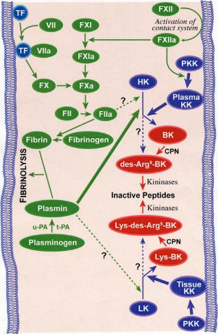
Classically, there are two main pathways by which kinins are generated (Fig. 1). The plasma kallikreinkinin system, by far the more complex, initiates activation of the intrinsic coagulation pathway. The second and simpler pathway of kinin generation involves tissue kallikrein and its substrate, LK. Each of these enzyme systems may play different pathophysiological functional roles.
2.1 The plasma kinin forming system
The plasma kinin forming system, also called the
Fig. 1. The kinin-forming systems. The kallikrein-kinin system and its interactions with both intrinsic and extrinsic coagulation cascades and fibrinolysis. Solid lines are established pathways, whereas dashed lines are speculative or experimental activation pathways. TF: tissue factor; PKK: prekallikrein; HK: high-molecular-weight kininogen; LK: low-molecular-weight kininogen; BK: bradykinin; CPN: carboxypeptidase; t-PA: tissue plasminogen activator; u-PA: urokinase plasminogen activator.
contact system of plasma, consists of 3 serine proenzymes (factor XII or Hageman factor, factor XI, and prekallikrein) and the kinin precursor HK.
2.1.1 Plasma prekallikrein
Chromosomal localization of the human plasma kallikrein gene was mapped to the q34 – q35 region of the long arm of chromosome 4 (15). Plasma kallikrein (EC 3.4.21.34), a serine protease, is encoded by a single gene, KLKB1, and synthesised in the liver. It is predominantly secreted by hepatocytes as an inactive molecule called prekallikrein that circulates in plasma as a heterodimer complex bound to HK with 1:1 molar stoichiometry (16, 17). Prekallikrein is a single chain α-globulin that is present in the plasma of humans and of other animal species at a concentration of 35 – 50 µg/mL. About 80 – 90% of prekallikrein is normally complexed to HK (16, 18).
2.1.2 Contact system activation of plasma
Contact of plasma with a negatively charged surface leads to the binding and autoactivation of factor XII (Hageman factor) to factor XIIa, activation of prekallikrein to kallikrein by factor XIIa, and cleavage of HK by kallikrein to release BK (19). Factor XII activation is not only a first step in the initiation of the intrinsic clotting cascade and the generation of kinins, but it also leads to the activation of the complement pathway (20).
In vitro, non-physiologic substances, such as glass (negatively charged silicates), carrageenan, kaolin, and a sulfated polysaccharide dextran sulfate (21) activate the contact system of plasma. In vivo, the physiologic surface remains unknown. Pathologic initiators may include proteoglycans (sulfate residues on heparin sulfate or chondroitin sulfate or mast-cell heparin). Endotoxins (lipopolysaccharide (LPS)) and crystals of uric acid or pyrophosphate (22) have also been hypothesized to be pathological activators. This intrinsic coagulation/kinin-forming cascade appears to be in equilibrium in plasma even in the absence of any exogenous surface. That is, activation occurs continuously at a finite rate, but is held in check by plasma inhibitors (19).
Plasma kallikrein cleaves human HK in a two-step process. First, HK is cleaved at the Arg389-Ser390 bond of the carboxy-terminal portion of the BK sequence, leaving the BK attached to the carboxy-terminal end of the heavy chain, and the sequence Leu378-Met-Lys- Arg381 is cleaved at the Lys380-Arg381 bond to liberate BK from the heavy chain (23). As a result of domain rearrangement, HKa acquires new properties. Recent observations suggest that HKa inhibits endothelial cell
10 |
ME Moreau et al |
proliferation and neovascularisation due to antiapoptotic properties. This property contrasts with the angiogenic effect of HK and LK, due to the release of BK (24).
2.2 Endothelial cells and kinin forming activity
Another mechanism for initiation of the activation of the kallikrein-kinin system depends on binding of components of the contact activation cascade on the surface of cells as leukocytes, platelets, endothelial cells, and myocytes (25).
HK specifically binds to platelets, granulocytes, and endothelial cells in a zinc-dependent, saturable and reversible reaction (26, 27). The dissociation constant equals 15 nM, indicating high affinity binding (21). This binding involves both the heavy (domain 3) and light (domain 5) chains of HK (28), which could be considered as a receptor for prekallikrein on endothelial cells (29 – 31). Binding of HK to endothelial cells leads to activation of prekallikrein to kallikrein (11, 25, 31) and presumably a release of BK from HK (31, 32).
The interaction of HK with endothelial cell membranes involves a multiprotein receptor complex comprising at least cytokeratin 1, gC1qR, and the urokinase plasminogen activator receptor (u-PAR) (33 – 38). These three proteins co-localize on the endothelial cell membrane (39). The same three proteins form a receptor complex for factor XII, but binding of factor XII in vivo is likely limited both by the low plasma concentration of free Zn2+, which is below the requirement for factor XII binding, and by the much higher plasma concentration of HK (40).
2.3 Tissue kallikrein-kinin system
Tissue (glandular) kallikrein (EC 3.4.21.35) is an acid glycoprotein which differs from plasma kallikrein. This tissue serine protease is encoded by one of the kallikrein gene family, KLK1 gene, located on chromosome 19 locus q13.2 – q13.4 (41, 42). Tissue kallikrein is widely distributed (kidney, blood vessels, central nervous system (CNS), pancreas, gut, salivary and sweat glands, spleen, adrenal, and neutrophils) (17, 42), and this wide distribution suggests a paracrine function (43). The origin of tissue kallikrein detected in plasma has been suggested to be the exocrine glands. Tissue kallikrein is synthesized as a proenzyme, prokallikrein, which is inefficiently activated by plasmin or plasma kallikrein (44).
Tissue kallikrein releases KD from LK (42), cleaving the Met379-Lys380 and Arg389-Ser390 bonds (45). Although LK is considered to be the main substrate of tissue kallikrein, tissue kallikrein is also capable of cleaving HK.
2.4 Other kinin forming enzymes
In addition to tissue and plasma kallikrein, other serum and tissue proteases have a kinin forming capacity (46). Plasmin, which is responsible for lysis of the fibrin clot, releases not only BK but also des-Arg9-BK from HK (47), circulates in plasma as an inactive zymogen, plasminogen. The presence of plasminogen activators and their inhibitors is essential in controlling fibrinolysis (48).
The major activator of plasminogen in vivo is tissue plasminogen activator (t-PA), a serine protease synthesized and secreted by endothelial cells as a single chain active enzyme (46). In the absence of fibrin, t-PA is an inefficient activator of plasminogen but the binding of t-PA to fibrin greatly accelerates the activation of plasminogen. Urokinase or urinary type plasminogen activator (u-PA) can also activate plasminogen and plays an important role in the degradation of the extracellular matrix. This facilitates the migration of cells which is important in wound healing and in tumour invasion and metastasis. u-PA can be of primary importance for cell-mediated activation of plasminogen in tissues, whereas t-PA, possessing a high affinity for fibrin, can be of primary importance for the lysis of fibrin clots in the circulation (49).
Factor XIIa, XIa, and kallikrein are also capable of converting plasminogen to plasmin in vitro. The contribution of these enzymes to the activation of plasminogen in vivo is uncertain; in fact, deficiencies in these proteins do not appear to lead to pathological states that could be explained by an impaired fibrinolysis (48).
3.Regulation of the kininogenase activity
Protease inhibitors regulate the contact activation of
plasma. The serpins of plasma are namely C1-inhibitor (C1INH), antithrombin III, α2-macroglobulin, α1-pro- tease inhibitor, and α2-antiplasmin (19, 50). However, C1INH is the major regulator of the intrinsic system, interfering with the activities of factor XIIa and of kallikrein (51). Both C1INH and α2-macroglobulin account for more than 90% of the kallikrein inhibitory activity of plasma.
The regulatory mechanism of tissue kallikrein remains partly unknown. Kallistatin and aprotinin are an example of serine protease inhibitors. Unlike many other serpins that are present only in plasma, kallistatin can be detected in various tissues, cells, and fluids, where it regulates the activity of tissue kallikrein (52, 53).
Plasmin, t-PA, and u-PA are also regulated by inhibitors in the serpin family. The primary inhibitor of active plasmin is α2-antiplasmin. Similarly, antithrombin, α1-antitrypsin, and C1INH have been shown to inhibit plasmin in vitro but have a minimal physio-
The Kallikrein-Kinin System |
11 |
logical effect in blood. Inhibitors of t-PA play an important role in regulating fibrinolysis. Currently, four distinct types of plasminogen activator inhibitors (PAI) have been described: PAI-1, PAI-2, PAI-3, and protease nexin. Of these, PAI-1 is the most important in inhibiting t-PA in plasma. PAI-1 is present in molar excess over t-PA; hence the majority of t-PA circulates bound to PAI-1, thereby preventing its interaction with plasminogen and thus a premature lysis of fibrin and a systemic fibrinolysis (54).
IIMetabolism of Kinins
The nature and properties of the various peptidases capable of metabolizing kinins have been extensively reviewed (55). We have experimental evidences that 4 metallopeptidases are mainly responsible for the metabolism of BK. These are angiotensin I-converting enzyme (ACE), aminopeptidase P (APP), neutral endopeptidase 24.11 (NEP, neprilysin), and carboxypeptidases M and N (CPM, CPN). The importance of these peptidases depends on the animal species, the analytical approach, the biological milieu, and the pathophysiological context. Interestingly, these peptidases are zinc metallopeptidases, that is, they all require zinc in their catalytic site to hydrolyse substrates, and are membrane-bound glycoproteins, except CPN, which is a soluble tetrameric glycoprotein. However, they are all present in a soluble form in biologic fluids.
1. Angiotensin I-Converting Enzyme (ACE)
1.1 Definition
ACE (EC 3.4.15.1) is a well-characterized type I ectoenzyme membrane anchored Zn2+-dependent dipeptidyl carboxypeptidase that regulates bioactivities of vasoactive peptides such as angiotensin I (Ang I) and BK, responsible for the control of blood pressure (56).
Two distinct forms of ACE are expressed in humans, a larger one (150 – 180 kDa) usually referred to as somatic ACE that is composed of approximately 1300 aminoacids and is present in most tissues (vascular endothelial surface of the lungs and on brush-border membranes of kidney, intestine, placenta, and choroid plexus) and a smaller isoenzyme referred to as the germinal form, testicular ACE (100 – 110 kDa), with only 730 aminoacids and is found exclusively in the testicles and appears to be involved in male fertility (56, 57).
The somatic form has two homologous metalloproteinase domains (N- and C-terminal domains) with an overall 60% homology in both nucleotide and aminoacid sequence, each containing a canonical Zn2+- binding sequence motif: HEXXH (His-Glu-X-X-His)
and bearing a functional active site (58, 59). The stoichiometry value of 1:1 for the complete inhibition of the enzyme indicates that both active sites would work in a cooperative manner (60).
The testicular ACE has a single domain corresponding to the C-terminal domain of somatic ACE (61) together with the hydrophobic membrane-anchoring domain and a small N-terminal region that has multiple O-linked oligosaccharides (62).
Although ACE is primarily a membrane-bound protein, a soluble form does exist in many body fluids and is the result of post-translational proteolytic cleavage in the juxtamembrane stalk by a membrane protein secretase or sheddase, itself a zinc metallopeptidase (63, 64). Although the ACE secretase has not yet been identified, studies with a range of hydroxamic acid-based inhibitors have shown that it has a remarkably similar inhibition profile to the amyloid precursor protein α-secretase, leading to the conclusion that the two secretases are, at the very least, closely related (65). Human plasma ACE originates from endothelial cells, while in other body fluids ACE originates from epithelial, endothelial, or germinal cells (66).
A number of proteins with sequences related to ACE have been described, but the only mammalian relative to be found is ACE2. Similar to ACE, ACE2 is also a type I integral membrane glycoprotein found on the surface of endothelial and epithelial cells, although it has a more limited tissue (testis, heart, kidney) distribution than ACE. ACE2 consists of a single active site domain that, by sequence comparison, more closely resembles the N-domain than the C-domain of somatic ACE. However, ACE2 differs from ACE in that it acts as a carboxypeptidase removing single aminoacid residues from its substrates, which include angiotensin II (Ang II) with high catalytic efficiency (kcat/Km = 1.9 × 10⁶ M−1 s−1) (67). ACE2 efficiently hydrolyzes des-Arg9-BK (kcat/Km = 1.3 × 10⁵ M−1 s−1), but fails to hydrolyze BK itself (68). Also, ACE2 is not inhibited by some inhibitors of ACE. Since the discovery of ACE2, some authors have begun to refer to ACE as ACE1 (69, 70).
1.2 Synthesis, regulation, and localization
Somatic and germinal ACE are transcribed from the same gene (17q23) using alternative promoters (71). Cloning of the gene that encodes ACE revealed the relationship between the two isoforms. Besides their different tissue specificity, expression of the two ACE transcripts is regulated by different developmental and hormonal controls; for example, the endothelial enzyme is induced by glucocorticoids, whereas the testicular form is stimulated by androgens (71).
12 |
ME Moreau et al |
1.3 Properties
1.3.1 Angiotensinase vs kininase
ACE catalyzes the conversion of the inactive decapeptide Ang I to the potent vasopressor octapeptide Ang II by the removal of the C-terminal dipeptide His9- Leu10.
Ang I is originally considered the main physiological substrate for ACE (Km, approximately 16 µM). Because of its higher affinity (Km, approximately 0.18 µM) for BK, ACE could also be now considered as a kininase (kininase II) (72, 73). As a peptidyl dipeptidase, ACE inactives BK by hydrolyzing two separate bonds on its C-terminal end. It removes sequentially the dipeptide Phe8-Arg9 and next cleaves the Phe5-Ser6 bond to generate the second dipeptide Ser6-Pro7, transforming BK into its inactive final BK[1–5] product (74, 75). ACE also metabolizes des-Arg9-BK by removing the carboxyterminal tripeptide Ser6-Pro7-Phe8, yielding the same final pentapeptide.
1.3.2 ACE GPIase activity
Recently, Kondoh et al. reported that ACE has a GPIase activity which allows the release of a membrane glycosylphosphatidylinositol (GPI)-anchored protein. They have found that ACE-specific inhibitors, such as captopril and lisinopril, which bind to the catalytic center and completely inhibit the peptidase activity, had only a minor inhibitory effect on this GPIase activity which does not require the zinc ion (76). This new activity of ACE which is different from the GPIase activity of GPI-specific phospholipase D (GPI-PLD) (76, 77) and of phosphatidylinositol-specific phospholipase C (PI-PLC)-like could be of physiological importance in fertilization.
1.3.3 ACE: a signal transduction molecule
By its short cytoplasmic domain (29 aminoacids), ACE could also play a role as a signal transduction molecule. In fact, the cytoplasmic tail of ACE is phosphorylated in endothelial cells (78). ACE inhibitors as well as BK elicit outside-in signalling in these cells. They enhance the activity of ACE-associated protein kinase CK2, increasing its phosphorylation of ACE, and lead to the activation of c-Jun N-terminal kinase (JNK) as well as the accumulation of phosphorylated c-Jun in the nucleus (79).
1.3.4 ACE insertion/deletion polymorphism
Population studies indicated that the large interindividual variability in plasma ACE levels is genetically determined. Regulation, as well as tissue ACE activity are under strong genetic control. An insertion /deletion (I/D) polymorphism located in the noncoding
region of the gene is associated with differences in the level of ACE in plasma and cells. The insertion that gives rise to the I allele is an alu repeat sequence (287 bp) in intron 16 of the ACE gene; the D allele results from the absence of the above insertion. There is a relationship between the ACE I/D polymorphism and circulating ACE activity, such that ACE activity is highest in individuals homozygous for the D allele, lowest in those homozygous for the I allele and intermediate in heterozygotes.
Mean serum ACE level in DD homozygotes are nearly twice the value measured in II plasma, while DI heterozygous have intermediate activities (80 – 82). Although some association between the I/D polymorphism with the incidence of cardiovascular and Alzheimer diseases, the results remain inconsistent (83 – 87). However, ACE genotype determines BK degradation and suggests another mechanism whereby the ACE D allele could be associated with deleterious cardiovascular effects (82).
2. Neprilysin (NEP)
2.1 Definition
NEP (EC 3.4.24.11, neprilysin) is a type II surface protein with a short membrane-proximal stalk region that is not sensitive to proteolytic activity of any particular secretase (88). It is the prototype zinc peptidase of the M13 membrane metalloendopeptidase (MME) family. This family includes other peptidases of potential pathophysiological interest: ECE-1 and ECE-2, which generate the vasoconstrictor endothelins from big endothelin; the erythrocyte cell-surface antigen KELL; PHEX, associated with congenital X-linked hypophosphatemic ricket; ECEL1, present in CNS; SEP (MML1 or NL1) present in testis; and MMEL2 (89 – 91).
2.2 Synthesis, localization, and properties
Human NEP gene (MME) is localized to 3q21 – q27 (92). First identified in the brush border of the kidney epithelial cells, where it represents 4 – 5% of the protein content, its immunoreactivity or activity has also been detected in cells or tissues as different as the CNS, the endothelium, testis, lungs, salivary glands, and bone marrow (93). NEP preferentially cleaves bonds on the amino-side of hydrophobic aminoacids residues. However, the physiological role of NEP depends on its tissue localization and the presence of the substrate.
Like ACE, NEP inactivates BK by sequentially removing dipeptide Phe8-Arg9 and tripeptide Phe5-Ser6- Pro7 to yield BK[1–4], an inactive metabolite. However, a prolonged incubation also results in the hydrolysis of the Gly4-Phe5 bond (94). In contrast to ACE, it is likely that NEP cleaves the Gly4-Phe5 bond of des-Arg9-BK
The Kallikrein-Kinin System |
13 |
to generate the same BK[1–4]-inactive peptides. NEP is the main enzyme responsible for the metabolism of kinins in the kidney and plays an important role in the metabolism of BK at the endothelium. In plasma, unlike ACE, NEP does not play a significant role in the metabolism of kinins (95).
Besides kinins, NEP also inactivates other peptides like enkephalins, neurokinins, and amyloid-β peptide, a marker for Alzheimer disease of the CNS (96). In the kidney, it regulates the degradation of natriuretic peptides (97).
3. Aminopeptidase P (APP)
3.1 Definition
Human APP (X-prolyl aminopeptidase, EC 3.4.11.9) exists at least in two forms: a soluble cytosolic (cAPP) and membrane-bound (mAPP) APP forms. Both forms have an X-prolyl aminopeptidase activity (98). cAPP and mAPP share 43% of homology. cAPP is a homodimer of 70 kDa subunits and the mature form APP has a predicted subunit molecular weight of 71 kDa (99, 100). mAPP is a heavily N-glycosylated, Zn2+-contain- ing, peptidase and contains a C-terminally attached GPI membrane anchor that increases the overall subunit molecular weight to approximately 90 kDa. A secreted form of mAPP has recently been produced and characterized in vitro in a transfected HEK-293 cell line. This protein exhibits a molecular mass of 85 kDa (101).
The effects of divalent metal ions on the activity of mammalian APPs appear to differ between the cytosolic and membrane-bound form of the enzyme (102). Indeed, whereas mAPP uses Zn2+ at the active site, human cAPP contains Mn2+ and is activated in vitro by the presence of Mn2+, but not Zn2+, which is inhibitory (99, 103).
There is evidence for the existence of another form of APP in mammals. The third isoform is hypothetical and has been identified on the basis of sequence homology. The chromosomal location of this isoform is 22q13.31 – q13.33. This isoform is predicted to have a subunit molecular weight of 57 kDa and is therefore smaller than cAPP and mAPP (104).
3.2 Synthesis, regulation, and localization
The chromosomal location of the human cAPP gene (XPNPEPL) is 10q25.3 (105) and the XPNPEP2 gene of mAPP localizes to chromosome Xq25-26.1 (106).
mAPP is located on the external side of the plasma membrane of vascular endothelial cells and on brush border membranes of epithelial cells in the intestine and the renal proximal tubule (107 – 112).
3.3 Properties
cAPP exhibits broad substrate specificity and can
cleave X-Pro dipeptides as well as longer peptides of the form X-Pro-Y-, where X and Y are of the common aminoacids (113 – 115). Compared to cAPP, mAPP has much more restricted substrate specificity; it fails to hydrolyze X-Pro dipeptides and cleaves X-Pro-Y- peptides poorly when X is Pro or Gly or when Y is an aminoacid with a bulky side chain (116 – 118).
Kinins are the best substrates for mAPP. In fact, the secreted form of mAPP exhibits a Km of 75 ± 15 µM for BK and a Km of 56 ± 13 µM for des-Arg9-BK. In human plasma, APP transforms BK and des-Arg9-BK into the inactive peptide BK[2–9] and BK[2–8], respectively, and is the major inactivating pathway for des-Arg9-BK in plasma (55, 95, 119).
4. Carboxypeptidase N and M (CPN, CPM)
Currently known as kininase I, both CPN and CPM are zinc metallopeptidases that exhibit a 41% sequence identity (120).
CPN (kininase I, EC 3.4.17.3) is a tetrameric protein synthetized in the liver and secreted in blood, although CPM (membrane-bound kininase I) is a GPI peptidase anchored at the membrane of lung and kidney epithelial cells. They cleave a variety of peptides containing a carboxy-terminal Arg or Lys. CPN inactivates complement anaphylatoxins (C3a, C4a, and C5a) by cleaving their carboxy-terminal Arg residue (121). Although likely, definitive evidence for such a metabolic role does not exist for CPM. Both carboxypeptidases transform BK and KD into des-Arg9-BK and des-Arg10-KD, which are active metabolites via B1 receptor (B1R). This kininase I activity constitutes a minor metabolic pathway unless ACE is inhibited (55, 121).
5. Other peptidases
The properties of dipeptidyl peptidase IV (DPP IV) have been extensively reviewed (122). DPP IV (CD26; EC 3.4.14.5) is a 110 kDa plasma membrane glycoprotein ectopeptidase (119, 122). The most important physiological actions of DPP IV is on regulatory peptides in mammals. DPP IV degrades the inactive metabolite BK[2–9] generated by APP and leads to the final BK[4–9] product (122). Substance P is another example of a regulatory peptide for which DPP IV plays an additional role in the inactivation (123).
Aminopeptidase N (APN, CD13) is an ectoenzyme (EC 3.4.11.2) of the superfamily of zinc metalloproteases widely used as a marker of myelomonocytic cells in the diagnosis of hematopoietic, malignant disorders (88, 124). Because of its ectopeptidase activity, APN has also been implicated in the regulation of vasoactive peptides, neuropeptide hormones, and immunomodulating peptides such as interleukin-6 and -8 (IL-6
14 |
ME Moreau et al |
and IL-8, respectively) (125, 126). Its importance in the field of kinins is related to its ability to hydrolyze the N- terminal Lys residue in KD and des-Arg10-KD; this reaction is functionally significant for the latter peptide which is the optimal agonist of the B1R in humans and other species. Indeed, the reaction product des-Arg9-BK has a much lower affinity at the human, rabbit, porcine, and bovine B1R. APN mediates the major inactivation pathway for des-Arg10-KD in the human isolated umbilical artery (127) and rabbit aorta (F. Marceau and A. Adam, unpublished observations). APN blockade with amastatin potentiates the hypotensive effect of the B1R agonist des-Arg10-KD in the LPS-pretreated rabbit (128).
IIIKinin Receptors
Two types of G-protein-coupled receptors (GPCRs) mediate the cellular effects of kinins, the B1R and B2 receptor (B2R) (129, 130). The various pharmacological effects of the BK-related peptides, such as vasodilatation, increased vascular permeability, stimulation of sensorial and sympathetic nervous connections, and smooth muscle contraction, derive from the presence of these receptors on various cell types such as the vascular endothelium, primary sensory afferent neurons, vascular and nonvascular smooth muscle, epithelial cells, and perhaps some types of leukocytes. The two types of receptors were initially defined using pharmacological criteria, before their molecular characterization.
1. Pharmacological classification
The potency order of agonists and the affinity of antagonists and their properties are summarized in Table 1 for B1R and Table 2 for B2R.
1.1 Potency order of agonists
The B2R has a high affinity for “native” kinins (generated by either plasma or tissue kallikreins), BK, and KD in all mammalian species. None of the fragments of BK retain a significant affinity for the B2R. KD has also a significant activity for the human and rabbit B1R (e.g., in radioligand binding assays), but in complex bioassay systems, BK or KD effects at B1R are often mediated by their des-Arg metabolites generated in situ (131). Indeed, the B1R is specialized across species to respond to kinin metabolites generated by arginine carboxypeptidases (either des-Arg9-BK, Lys-des-Arg9-BK or des-Arg10-KD) (129, 130). Des- Arg9-BK is a highly selective agonist of the mammalian B1R, but of high affinity (nanomolar) only in rodents (the rat and mouse); the only natural kinin sequence with a subnanomolar affinity for the human, rabbit,
porcine, and bovine B1R is Lys-des-Arg9-BK, suggesting that the B1R works in concert with the tissue kallikrein that generates its parent native peptide, KD (130).
1.2 Affinity of antagonists
Antagonists for the B1R were discovered almost 10 years before the antagonists for the B2R; thus, the receptor nomenclature is justified by the fact that it was the first to be pharmacologically fully defined. The first series of compounds capable of antagonizing BK and des-Arg9-BK with specificity for B1R included the prototype [Leu8]des-Arg9-BK (132). However, this prototype exhibits fairly high partial agonist behavior in some species, especially in the rat and mouse (130). This has practical implications because one of the emerging therapeutic applications of B1R antagonists, analgesia, is commonly evaluated using models involving these species.
The first generation of B2R antagonists was based on [D-Phe7]-BK (133), but these early peptidic compounds showed an antagonist/partial agonist activity and a low potency. This problem was progressively solved with the second generation of B2R antagonists in which rigidity was added to the peptide backbone by introducing non-natural aminoacid residues; thus, the spatial orientation of the C-terminal region of the peptide molecule critical for antagonism was more precise. Optimal peptide B2R antagonists retain both Arg1 and Arg9 residues and B1R agonists or antagonists typically lack Arg9 (as in [Leu8]des-Arg9-BK), just like agonists; furthermore, the N-terminal Lys residue present in KD also confers high affinity to peptide B1R antagonists in human, rabbit, and other species (as in Lys-[Leu8]des- Arg9-BK).
HOE 140 (D-Arg-[Hyp3, Thi5, D-Tic7, Oic8]-BK, Icatibant) is a representative of the second generation of peptide B2R antagonists (134) and has been exploited in more than 700 research papers and several clinical studies. Significant species-related differences in potency and competitive behavior are typically seen with kinin receptor antagonists; thus HOE 140 is potent and competitive at the human B2R, but insurmontable and atypically induces a slow B2R endocytosis of the rabbit B2R (135). The selectivity of HOE 140 for the human B2R is fair, but not complete; the fragment des-Arg10- HOE 140 is predominantly a B1R antagonist (130). Second generation B1R antagonists include the N- terminal Lys residue or its analog Orn that affords high affinity for the human B1R (as in B-9858 = Lys- Lys-[Hyp3, Igl5, D-Igl7, Oic8]des-Arg9-BK or R-954 = Ac- Orn-[Oic2, α-MePhe5, D-βNal7, Ile8]des-Arg9-BK) (136). The presence of D-Arg instead of Lys, as in des-Arg10-

|
The Kallikrein-Kinin System |
|
15 |
Table 1. Pharmacological and clinical application of kinin B1-receptor ligands |
|
||
|
|
|
|
Ligands |
Application |
Studies |
References |
|
|
|
|
Agonists |
|
|
|
R-838 (Sar-[D-Phe8]des-Arg9-BK) |
Metabolically stable |
Rabbit |
128 |
|
High affinity and selectivity |
Rodent |
130, 281 |
|
Hypertension |
|
|
|
Stimulation of vasculature formation |
|
|
|
(following ischemia) |
|
|
Antagonists
[Leu8]des-Arg9-BK |
Pain |
|
Ischemic vascular disease |
Lys-[Leu8]des-Arg9-BK |
Optimal B1R antagonist |
Ac-Lys-[MeAla6, Leu8]des-Arg9-BK |
Metabolically stable (not very potent |
|
compared with the affinity of the |
|
reference compound Lys-[Leu8]des-Arg9-BK) |
R-715 (Ac-Lys-[βD-Nal7, Ile8]des-Arg9-BK) |
High affinity |
|
Allergic lung inflammation |
B9858 (Lys-Lys-[Hyp3, Igl5, D-Igl7, |
Fairly high selectivity for B1R due to Lys0 |
Oic8]des-Arg9-BK) |
Metabolically stable residue |
|
|
des-Arg10-HOE 140 |
Residual antagonistic effects on B2R |
|
Moderate affinity |
B9430 (D-Arg-[Hyp3, Igl5, D-Igl7, Oic8]-BK) |
Mixed B1R and B2R antagonist even if |
|
desArg9 fragment has substantial |
|
selectivity for B1R |
R-954 (Ac-Orn-[Oic2, α-MePhe5, D-βNal7, |
Allergic lung inflammation |
Ile8]des-Arg9-BK) |
Airway allergy |
|
|
PS020990 |
Potent and competitive B1R antagonist |
|
High affinity |
Compound 12 (benzodiazepine-based |
Selective antagonist |
structure) |
|
Benzo-sulfonylamide componds |
Powerful and selective antagonists |
Compound 12 |
Hyperalgesia |
Compound 11 |
Speculative on pain, inflammation and sepsis |
SSR240612 |
Inflammation and hyperalgesia |
Rat |
132 |
Mice |
302, 317 |
Mice |
130, 281 |
Human B1R |
130 |
Rabbit |
318 |
Human and rabbit B1R |
130 |
Mice |
319 |
Human and rabbit B1R |
320 |
Rabbit jugular vein, guinea pig ileum, |
321 |
rabbit aorta |
|
Demonstration of compatibility of B1R |
322 |
and B2R structure by the accommodation |
|
of a single antagonist pharmacophore |
|
Mice |
319, 323 |
Rat model, speculative on human |
324 |
Human receptor (no in vivo data) |
139 |
Human and rat B1R in vitro (equal activity) |
140 |
Rat, dog, orally bioavailable |
142 |
Rabbit aortic preparations, |
141 |
rabbit jugular vein |
|
Mice, rat, oral activity |
143 |
HOE 140 and D-Arg-[Hyp3, Igl5, D-Igl7, Oic8]-BK, is associated with a strong decrease in affinity in human and rabbit, but not in the mouse B1R, corroborating the previous interpretation.
Efforts to create a third generation of kinin receptor antagonists have evolved towards conventional nonpeptide drug development programs, with oral bioavailability, higher lipophilicity, and lower molecular weight increasingly represented. Various chemical subclasses of nonpeptide B2R antagonists have been discovered: phosphonium compounds, as in WIN64338, WIN62318 (136 – 138); quinoline and imidazol [1,2-a]
pyridine family, such as FR165649, FR167344, FR173657, and FR184280; compound 38; substituted 1,4-dihydropyridines; CP2522; and CP0597 (138). Several nonpeptide B1R antagonists have also been synthesized: PS020990 (139), benzodiazepine-based compounds, such as compound 11 and 12 (140); powerful oxo-sulfonyl agents (141 – 143).
Some kinin antagonists have also been discovered as natural compounds: martinelline, a pyrroloquinoline alkaloid isolated from the plant Martinella iquitosensis, is the most remarkable example (144).