

Учебники / Textbook and Color Atlas of Salivary Gland Pathology - DIAGNOSIS AND MANAGEMENT Carlson 2008
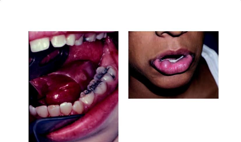
.pdfFigure 3.11a. 9-year-old girl with a left parotid swelling with overlying erythema of skin but no signs of acute infection. Reprinted from: J. Cranio-Max.-Fac. Surg volume 16, Mitchell DA, Ord RA, Atypical mycobacterial infection presenting as a parotid mass in a child, 221–223, 1988, Georg Thieme Verlag Stuttgart, New York.
Figure 3.11c. Two months following the parotidectomy, a left submandibular lymph node became enlarged and was treated with medical therapy. Reprinted from: J. Cranio- Max.-Fac. Surg volume 16, Mitchell DA, Ord RA, Atypical mycobacterial infection presenting as a parotid mass in a child, 221–223, 1988, Georg Thieme Verlag Stuttgart, New York.
Figure 3.11b. The patient underwent left superficial parotidectomy and excision of a submandibular lymph node. Histopathology showed non-caseating granulomas, and cultures showed mycobacterium avium intracellulare. Reprinted from: J. Cranio-Max.-Fac. Surg volume 16, Mitchell DA, Ord RA, Atypical mycobactevial infection presenting as parvotid mass in a child, 221–223, 1988, Georg Thieme Verlag Stuttgart, New York.
86

Viral Salivary Gland Infections
MUMPS
Viral mumps is an acute, nonsuppurative communicable disease that often occurs in epidemics during the spring and winter months. The term “mumps” is derived from the Danish “mompen,” which refers to mumbling, thereby describing the difficulty with speech because of inflammation and trismus (Arrieta and McCaffrey 2005; McQuone 1999). The nearly routine administration of the measles-mumps-rubella (MMR) vaccination has decreased the incidence of mumps in industrialized nations. Since the introduction of the live attenuated vaccine in the United States in 1967 and its administration as part of the MMR vaccine, the yearly incidence of mumps cases has declined from 76 to 2 cases per 100,000 (Murray, Kobayashi, and Pfaller 1994). Mumps characteristically occurs in the parotid glands. Although the disease is typically seen in children between 6 and 8 years of age, it may occur in adults who have avoided childhood infection as well, and displays an equal sex predilection. The disease is caused most commonly by a paramyxovirus, a ribonucleic acid virus related to the influenza and parainfluenza virus groups. Several other nonparamyxoviruses may cause mumps, including coxsackie A and B viruses, Epstein-Barr virus, influenza and parainfluenza viruses, enteric cytopathic human orphan (ECHO) virus, and human immunodeficiency virus (HIV). Mumps is transmitted by infected saliva and urine. The incubation period between exposure and the development of signs and symptoms is 15–18 days. A prodromal period occurs that lasts 24–48 hours and involves fever, chills, headache, and preauricular tenderness. Following the prodromal period, rapid and painful unilateral or bilateral swelling of the parotid glands occurs. Features that distinguish sialadenitis due to mumps vs. bacteria include a lack of purulent discharge, positive serum titers for mumps, and a relative lymphocytosis in mumps. The diagnosis is made by demonstrating complement-fixing soluble (S) antibodies to the nucleoprotein core of the virus, which are the earliest antibodies to appear. These antibodies peak at 10 days to 2 weeks and disappear within 8–9 months. The S antibodies are therefore associated with active infection. The complement-fixing viral (V) antibodies are against outer surface hemagglutinin and appear later than S antibodies but
Infections of the Salivary Glands |
87 |
persist at low levels for years. The diagnosis may also be made by isolating the virus from urine, which is possible up to 6 days prior and 13 days after the salivary gland symptoms occur (Rice 1998). Serum amylase levels may be elevated regardless of an associated pancreatitis. Abdominal pain is often indicative of mumps pancreatitis. Mumps orchitis occurs in 20% of adult males who have mumps parotitis (Goldberg and Bevilacqua 1995). Approximately half of these males will experience secondary testicular atrophy that may result in sterility if the testicular atrophy occurs bilaterally. Other rare complications of mumps include mumps hepatitis, mumps myocarditis, and mumps thyroiditis.
Treatment of mumps is supportive as spontaneous resolution of the disease occurs within 5– 10 days. Such supportive care includes bedrest, proper hydration, and dietary modifications to minimize glandular activity. Persistent or recurrent parotid swelling may indicate the presence of sialadenitis.
HUMAN IMMUNODEFICIENCY VIRUS
HIV infection is associated with numerous pathologic processes involving the salivary glands, with the parotid gland being the most common. “HIVassociated salivary gland disease” (HIV-SGD) is a term used to describe the diffuse enlargement of the salivary glands. HIV-SGD may affect patients throughout all stages of the infection, and may be the initial manifestation of HIV infection (Schiodt, Dodd, and Greenspan et al. 1992).
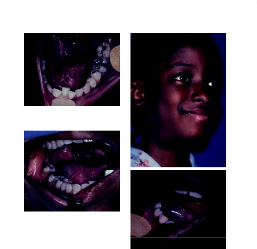
Patients with HIV-SGD present with a history of nontender swelling of one or more of the salivary glands (Figure 3.12). These swellings may fluctuate but are generally persistent. Imaging studies are generally beneficial so as to diagnose lymphoepithelial cysts in this patient population that may clinically resemble the nontender swellings of the parotid glands in this population. Decreased salivary gland function results in xerostomia and sicca symptoms. This sicca symptom complex mimics Sjogren’s syndrome and has resulted in the classification of another HIV-related salivary gland process known as the diffuse infiltrative lymphocytosis syndrome (DILS). This pathologic process is characterized by the presence of persistent circulating CD8 lymphocytes and infiltration of organs by CD8 lymphocytes that occur predominantly in the salivary glands and

88 Infections of the Salivary Glands
Figure 3.12. A 6-year-old African female with AIDS showing involvement of the right parotid gland by diffuse infi ltrative lymphocytosis syndrome (DILS).
lungs. While DILS appears similar to Sjogren’s syndrome, it can be differentiated by the presence of extraglandular involvement of the lungs, kidneys, and gastrointestinal tract. In addition, Sjogren’s autoantibodies will be absent in patients with DILS.
Medical management of HIV-SGD involves the use of antiretrovirals, observing meticulous oral hygiene, and the use of sialogogues. Corticosteroids may also be of use.
Collagen Sialadenitis
All of the collagen vascular diseases may affect the salivary glands, including polymyositis, dermatomyositis, and scleroderma; however, systemic lupus erythematosus is most commonly responsible. This disease is most frequently seen in fourthand fifth-decade women. Any of the salivary glands may become involved, and a slowly enlarging gland is the presentation. The diagnosis is made by identification of the underlying systemic disorder, and salivary chemistry levels will reveal sodium and chloride ion levels that are elevated two to three times normal levels (Miloro and Goldberg 2002). The treatment of collagen sialadenitis involves treatment of the responsible systemic disease.
Summary
•Sialadenitis is an infection of salivary glands that has numerous etiologies including microorganisms and autoimmune diseases.
•Staphylococcal and streptococcal species are involved in community acquired acute bacterial parotitis, and Pseudomonas, Klebsiella, Prevotella, Fusobacterium, Hemophilus, and Proteus species are cultured from hospital acquired cases of acute bacterial parotitis. Methicillin resistant Staphylococcal aureus may be cultured from cases of community acquired and hospital acquired acute bacterial parotitis.
•The clinician must rule out a neoplastic process in a prompt fashion during the course of treating the sialadenitis.
•The presence of a sialolith must be considered in the initial workup of patients with a clinical diagnosis of sialadenitis. A screening panoramic radiograph or occlusal radiograph should be obtained.
•The parotid and submandibular glands are the most commonly affected salivary glands by sialadenitis.
•The purpose of initial treatment for sialadenitis is to provide medical therapy for the disorder, with surgical therapy being introduced if the disorder becomes refractory to medical treatment.
•Minimally invasive strategies have a role to play in the surgical treatment of sialadenitis, as well as surgical removal of the salivary gland.

References
Andrews JC, Abemayor E, Alessi DM et al. 1989. Parotitis and facial nerve dysfunction. Arch Otolaryngol Head Neck Surg 115:240–242.
Arrieta AJ, McCaffrey TV. 2005. Inflammatory disorders of the salivary glands. In: Cummings CW (ed.), Cummings Otolaryngology: Head and Neck Surgery (4th ed.). Philadelphia: Elsevier Mosby, pp. 1323–1338.
Baurmash HD. 2004. Chronic recurrent parotitis: A closer look at its origin, diagnosis, and management. J Oral Maxillofac Surg 62:1010–1018.
Brodie, BC. 1834. Inflammation of the parotid gland and salivary fistulae. Lancet 1:450–452.
English CK, Wear DJ, Margileth AM et al. 1988. Cat-scratch disease. JAMA 259:1347–1352.
Galili D, Marmary Y. 1985. Spontaneous regeneration of the parotid salivary gland following juvenile recurrent parotitis. Oral Surg 60:605–606.
Goldberg M, Harrigan W. 1965. Acute suppurative parotitis. Oral Surg 20:281–286.
Goldberg MH, Bevilacqua RG. 1995. Infections of the salivary glands. In: Carlson, ER (ed.), The Comprehensive Management of Salivary Gland Pathology. Philadelphia: W.B. Saunders, pp. 423–430.
Guralnick W, Donoff R, Galdabini J. 1968. Parotid swelling in a dehydrated patient. J Oral Surg 26:669–675.
Hasson O. 2007. Sialoendoscopy and sialography: Strategies for assessment and treatment of salivary gland obstructions. J Oral Maxillofac Surg 65:300–304.
Katz J, Fisher D, Levine S. 1990. Bacterial colonization of the parotid duct in xerostomia. Int J Oral Maxillofac Surg 19:7–9.
Infections of the Salivary Glands |
89 |
McQuone SJ. 1999. Acute viral and bacterial infections of the salivary glands. Otolaryngol Clin North Am
32:793–811.
Miloro M, Goldberg MH. 2002. Salivary gland infections. In: Topazian RG, Goldberg MH, Hupp JR (eds.), Oral and Maxillofacial Infections (4th ed.). Philadelphia: W.B. Saunders, pp. 279–293.
Mitchell DA, Ord RA. 1988. Atypical mycobacterial infection presenting as a parotid mass in a child. J. Cranio- Max.-Fac. Surg. 16:221–223.
Murray PR, Kobayashi GS, Pfaller KS. 1994. Paramyxoviruses. In: Medical Microbiology (2nd ed.). St. Louis: Mosby, pp. 629–640.
Nahlieli O, Nakar LH, Nazarian Y, Turner MD. 2006. Sialoendoscopy: A new approach to salivary gland obstructive pathology. JADA 137:1394–1400.
Patey DH. 1965. Inflammation of the salivary glands with particular reference to chronic and recurrent parotitis.
Ann R Col Surg Engl 36:26–44.
Petersdorf R, Forsyth B, Bernanke D. 1958. Staphylococcal parotitis. N Engl J Med 259:1250–1254.
Qi S, Liu X, Wang S. 2005. Sialoendoscopic and irrigation findings in chronic obstructive parotitis. Laryngoscope 115:541–545.
Rice DH. 1998. Diseases of the salivary glands—nonneo- plastic. In: Bailey, BJ (ed.), Head and Neck Surgery— Otolaryngology (2nd ed.). Philadelphia: Lippincott Raven Publishers, pp. 561–570.
Robinson JR. 1955. Surgical parotitis, vanishing disease. Surgery 38:703–707.
Schiodt M, Dodd C, Greenspan D, et al. 1992. Natural history of HIV-associated salivary gland disease. Oral Surg Oral Med Oral Pathol 74:326–331.


Chapter 4
Cysts of the Salivary Glands
Outline
Introduction
Mucous Escape Reaction
Clinical Features and Treatment of the Mucous Escape Reaction
Mucocele Ranula
Cyst of Blandin and Nuhn’s Gland Mucous Retention Cyst
Parotid Cysts Associated with Human Immunodeficiency Virus Infection
Branchial Cleft Cysts Summary References
Introduction
Cysts of the salivary glands may originate as benign non-neoplastic entities or in association with benign and malignant tumors of the salivary glands. Cystic development as part of specific neoplasms of the salivary glands is well recognized, including those that occur in the pleomorphic adenoma, Warthin’s tumor, mucoepidermoid carcinoma, acinic cell carcinoma, and the adenoid cystic carcinoma. The histologic features of these neoplasms are sufficiently distinctive; however, non-neoplastic salivary gland cysts do require differentiation from cystadenoma, mucoepidermoid carcinoma, and acinic carcinoma (Dardick 1996). Many cysts of the salivary glands may be generically attributed to an obstructive process. They can occur as a result of traumatic severance of salivary gland ducts, partial or complete blockage of the excretory ducts, or stasis of salivary flow in ducts. For the purpose of this chapter, salivary cysts are categorized in many ways, including those that originate directly from the salivary gland and those
entities that are associated with the salivary glands. In addition, there are those salivary cysts that exhibit a true cystic epithelium and those that are lined with a non-epithelial lining, that is, pseudocysts. Finally, it is possible to categorize these lesions as acquired (obstructive due to stricture, neoplasms, sialoliths, or trauma) and developmental (dermoid, branchial cleft, branchial pouch, and ductal). It is the purpose of this chapter to discuss those salivary gland cysts that are developmental and acquired in a non-neoplastic nature (see Table 4.1).
Mucous Escape Reaction
The mucous escape reaction can be defined as a pooling of salivary mucus within a connective tissue lining. This concept is defined by a number of names including mucocele, ranula, mucous retention phenomenon, and mucous retention cyst. Of these, mucocele and ranula are the two bestknown entities to clinicians diagnosing and managing pathology in the head and neck region. It was once believed that the lesion developed as a result of obstruction of a salivary gland’s excretory duct with the subsequent formation of an epithelially lined cyst (Thoma 1950). Early studies investigated the result of ligation of the excretory ducts of the submandibular and sublingual glands (Bhaskar, Bolden, and Weinmann 1956a). A mucous escape reaction did not result, thereby leading to further investigation. The complete obstruction of a salivary duct by the presence of a sialolith without the development of a mucous escape reaction substantiates the lack of a cause and effect relationship. Subsequent studies determined that severance of the excretory duct was required to produce extravasation of salivary mucin into the surrounding tissues with the development
91

92 Cysts of the Salivary Glands
Table 4.1. Cystic lesions of the salivary glands—nomen- clature and classification.
Nomenclature
Mucous escape reaction
Mucous retention cysts
Lymphoepithelial cysts
HIV-associated lymphoepithelial cysts
Developmental cysts
Branchial cleft cysts
Dermoid cysts
Polycystic (dysgenetic) disease
Classifi cation
I.Etiology
a.Origination from salivary gland tissue
i.Mucous escape reaction
ii.Mucous retention cyst
b.Association with salivary gland tissue II. Lining
a.True cystic lining
i.Mucous retention cyst
b.Non-epithelial lining (pseudocyst)
i.Mucous escape reaction III. Occurrence
a.Acquired
i.Mucous escape reaction
ii. Mucous retention cyst b. Developmental
of a lesion histologically identical to the mucous escape reaction observed in humans (Bhaskar, Bolden, and Weinmann 1956b). It is now accepted that severance of a salivary duct with resultant pooling of mucus into surrounding tissues is the pathophysiology of the mucous escape reaction. The fibrous connective tissue encasing the pooled saliva is presumably due to the foreign body nature of the saliva. The occasional report of an epitheliallike lining can be explained as a misinterpretation of compressed macrophages resembling a layer of cuboidal-shaped cells (van den Akker, Bays, and Becker 1978). When these lesions occur in the floor of the mouth, a designation of ranula is given, while a similar lesion in the lower lip carries a designation of mucocele.
CLINICAL FEATURES AND TREATMENT OF THE MUCOUS ESCAPE REACTION
The mucous escape reaction may develop from a major or minor salivary gland but seems to be
Figure 4.1. The typical appearance of a ranula of the fl oor of the mouth. The characteristic raised nature of the lesion, as well as its blue hue, are appreciated.
more commonly observed in the minor glands. Armed Forces Institute of Pathology data of 2,339 cases indicates that the minor glands are the site of predilection of this lesion, with 2,273 (97.2%) of the 2,339 lesions occurring in these glands. The lip accounted for 1,502 of these lesions (64.2%), with the lower lip being the most common site (98.8% when the site was specified). This figure is consistent with other series that indicate a predilection of lower lip lesions (Cataldo and Mosadomi 1970). The major glands showed a nearly equal distribution of occurrence in the parotid, submandibular, and sublingual glands, and collectively accounted for only 2.9% of the 2,339 cases.
Most investigators consider these lesions to be most common in children and young adults, with a mean age of 25 years. No significant sex predilection has been offered. The clinical appearance of these lesions differs depending on their depth within surrounding soft tissues. Superficial lesions present as blue, raised soft tissue swellings with a fluctuant character on palpation (Figure 4.1). The blue hue is generally reflective of the color of pooled saliva at the mucosal surface. Lesions that are located more deeply in the soft tissues take on the color of the surrounding soft tissues; however, they may retain their fluctuant character. The most common clinical course of mucous escape reactions is that of a painless mucosal swelling that develops during a period of between a few days to 1 week and ruptures with
Cysts of the Salivary Glands |
93 |
Figure 4.2. This ranula has resulted in significant pain experienced by the patient. The size of the ranula has resulted in obstruction of the sublingual gland.
apparent resolution with subsequent recurrence occurring within 1 month later. Mild symptoms of pain may accompany mucous escape reactions if secondary trauma or inflammation occurs. Pain may also occur in the rare event that the mucous escape reaction impedes the flow of saliva due to obstruction (Figure 4.2).
Mucocele
Mucoceles are common lesions of the oral mucosa, and perhaps the most common benign salivary gland lesion in the oral cavity. The incidence of mucoceles is understandable due to the prevalence of minor salivary gland tissue in the oral cavity and the frequent occurrence of trauma to these tissues, which results in their formation. These lesions are painless, freely movable, smooth, and fluctuant. Their appearance is so characteristic that the clinical diagnosis is most frequently confirmed by subsequent histopathologic diagnosis following removal (Figure 4.3). As such, an incisional biopsy is not required for proper surgical treatment of
Figure 4.3. The classic appearance of a mucocele of the lower lip. Similar to a ranula of the floor of the mouth, it shows an elevated blue lesion.
the mucocele. Clearly, the most common location for these lesions is the lip, and specifically the lower lip. This notwithstanding, mucoceles occur on the buccal mucosa, tongue, and palate. Patients often give a history of the lesions spontaneously bursting with predictable recurrence. Mucoceles occur most commonly in children and young adults, probably due to the relatively high incidence of oral trauma in younger patients. Treatment with surgical excision of the mucocele and its associated minor salivary gland tissue is highly curable.
Ranula
The ranula represents the prototypical mucous escape reaction occurring in the floor of the mouth. Its nomenclature stems from its derivation from the Latin diminutive “rana,” or frog, which refers to its resemblance to the belly of a frog (Figure 4.4). The lesion has a characteristic appearance and history, commonly exhibiting a blue color and displaying periods of bursting of the lesion with liberation of saliva, only to relapse some time thereafter (Figure 4.5). The development of a cervical component of the ranula has been a subject of fascination for centuries (Catone 1995). The oral and cervical mucous escape reactions may exist simultaneously (Figure 4.6), or they may occur independently of one another. As such, it was once considered possible that they had different etiologies, with the oral lesion being derived from the sublingual gland and the cervical lesion being derived from the submandibular gland. Some
94 Cysts of the Salivary Glands
Figure 4.4. A ranula of the left floor of the mouth. While the lesion is clearly elevated, only subtle signs of its blue color exist.
a
Figure 4.5. A ranula of the right floor of the mouth. Classic signs of elevation and the blue discoloration are present.
observed that the neck mass was often preceded by repeated spontaneous evacuations or surgical drainages of the oral lesion. This was perhaps the
first explanation that scar tissue formation in the b mucosa of the floor of the mouth was responsible
for the development of the cervical mass, as it descended through the cleft of the posterior extent of the mylohyoid muscle as a path of least resistance (Figure 4.7) (Braun and Sotereanos 1982). It has also been demonstrated that approximately one-third of the population has discontinuities of the mylohyoid muscle such that direct invasion of
Figures 4.6a and 4.6b. An 8-year-old girl with obvious right submandibular swelling (a), as well as simultaneous clinical evidence of a ranula in the right floor of the mouth (b).

Cysts of the Salivary Glands |
95 |
the pseudocyst through these defects of the muscle permits extension into the neck (McClatchey et al. 1984). Perhaps equally controversial is the concept of how it is most appropriate to treat the ranula.
If anything has been learned by reading the scientific literature on this topic, it is the common pathogenesis of three clinical entities: the mucocele, the oral ranula, and the plunging ranula. Specifically, it is their lack of an epithelial lining and their association with a salivary gland, whether major or minor. If the offending salivary gland is not removed, the lesion has a statistical likelihood of recurrence (Catone, Merrill, and Henny 1969). This notwithstanding, there are several published papers adamantly recommending that more conservative procedures be performed as first-line therapy (Baurmash 1992, 2007). One such proce-
Figure 4.7b. Computerized tomograms of the neck show a fluid-fi lled lesion of the submandibular region.
Figure 4.7a. This elderly woman shows left submandibular swelling. Her history includes numerous aspirations of fl uid within a ranula of the left floor of the mouth.
Figure 4.7c. A diagnosis of plunging ranula was made and the patient underwent left sublingual gland excision. Examination of the left floor of the mouth did not show signs of ranula in this region. Scar tissue formation from her previous aspirations resulted in the development of a plunging ranula.