

2 КУРС (БИОХИМИЯ) / Модель макета
.pdfPaper Model of Green Fluorescent Protein (GFP)
www.rcsb.org • info@rcsb.org GFP is a protein that can exhibit light. It is widely used in biotechnology.
Step1
Cut out the 12 strips of paper (outlined in dotted line) representing the 11 beta strands and 1 helical region of the GFP.
Step 3
Lay the strand labeled A on the table so that the arrow is pointing upwards, place strand B to the right of A and orient it so that the arrow points to the bottom. Align the grey areas between the strands (representing the hydrogen bonds between them), and tape top to bottom on the aligned grey area.
Step 5
Close the GFP beta barrel by aligning the grey area between strands A and K and taping them together. The chromophore should be inside the barrel.
align
&
tape
1
strand number
Step2
Tape together the strips so that the lowercase letter on the white background (e.g., a) is taped on top of the same letter on the
shaded background. Repeat this for letters b through k to create a long strip.
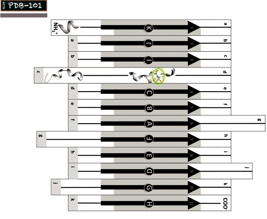
The Primary Structure
The long strip of paper from amino (NH3+) to carboxy (COO-) represents the primary structure of GFP. Regions of secondary structure (beta strands and alpha helices) are marked.
Step 4
Next tape strand C next to B, D next to C and so on till strand K, in each case aligning the grey areas between the strands and making sure the arrows in these strands point up and down
alternately as shown in the diagram on the right.
At the end of this step the beta sheet should have strands labeled A-K reading from
left to right. |
A |
At this point make sure the chromophore |
|
strip is behind the printed side |
|
of the beta sheet. |
|
alternate the arrows |
I J K |
|||||
|
||||||
|
|
|
|
F |
G H |
|
|
|
|
E |
|
align |
|
|
|
|
|
|
||
|
|
|
|
|
|
|
B |
C |
D |
0.8035 in |
|
|
|
|
|
|
|
|||
move the chromophore strip from the front (shown)
to the back of the printed side
Exploring the Model
1.Can you trace the polymer chain from the amino to carboxy terminus? Hint: the order of the strands from amino to carboxy terminal are marked with the numbers 1-11 (in the arrowheads of each strand).
2.What is the relationship between the strand labels A-K and 1-11? Comment also on the loops between the strands and location of the chromophore.
3.Identify one example each of parallel and antiparallel strands in the GFP barrel.
The GFP model was created using data from the PDB archive
PDB ID: 1EMA
M. Ormo, A. B. Cubitt, K. Kallio, L. A. Gross, R. Y. Tsien, S. J. Remington (1996)
Crystal structure of the Aequorea victoria green fluorescent protein.
Science 273: 1392-1395
&To learn more, read the
Molecule of the Month feature on (GFP) at dx.doi.org/10.2210/rcsb_pdb/mom_2003_6
Paper Model of Green Fluorescent Protein (GFP)
www.rcsb.org • info@rcsb.org
K
chromophore
www.rcsb.org