

МРТ
.pdf
|
Hundley et al |
Expert Consensus on Cardiovascular Magnetic Resonance |
2471 |
||
Table 2. Cardiovascular Magnetic Resonance–Derived Parameters in Patients With Suspected Heart Failure |
|
||||
|
|
|
|
|
|
|
Parameters |
|
Acronym |
Units |
Reference |
|
|
|
|
|
|
Systolic function |
LV and RV end-diastolic volumes and indices |
LVEDV(I), RVEDV(I), |
mL, mL/cmheight, mL/m2BSA |
16,79,81 |
|
|
LV and RV end-systolic volumes and indices |
LVESV(I), RVESV(I) |
mL, mL/cmheight, mL/m2BSA |
|
|
|
LV and RV stroke volume and index |
LVSV(I), RVSV(I) |
mL, mL/cmheight, mL/m2BSA |
|
|
|
LV and RV ejection fraction |
|
LVEF, RVEF |
% |
|
|
Cardiac output and cardiac index |
CO, CI |
mL/min, mL/min/m2BSA |
|
|
|
Regional and global systolic wall thickening |
|
% |
|
|
|
Regional or global measures of myocardial strain |
Ecc |
(%), (%)/s |
|
|
Morphology |
LV mass and indices |
|
LVM |
g, g/cmheight, g/m2BSA |
16,79,81 |
|
Mean and maximum myocardial wall thickness |
MWT |
mm |
|
|
|
Assessment of pericardium |
|
|
mm |
|
Wall stress |
End-systolic wall stress |
|
ESWS |
N/m2 1000 |
30 |
Diastolic function |
Circumferential strain and strain rate |
Ecc |
(%), (%)/s |
82 |
|
|
Peak untwisting rate |
|
|
°/s |
|
|
End-diastolic forward flow in pulmonary veins |
|
|
|
|
|
E/A ratio |
|
E/A, Ea |
|
83 |
Reversible acute injury |
Edema (regional or global high signal intensity in |
|
|
84 |
|
|
T2-weighted images) |
|
|
|
|
Irreversible injury, prognosis |
Myocardial fibrosis (late enhancement) |
|
% of LV mass or myocardial |
85 |
|
|
|
|
|
segment |
|
|
|
|
|
|
|
BSA indicates body surface area; E/A, early/atrial (late) ratio for ventricular filling; LV, left ventricular; N, Newton; and RV, right ventricular.
emic versus nonischemic disease) of LV or RV dysfunction, and identifying prognostic factors related to patient outcomes. Often, follow-up studies are required during or after therapeutic interventions. CMR offers more accurate assessment of function and morphology than most available imaging modalities, providing reliable volumetric data with high diagnostic image quality in nearly all patients. Table 2 displays quantitative and qualitative parameters, each of which can be used as diagnostic markers or descriptors in patients with suspected heart failure.
In general, cine SSFP sequences are used to visualize and quantify global left and right atrial and ventricular systolic function with reference data sets for normal subjects.16,81,86 Regional LV and RV systolic function can be assessed in great detail using myocardial tagging, with circumferential strain the most widely described parameter.82,87
Diastolic LV function has also been assessed with CMR. For this purpose, analogous echocardiographic parameters such as transmitral flow pattern or the presence of enddiastolic pulmonary vein forward flow can be utilized.83 In addition, CMR provides approaches for quantifying LV myocardial tissue velocity and strain/strain rates. Indicating its usefulness, strain analysis has been used for detecting regional abnormalities in patients with LV hypertrophy despite normal systolic function and lack of clinical evidence for heart disease.33
CMR may also provide important information regarding tissue abnormalities (see Section 2.7, Tissue Characterization). Focal fibrosis defined by LGE has provided novel insights into etiology and risk assessment of patients with LV dysfunction. Of great importance, the regional distribution of scarring allows an accurate discrimination of ischemic from nonischemic cardiomyopathies.88 In contrast to subendocardial involvement, patients with nonischemic etiologies of
heart failure either do not have detectable focal scars or have a nonsubendocardial distribution that is very distinct from ischemic subendocardial and transmural patterns. Even within the group of nonischemic cardiomyopathies, the regional distribution may help to identify the underlying etiology. In hypertrophic cardiomyopathy (HCM), the LGE is typically found in hypertrophied regions and in the interventricular septum close to the RV insertion areas. In dilated cardiomyopathy, an intramural layer of septal fibrosis has been described as a typical feature and is of strong prognostic value.85,89 Typical regional patterns of LGE in various etiologies have been reviewed elsewhere.90
In patients with acute heart failure, T2-weighted CMR may be useful to detect myocardial inflammation due to acute myocarditis.91 In cardiac iron overload, quantification of T2* relaxation times92 have proven useful for estimating intramyocardial iron content.
Abnormal high-energy phosphate metabolism has been studied by 31P-CMR spectroscopy in patients with dilated cardiomyopathy93 and HCM.94 31P-CMR spectroscopy, however, is limited by a strong signal from water-bound protons and difficulties in spectral interpretation due to the weak 31P signal. Due to these limitations, 31P-CMR spectroscopy does not yet have a clinical role in the management of heart failure.
3.1.1. Potential Advantages of CMR Relative to Other Imaging Modalities
CMR measurements of biventricular function and volumes are highly reproducibile, accurate, and can be acquired with a high temporal resolution, thereby allowing precise identification of the point in time in which end-systole and end-diastole occurs. High precision and avoidance of ionizing radiation allows CMR to be used in longitudinal serial evaluations of patients with heart failure and to assess response to medical

2472 |
Circulation |
June 8, 2010 |
intervention or to evaluate disease progression.26,95 Furthermore, CMR has unique approaches to visualize tissue pathology, such as fibrosis, and therefore provides important diagnostic information. Importantly, CMR is highly advantageous in patients that may have body habitus limitations with other imaging techniques (ie, acoustic window limitations or attenuation artifacts).
3.1.2. Summary of Existing Guidelines and Appropriate Use Criteria
The ACC/AHA 2005 Guideline Update for the Diagnosis and Management of Chronic Heart Failure in the Adult indicates that CMR may be useful in evaluating chamber size and ventricular mass, as well as assessing cardiac function and wall motion.96 CMR may also be used to identify myocardial viability and scar tissue in patients with heart failure. CMR of the heart or liver may be useful for confirming the presence of iron overload.96
The ACCF/ACR/SCCT/SCMR/ASNC/NASCI/SCAI/SIR 2006 Appropriateness Criteria for Cardiac Computed Tomography and Cardiac Magnetic Resonance Imaging lists CMR evaluation of LV function as an appropriate indication in heart failure patients or those with technically limited echocardiograms.97
3.2. Coronary Artery Disease
CMR may be useful for identifying coronary artery anomalies and aneurysms, and for determining coronary artery patency. In specialized centers, CMR may be utilized to identify patients with multivessel CAD without exposure to ionizing radiation or iodinated contrast medium.
Over the past decade, CMR has evolved into an important diagnostic modality for patients with suspected anomalous CAD and coronary artery aneurysms. In specialized academic centers of excellence, CMR has reached sufficient maturity for discrimination of patients with multivessel CAD. This may be especially helpful among patients presenting with a dilated cardiomyopathy in the absence of a clinical history of myocardial infarction.
Coronary CMR is more technically challenging than CMR of other vascular beds due to several unique issues including: the small caliber of the coronary arteries (3- to 6-mm diameter), the near constant motion of the coronary arteries (during both the respiratory and the cardiac cycles), the high level of tortuosity of the coronary arteries, and the surrounding signal from adjacent epicardial fat and myocardium.98 –105 To overcome these obstacles, CMR approaches employ 1) cardiac triggering (eg, vector electrocardiogram [ECG]) to suppress bulk cardiac motion; 2) respiratory motion suppression (eg, breath-hold, CMR navigators); 3) prepulses to enhance contrast-to-noise ratio of the coronary arterial blood (eg, fat saturation, T2 preparation); and 4) 3D acquisition that offers superior postprocessing capabilities. Bright blood (segmented k-space GRE and SSFP) are most commonly used without an exogenous contrast agent (eg, Gd diethylene triamine pentaacetic acid). A special consideration in this population is intracoronary stents (see Section 4, CMR Safety),
Table 3. CMR Identification of Anomalous Coronary Vessels
|
|
Correctly Classified |
|
Investigators |
n |
Anomalous Vessels |
|
|
|
|
|
McConnell et al.101 |
15 |
14 |
(93%) |
Post et al.102 |
19 |
19 |
(100%)* |
Vliegen et al.105 |
12 |
11 |
(92%)† |
Taylor et al.104 |
25 |
24 |
(96%) |
Bunce et al.98 |
26 |
26 |
(100%)‡ |
*Includes 3 patients originally misclassified by x-ray angiography. †Includes 5 patients unable to be classified by x-ray angiography. ‡Includes 11 patients unable to be classified by x-ray angiography.
which are generally CMR compatible but demonstrate a local signal void/image distortion that is dependent on both the stent material and the CMR sequence, thereby precluding direct evaluation of intrastent and peristent coronary integrity.
3.2.1.Anomalous Coronary Artery Identification
Although unusual (less than 1% of the general population106) and usually benign, congenital coronary anomalies in which the anomalous segment courses between the aorta and pulmonary artery are a well-recognized cause of myocardial ischemia and sudden cardiac death, especially among young adults.99 Catheter-based x-ray angiography has traditionally been the diagnostic imaging test to identify these anomalies, but the presence of an anomalous vessel is sometimes only suspected after the procedure, particularly in a situation where there was unsuccessful engagement or visualization of a coronary artery.
3.2.2.Potential Advantages of CMR Relative to
Other Imaging Modalities
CMR has several advantages for diagnosing coronary artery anomalies. CMR does not require ionizing radiation (likely to be an important consideration among adolescents and younger adults with suspected anomalous CAD) or iodinated contrast agents. Both 2D breath-hold and targeted 3D or whole-heart free-breathing navigator coronary CMR methods have been used with similar excellent results (Table 3), (Figures 2 and 3),98,100 –105 including several instances in which the 3D aspects of coronary CMR were of marked utility relative to 2D projection techniques (Table 3). The use of coronary CMR for suspected anomalous coronary disease is also very helpful when an intramural course is suspected or present.107
3.2.3. Coronary Artery Aneurysms
In the absence of a percutaneous intervention, the vast majority of acquired coronary aneurysms are due to mucocutaneous lymph node syndrome (Kawasaki’s disease). These aneurysms are the source of both shortand longterm morbidity and mortality.108 Coronary CMR (Figure 4) studies have confirmed the high accuracy of coronary CMR for both the identification and the characterization (diameter/ length) of these aneurysms.109 –111 Similar data have been reported for ectatic coronary arteries and fistulas.112

Hundley et al |
Expert Consensus on Cardiovascular Magnetic Resonance |
2473 |
Figure 2. Cardiovascular magnetic resonance of a coronary artery anomaly. An oblique axial reconstruction is presented from a “whole-heart coronary MRA” sequence. The white arrow notes the normally arising left main coronary artery from the left sinus of Valsalva. The black arrowhead highlights the right coronary artery arising anomalously from the anterior aspect of the left sinus of Valsalva superior to the left main origin and then coursing between the aortic root and the outflow tract of the right ventricle. MRA indicates magnetic resonance angiography.
3.2.4. Coronary CMR for Identification of Native Vessel Coronary Stenoses
Data regarding the clinical utility of coronary CMR for native vessel integrity are based on high-risk populations referred for x-ray angiography. No data are available regarding the use of coronary CMR for patients presenting with chest pain or for screening purposes of even high-risk patients. In addition,
Figure 3. Cardiovascular magnetic resonance of a single coronary artery. A 3-dimensional volume-rendered reconstruction from a “whole-heart coronary MRA” sequence in a patient with single ventricle and a single coronary artery. The white arrow denotes the proximal right coronary artery, whereas the black arrow highlights the elongated left main coronary artery arising from a common origin with the right coronary artery. MRA indicates magnetic resonance angiography.
Figure 4. Cardiovascular magnetic resonance of a proximal aneurysm. Transverse targeted 3-dimensional T2 prepulse coronary MRA of a subject with a proximal right coronary artery aneurysm. Ao indicates aorta; L, left coronary artery; and MRA, magnetic resonance angiography.
the majority of CMR data has been generated in a few highly specialized centers.
Using modern free-breathing, navigator-gated 3D-segmented GRE methods, good results have been shown, especially for the proximal coronary segments and in subjects with high image quality scans (Table 4).113–123 Focal disease is depicted as local signal attenuation. An international multicenter, free-breathing, 3D volume-targeted coronary CMR study of patients without prior x-ray angiography using common hardware and software demonstrated a very high sensitivity (100%) and modestly high specificity (85%) with very high negative-predictive value (100%) of coronary CMR for the identification of left main and multivessel CAD (greater than or equal to 50% diameter stenosis by quantitative coronary angiography) (Table 4).118 The results were not as useful for identifying single-vessel disease. Accordingly, coronary CMR is especially valuable for patients who present with a dilated cardiomyopathy in the absence of clinical infarction. Data suggest it is useful and can supplement LGE methods for determining the underlying etiology (ischemic versus nonischemic) of the cardiomyopathy.124
Increasing data are now available on whole-heart SSFP coronary CMR methods. Although the technique utilizes an inferior in-plane spatial resolution, data appear to be at least as accurate as free-breathing methods (Table 4).117,121,122 This type of data may be useful in heavily calcified lesions.107 Coronary MRA may also be useful for assessing heavily calcified arteries on computed tomography where blooming artifact may obscure the vessel lumen.125
3.2.5. Coronary CMR for Coronary Artery Bypass Graft Assessment
In comparison with the native coronary arteries, reverse saphenous vein and internal mammary artery grafts are relatively easy to image due to their minimal motion during the cardiac and respiratory cycles and the larger lumen of reverse saphenous vein grafts. With schematic knowledge of

2474 |
Circulation |
June 8, 2010 |
Table 4. Free-Breathing 3D Gradient Echo Coronary CMR Using Prospective Navigators for Identification of Focal >50% Diameter
Coronary Stenoses
|
|
|
For 50% Diameter Stenosis |
|
|
|
|
|
|
Investigators |
n |
Technique |
Sensitivity (%) |
Specificity (%) |
Prospective navigators with real-time correction-targeted 3D |
|
|
|
|
Bunce et al.98* |
34 |
|
88 |
72 |
Sommer et al.123† |
112 |
|
74 |
63 |
|
|
|
88 (good quality) |
91 (good quality) |
Bogaert et al.113 |
19 |
|
85–92 |
50–83 |
Dewey et al.115 |
15‡ |
SSFP |
86 |
98 |
Maintz et al.119 |
|
TFE |
92 |
67 |
|
|
SSFP |
81 |
82 |
Ozgun et al.120 |
20 |
SSFP |
82 |
82 |
Jahnke et al.116 |
21 |
SSFP |
79 |
91 |
Prospective navigators with real-time correction whole-heart SSFP |
|
|
|
|
Sakuma et al.121 |
101 |
|
82 |
91 |
Jahnke et al.117 |
55 |
|
78 |
91 |
Sakuma et al.122 |
106 |
|
82 |
90 |
Kim et al.118 |
109 |
|
93 (patient) |
59 (patient) |
|
|
|
100 (LM/3VD) |
85 (LM/3VD) |
|
|
|
|
|
3D indicates 3-dimensional; CMR, cardiovascular magnetic resonance; LM/3VD, left main coronary artery or 3-vessel disease; SSFP, steady-state free precession; and TFE, turbo fast-echo.
*Excludes 5 patients for “lack of cooperation” and 15 segments for being uninterpretable. †Based on 74% of coronary artery segments analyzable by CMR.
‡Based on 60% of patients with good free breathing CMR images.
the origin and touchdown site of each graft, a variety of CMR sequences have been used to identify graft patency.126 –131
Limitations of coronary CMR bypass graft assessment include difficulties related to local signal loss/artifact due to implanted metallic objects (hemostatic clips, ostial stainless steel graft markers, sternal wires, coexistent prosthetic valves and supporting struts or rings, and graft stents). Imaging strategies used to image coronary arteries have also been applied to saphenous vein grafts132 and reported to be quite accurate for assessment of saphenous vein graft stenoses, with very good agreement between quantitative x-ray angiography for assessment of both graft occlusion (sensitivity 83% [36% to 100%]; specificity 100% [92% to 100%]) and graft stenosis (greater than or equal to 50%; sensitivity 82% [57% to 96%]; specificity 88% [72% to 97%]).133 Saphenous vein and internal mammary artery bypass graft CMR can also be combined with rest and adenosine stress graft flow assessment using phase velocity CMR techniques133 and suggest superior results.
3.2.6. Potential Advantages of CMR Relative to Other Imaging Modalities
In addition to coronary artery anomalies, CMR is highly advantageous for identifying aneurysms or fistula without the use of contrast materials or exposing patients to ionizing radiation. These particular advantages are well suited for assessing both children and relatively young women that experience an increased risk of adverse events associated with exposure to ionizing radiation. At expert centers, early data suggest CMR may have a role in identifying coronary arterial stenoses in arterial bypass
grafts, as well as excluding the presence of left main or 3-vessel coronary arterial disease.
3.2.7. Summary of Existing Guidelines and Appropriate Use Criteria
The ACC/AHA 2002 Guideline Update for the Management of Patients With Chronic Stable Angina indicates that coronary CMR is a suitable method to identify anomalous origins of coronary arteries. It may be particularly useful in younger individuals with signs or symptoms of myocardial ischemia for the purpose of identifying coronary artery anomalies and in individuals with the presence of a continuous murmur for identifying an anomalous origin of the left anterior descending or circumflex artery from the pulmonary artery or coronary arterial venous fistulas.134
Similarly, the ACCF/ACR/SCCT/SCMR/ASNC/NASCI/ SCAI/SIR 2006 Appropriateness Criteria for Cardiac Computed Tomography and Cardiac Magnetic Resonance Imaging indicates that it is appropriate to use CMR to evaluate patients suspected of exhibiting coronary anomalies.97
3.3. Ischemic Heart Disease
The combination of CMR stress perfusion, function, and LGE allows the use of CMR as a primary form of testing for: 1) identifying patients with ischemic heart disease when there are resting ECG abnormalities or an inability to exercise; 2) defining patients with large vessel CAD and its distribution who are candidates for interventional procedures; or 3) determining patients who are appropriate candidates for interventional procedures. Assessment of LV wall motion after low-dose dobutamine in patients

Hundley et al |
Expert Consensus on Cardiovascular Magnetic Resonance |
2475 |
Figure 5. Myocardial perfusion imaging. First-pass contrast-enhanced perfusion images from a 73-year-old diabetic man using a hybrid gradient echo– echo planar pulse sequence with parallel imaging during infusion of 0.075 mM/kg of gadolinium chelate at 4 cc/s. The top panel of short-axis images was obtained during adenosine stress, a 4-minute infusion at 0.14 mg/kg, and the bottom panel obtained in the same short-axis slices 10 minutes later at rest. The base of the left ventricle on the left demonstrates an inferior wall perfusion abnormality seen at both stress and rest, consistent with myocardial infarction. The mid left ventricle demonstrates a large perfusion defect only at stress in the anterolateral and inferior walls. The apical left ventricle shows an inferolateral perfusion defect at stress but is normal at rest. cc indicates cubic centimeter; and mM, millimolar.
with resting akinetic LV wall segments is useful for identifying patients that will develop improvement in LV systolic function after coronary arterial revascularization. The writing committee recognizes the potential advantages of spectroscopic techniques for identifying early evidence of myocardial ischemia that may or may not be evident using existing non-CMR methods.
CMR is well suited to detect many of the physiologic consequences of ischemia through the assessment of myocardial abnormalities of perfusion, diastolic and systolic performance, and metabolism.
3.3.1. Myocardial Perfusion Imaging
CMR perfusion imaging is performed using a T1-weighted sequence to visualize first passage of a Gd-based contrast agent in transit through the heart. Following peripheral injection, the contrast is detected against the background of nulled (dark) myocardium with rapid enhancement during vasodilation stress. Signal intensity correlates with contrast concentration and analysis can be performed in a quantitative, semiquantitative, or qualitative fashion. Qualitatively, an experienced observer examines the myocardium for regions of low signal or hypoperfusion relative to normally perfused segments (Figure 5). Because the contrast agents rapidly redistribute into the extracellular space, quantitative analysis is limited to the initial upslope in the tissue intensity curve, which has been shown to correlate well with measures of microsphere blood flow.135
Validation of CMR perfusion in humans has been performed in a number of clinical studies employing a variety of contrast agents, analysis techniques, and reference standards136 (Table 5). One study examined signal-intensity time curves in both patients and controls following dipyridamole infusion and bolus injection of a Gd chelate.55 Using a linear fit to determine the upslope, a threshold value was defined to distinguish between normal and ischemic myocardium. Di-
agnostic accuracy was 87% with a high level of interobserver agreement. CMR perfusion, 13N-ammonia positron emission tomography, and quantitative coronary angiography were compared in a study using calculation of regional signal intensity upslopes.67 Analysis of the subendocardial upslope data showed a sensitivity and specificity of 91% and 94%, respectively, when compared to 13N-ammonia positron emission tomography and greater than 85% when compared to quantitative angiography. A study combining qualitative analysis of CMR perfusion images with LGE identification of myocardial infarction yielded a sensitivity of 89%, specificity of 87%, and overall accuracy of 88% compared to x-ray angiography.142 A meta-analysis of all CMR perfusion studies demonstrated a sensitivity of 91% and specificity of 81% for the diagnosis of CAD on a per-patient level.146 A multicenter study comparing CMR perfusion to SPECT suggests a higher specificity of CMR perfusion but similar overall accuracy.65 Clinically, it is important to note that to accomplish results associated with these multicenter results, appropriate physician and staff training is required, and a facility capable of performing the stress testing is required.
3.3.2. Stress Imaging of Ventricular Function
Dobutamine is commonly administered to evaluate stressfunction CMR with a qualitative evaluation of wall motion as the dose of dobutamine is increased, an application similar to DS echocardiography. CMR safety and efficacy have been assessed extensively. CMR exhibits major complications (ie, the development of sustained ventricular tachycardia) in less than 0.1% of subjects, findings that are similar to those observed with DS echocardiography.147
Studies have shown breath-hold GRE DS CMR to have a high accuracy for detecting ischemia, related in part to excellent LV endocardial visualization throughout dobut-

2476 |
Circulation |
June 8, 2010 |
Table 5. Sensitivity and Specificity of Recent CMR Perfusion Studies on a Per-Patient Basis for Detecting Coronary Arterial Luminal Narrowings >50%
Investigators |
n |
Stress Agent |
Sensitivity (%) |
Specificity (%) |
Cury et al.137 |
46 |
Dipyridamole |
97 |
75 |
Doyle et al.138 |
184 |
Dipyridamole |
57 |
78 |
Giang et al.139 |
44 |
Adenosine |
93 |
75 |
Ishida et al.140 |
104 |
Dipyridamole/isometric handgrip exercise |
90 |
85 |
Kawase et al.141 |
50 |
Nicorandil |
94 |
94 |
Klem et al.142 |
92 |
Adenosine |
89 |
87 |
Nagel et al.55 |
84 |
Adenosine |
88 |
90 |
Pilz et al.143 |
171 |
Adenosine |
96 |
83 |
Plein et al.144 |
68 |
Adenosine |
96 |
83 |
Plein et al.144 |
82 |
Adenosine |
88 |
74 |
Sakuma et al.66 |
40 |
Dipyridamole |
81 |
68 |
Schwitter et al.67 |
47 |
Dipyridamole |
86 |
70 |
Takase et al.145 |
102 |
Dipyridamole |
93 |
85 |
CMR indicates cardiovascular magnetic resonance.
Modified from Nandalur et al.146
amine/atropine stress protocols.148 DS CMR appears to be particularly valuable for patients who are poor candidates for DS echocardiography.149 A list of DS cine CMR studies is shown in Table 6. A meta-analysis of stress-functional CMR studies demonstrated a sensitivity of 83% and specificity of 86% for the demonstration of CAD on a per-patient level.146
CMR tagging may further improve the accuracy of DS CMR for detecting ischemia.161 In addition, in patients with resting LV wall motion abnormalities, low-dose dobutamine CMR is useful for identifying contractile reserve indicative of potential for recovering systolic thickening after coronary arterial revascularization.162 In summary, DS CMR is useful for identifying inducible myocardial ischemia and identifying
contractile reserve of LV wall motion after coronary artery revascularization.
3.3.3. Stress Perfusion and Functional Imaging for Prognosis Assessment
Prognostic data are now available using both vasodilator and DS CMR methods.52 Three-year event-free survival has been reported at 99.2% for patients with normal stress perfusion CMR or DS CMR and 83.5% for those with abnormal stress perfusion or DS CMR. Ischemia suggested by stress perfusion CMR or DS CMR is predictive of cardiac events over the 3-year time period with hazard ratios of 12.5 and 5.4, respectively, compared with those without evidence of myocardial ischemia. In summary, abnormalities observed during
Table 6. Sensitivity and Specificity of Recent CMR Wall Imaging Studies on a Per-Patient Basis in Detecting Coronary Arterial Luminal Narrowings >50%
Investigators |
n |
Stress Agent |
Sensitivity (%) |
Specificity (%) |
Baer et al.150 |
23 |
Dipyridamole |
78 |
NA |
Baer et al.151* |
32 |
Dobutamine |
84 |
NA |
Hundley et al.149 |
41 |
Dobutamine and atropine |
83 |
83 |
Jahnke et al.152 |
40 |
Dobutamine |
89 |
75 |
Nagel et al.148 |
172 |
Dobutamine |
86 |
86 |
Paetsch et al.153 |
79 |
Adenosine |
91 |
62 |
Paetsch et al.153 |
79 |
Dobutamine and atropine |
89 |
81 |
Paetsch et al.154 |
150 |
Dobutamine |
78 |
88 |
Pennell et al.155 |
40 |
Dipyridamole |
62 |
100 |
Pennell et al.156 |
25 |
Dobutamine |
91 |
100 |
Rerkpattanapipat et al.157 |
27 |
Exercise |
79 |
85 |
Schalla et al.158 |
22 |
Dobutamine |
81 |
83 |
van Rugge et al.159 |
45 |
Dobutamine |
81 |
100 |
van Rugge et al.160 |
39 |
Dobutamine |
91 |
0.83 |
CMR indicates cardiovascular magnetic resonance; and NA, not available.
*Utilized 2 perfusion territories (left anterior descending coronary artery and combined left circumflex artery/right coronary artery). Modified from Nandalur et al.146
Hundley et al |
Expert Consensus on Cardiovascular Magnetic Resonance |
2477 |
stress CMR serve as independent predictors of adverse cardiac events.
3.3.4. Magnetic Resonance Spectroscopy
Spectroscopy provides the CMR basis for the assessment of myocardial metabolism without the need for contrast agents or radionuclides.93,163,164 Hydrogen spectroscopy may be useful for assessing myocardial cellular triglyceride levels. Phosphorus spectroscopy has been used to measure myocardial energetics. In an early clinical application of Neubauer and his colleagues, 39 patients with dilated cardiomyopathy underwent 31P myocardial spectroscopy and were followed up at approximately 30 months.93 Kaplan-Meier analysis showed significantly reduced total and cardiovascular mortality for patients with greater than 1.6 versus patients with low or less than 1.6 PCr/ATP; a Cox model for multivariate analysis showed that the PCr-to-ATP ratio offered significant independent prognostic information on cardiovascular mortality. In patients with left anterior descending CAD, Weiss et al164 used spatially localized 31P magnetic-resonance spectra from the anterior myocardium before, during, and after isometric hand-grip stress. In patients with significant LAD or left main CAD (n 16), the ratio decreased from 1.45 0.31 at rest to 0.91 0.24 during stress (P 0.001) and recovered to 1.27 0.38 two minutes after exercise.
In a more recent study, handgrip stress was used in association with 31P spectroscopy in women with cardiac symptoms but without significant angiographic CAD. Of 35 women studied, 20% demonstrated an abnormal reduction in PCr/ATP with stress.163 In a follow-up study, the women with an abnormal PCr/ATP had a significantly greater incidence of recurrent symptoms and rehospitalizations compared with patients with a normal PCr/ATP response to exercise.165
3.3.5.Potential Advantages of CMR Relative to Other Imaging Modalities
CMR provides high spatial and temporal resolution images of myocardial perfusion, myocardial function, and identification of infarcts using LGE techniques. This unique combination offers the ability to reliably identify subendocardial ischemic processes. There is future promise of potentially incorporating spectroscopic techniques that may provide informative information regarding myocardial metabolism.
3.3.6.Summary of Existing Guidelines and Appropriate Use Criteria
The ACC/AHA 2002 Guideline Update for the Management of Patients With Chronic Stable Angina indicates that CMR may be used to assess LV performance, including ejection fraction.134 In the ACC/AHA Guidelines for the Management of Patients With ST-Elevation Myocardial Infarction, CMR is recommended for differentiating ST-segment elevation myocardial infarction from aortic dissection in patients for whom this distinction is initially unclear (Class I, Level of Evidence: B).166 Within the ACC/AHA/ESC 2006 Guidelines for Management of Patients With Ventricular Arrhythmias and the Prevention of Sudden Cardiac Death, CMR is probably indicated in patients with ventricular arrhythmias when echo-
cardiography does not provide accurate assessment of LV and RV function and/or evaluation of structural changes (Class IIa, Level of Evidence: B).167
The ACCF/ACR/SCCT/SCMR/ASNC/NASCI/SCAI/SIR 2006 Appropriateness Criteria for Cardiac Computed Tomography and Cardiac Magnetic Resonance Imaging indicates that the use of CMR stress testing (vasodilator or dobutamine) is appropriate in individuals with intermediate pretest probability of CAD or those with an uninterpretable ECG or those who are unable to exercise. CMR is also appropriate to determine viability prior to revascularization and establish the likelihood of recovery of systolic function with mechanical revascularization. CMR is appropriate to assess myocardial viability when determinations from other forms of noninvasive testing are equivocal or exhibit indeterminate results. The use of CMR stress testing is appropriate for identifying cardiac risk in patients with prior coronary angiography or stenoses of unclear significance.97
At present, ACCF/ACR/SCCT/SCMR/ASNC/NASCI/SCAI/ SIR 2006 Appropriateness Criteria for Cardiac Computed Tomography and Cardiac Magnetic Resonance Imaging is uncertain of the utility of CMR stress testing procedures in individuals with: 1) an interpretable ECG and better ability to exercise; 2) a high pretest probability of coronary disease; 3) acute chest pain with an intermediate pretest probability of coronary disease; 4) no ECG changes, with serial cardiac enzymes remaining negative; 5) a prior equivocal stress test from another modality; 6) intermediate CAD risk profile using Framingham criteria; 7) intermediate preperioperative cardiovascular risk; or 8) post percutaneous intervention myocardial necrosis.97
3.4. Myocardial Infarction/Scar
LGE-CMR can be used for identifying the extent and location of myocardial necrosis in individuals suspected of having or possessing chronic or acute ischemic heart disease.
3.4.1. Infarct Imaging
The spatial extent of LGE closely mirrors the distribution of myocyte necrosis in the early period following infarction and that of collagenous scar seen at 8 weeks,168 whereas in regions of the heart subjected to reversible injury, the retention of contrast does not occur.76 LGE accurately delineates infarction as defined by histology at various time points following injury169 (Figure 6). When compared with SPECT, LGE is more reliable in detecting subendocardial infarct scar.68,170 LGE also improves the detection of RV infarction.171
Transmural extent of infarct scar, as determined on LGE, is inversely related to the functional recovery of LV wall motion following acute infarction. Previous studies have noted an inverse relationship between transmural extent of LGE and segmental recovery of function.172 The best predictor of improved wall thickening and global function was the extent of dysfunctional myocardium that had either no LGE or less than 25% transmurality of LGE.
Investigators have exploited the enhanced sensitivity of LGE to study small infarctions after percutaneous intervention,173,174 demonstrating enzyme leak and discrete areas of
2478 |
Circulation |
June 8, 2010 |
Figure 6. Infarct imaging. Images from the same patient as in Figure 5. The panel of images demonstrates phase-sensitive inversion recovery gradient echo images in the same 3 short-axis locations obtained 10 minutes after 0.15 mM/kg of gadolinium was infused intravenously. The basal left ventricle shows a 50% transmural inferior infarction while the mid and apical left ventricle show a 25% to 50% transmural inferior infarction. Putting this data together with Figure 5, the findings are consistent with an inferior infarction with peri-infarct ischemia in the mid and apical inferior walls as well as mid anterolateral ischemia, consistent with multivessel coronary artery disease. mM indicates millimolar.
LGE in the target vessel territory. LGE persists at follow-up scans 3 to 12 months after initial procedures. Similar studies have been performed in patients undergoing coronary artery bypass surgery.175
Evidence suggests that the presence of any LGE may be a valuable tool for predicting major adverse cardiac events and cardiac mortality. In a study of patients evaluated for ischemic heart disease for various reasons, the presence of any LGE was found to be the strongest predictor of major or adverse cardiac events, independent of LV ejection fraction and other conventional clinical markers.78 A study of randomly chosen patients greater than 70 years old showed that more than 24% had evidence of LGE, over three fourths of which were unrecognized myocardial infarction.176 Thus, the finding of LGE is likely to become an important marker of silent infarction and prognosis.
Border zones of infarcts may have prognostic utility in patients sustaining prior infarction. These regions experience LGE at a level above the intensity of normal background intensity, but below the 2 standard deviations in intensity above background normal tissue that is used to identify infarcts. This “intermediate” intensity is due in part to a mixture of healthy and diseased myocytes and, in small studies, had been found associated with future incidences of ventricular arrhythmias.34,177 Investigators have also established the clinical importance of microvascular obstruction (MO) regions, sometimes referred to as no-reflow zones.178 Acutely, tissue edema, hemorrhage, and inflammation can increase infarct volume by as much as 25%.179 Beyond these necrotic regions, dysfunctional, non-necrotic tissue coexists, which has the potential for functional recovery.180 Thus, a region of systolic dysfunction following myocardial infarction will generally consist of a combination of reversibly injured (stunned) and irreversibly injured (infarcted) myocardium, with the severity of dysfunction a poor marker for the transmural extent of necrosis.181 With the development of LGE, these tissue states can be distinguished within the same segment of myocardium (Figure 7). Studies have demonstrated that regions with MO are nonviable with no recovery of function at 7 to 8 weeks post-myocardial infarction in these territories.182,183 MO, defined as hypoenhancement at 1 to 2 minutes after Gd injection, is also a prognostic marker of postinfarction complications even after controlling for absolute infarct size.62 Furthermore,
MO is a better predictor of major adverse cardiac events than LGE-defined infarct size.62
3.4.2. LV Remodeling After Acute Myocardial Infarction
The technique of LGE has enabled investigators to simultaneously chronicle changes in infarct scar and LV function and geometry following acute myocardial infarction. LGE infarct size and transmurality appear to slightly decline over the first 1 to 2 months from acute myocardial infarction, with involution of LGE contours,184 observed to a greater degree among patients with MO. Apoptosis and cellular loss likely play a role in this infarct involution.178
Figure 7. Microvascular obstruction of a patient after anteroseptal myocardial infarction. This figure is a short-axis late gad- olinium-enhanced inversion recovery gradient echo axis image obtained 10 minutes after gadolinium infusion in a patient on Day 3 after reperfused anteroseptal myocardial infarction. Note the transmural late gadolinium enhancement in the anteroseptum. The arrow points to a region of microvascular obstruction in the core of the infarction that represents a region of capillary damage to the extent that contrast is unable to fill this region even 10 minutes after contrast. MO is generally only seen in the first 7 to 10 days post-myocardial infarction and signifies an infarction and patient with poorer prognosis than those without MO. MO indicates microvascular obstruction.

Hundley et al |
Expert Consensus on Cardiovascular Magnetic Resonance |
2479 |
3.4.3.Potential Advantages of CMR Relative to Other Imaging Modalities
Due to high spatial resolution and few limitations imposed by body habitus, CMR provides a noninvasive mechanism to reliably identify subendocardial or transmural infarctions. Regions of microvascular obstruction can be identified within infarcts. This imaging may be combined with other structural or functional heart assessments to provide a comprehensive cardiac assessment of patients sustaining myocardial injury.
3.4.4.Summary of Existing Guidelines and Appropriate Use Criteria
The ACC/AHA 2005 Guideline Update for the Diagnosis and Management of Chronic Heart Failure in the Adult indicates the use of LGE to identify myocardial viability in scar tissue.96
The ACCF/ACR/SCCT/SCMR/ASNC/NASCI/SCAI/SIR 2006 Appropriateness Criteria for Cardiac Computed Tomography and Cardiac Magnetic Resonance Imaging indicates the appropriate use of LGE for determining the location and extent of myocardial necrosis, including no-reflow regions, assessments of patients post acute myocardial infarction, assessing viability prior to revascularization, establishing the likelihood of recovery of function with coronary artery revascularization, and to determine viability prior to revascularization, and assessing viability when low-dose dobutamine echocardiography has provided indeterminate results.97
3.5. Nonischemic Cardiomyopathy/Myocarditis
CMR may be used for assessment of patients with LV dysfunction or hypertrophy, or suspected forms of cardiac injury not related to ischemic heart disease. When the diagnosis is unclear, CMR may be considered to identify the etiology of cardiac dysfunction in patients presenting with heart failure including: 1) evaluation of dilated cardiomyopathy in the setting of normal coronary arteries; 2) patients with positive cardiac enzymes without obstructive atherosclerosis on angiography; 3) patients suspected of amyloidosis or other infiltrative diseases; 4) HCM; 5) arrhythmogenic RV dysplasia; or 6) syncope or ventricular arrhythmia.
Nonischemic cardiomyopathies include genetic forms (HCM, arrhythmogenic right ventricular cardiomyopathy [ARVC], LV noncompaction, and others), mixed forms (dilated cardiomyopathies [DCM], and restrictive cardiomyopathies), and acquired forms (myocarditis, stress-induced cardiomyopathy, peripartum cardiomyopathy, and others). Knowledge of the etiology of a cardiomyopathy is important for diagnosis, therapy, and prognosis. CMR provides a noninvasive measure to provide this knowledge through determination of cardiac chamber size and structure, LV and RV regional and global function, perfusion, metabolism, and tissue composition.
3.5.1. Hypertrophic Cardiomyopathy
Inappropriate myocardial hypertrophy, loss of diastolic function, development of intramyocardial fibrosis, and possible dynamic systolic obstruction of the LV outflow tract are hallmarks of HCM. CMR accurately quantifies myocardial mass and regional wall thickness in all myocardial segments. In obstructive HCM, systolic anterior movement of the anterior mitral valve apparatus
and a turbulent jet can be identified on long-axis cine bright blood imaging studies. The area of the obstructed LV outflow tract can be quantified for diagnosis and directing therapy longitudinally over time.185 Specific patterns of focal or regional LGE have been reported in HCM186 and found to be associated with regional hypertrophy, decreased systolic thickening, and perfusion deficits.187 These patterns can be scattered throughout the hypertrophied myocardium and are dissimilar to the endocardial based patterns of LGE seen after myocardial infarction. Preliminary data suggest a prognostic relevance of LGE in patients with HCM.89,186 CMR is also very sensitive for detecting HCM in the first-degree relatives of those with clinical HCM.188 During treatment, CMR can readily identify the effects of alcohol septal ablation.25 LGE in hypertrophied muscle has been shown to be associated with increased fibrosis within the LV myocardium.
3.5.2. Arrhythmogenic Right Ventricular Cardiomyopathy
Characteristics of ARVC include global or regional dilatation and dysfunction of the RV (and in some cases, the LV) myocardium. Furthermore, fatty and/or fibrous replacement may be found. Morphological and functional targets for CMR include regional or global wall motion abnormalities, aneurysms, and segmental or global dilation, as well as global hypokinesis, with quantitative analysis of RV volume and function.189,190 The role of CMR in ARVC has been recently reviewed.191 In contrast to earlier reports, the identification of myocardial fat is not the only structural wall abnormality associated with ARVC 192 and may be less specific for the disease.192 LGE of RV fibrosis has been reported as a useful marker (Figure 8).193 Combined protocols
Figure 8. Late gadolinium enhancement in ARVC in a patient with family history of ARVC. Upper panel: irregular silhouette of the free RV wall with microaneurysm. Lower panel: evidence for LGE of the RV wall (arrowheads) but also focal fibrosis of the interventricular septum (arrow). ARVC indicates arrhythmogenic right ventricular cardiomyopathy; LGE, late gadolinium enhancement; and RV, right ventricular.

2480 |
Circulation |
June 8, 2010 |
Figure 9. Late gadolinium enhancement in left ventricular noncompaction. Upper panels: systolic long-axis (left) and short-axis (right) still frames. Lower panels: left: short-axis late Gd enhancement image showing several areas of fibrosis. Right: late Gd enhancement study using a short inversion time (fibrosis appears with low SI). Confirmation of lesions in the myocardium (arrows) and in the trabecular tissue (arrowhead) are shown. Gd indicates gadolinium; LGE, late gadolinium enhancement; and SI, signal intensities.
involving determination of wall motion and RV tissue characteristics may provide an excellent diagnostic accuracy, as shown in patients with genetically defined disease.194 Recent studies in gene carriers have also emphasized the important role of LV involvement in ARVC.195
3.5.3. Noncompaction Cardiomyopathy
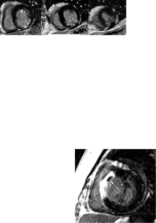
Noncompaction cardiomyopathy is described as a cardiomyopathy that occurs due to an autosomally dominant inherited trait in which the middle and apical segments exhibit a thin compact wall with regional dilatation, dysfunction, and significant hypertrabeculation (Figure 9). An end-diastolic ratio of noncompacted to compacted LV myocardium of greater than or equal to 2.3 defines the condition.196 Also, LV wall motion abnormalities, global dysfunction, or coronary intraventricular thrombi are often present in the disorder. Refined diagnostic criteria may be forthcoming as this disorder becomes recognized with greater frequency.
3.5.4. Dilated Cardiomyopathy
Diagnostic targets for CMR in DCM include progressive LV dilation, LV systolic dysfunction, and regional midwall myocardial fibrosis.88 Recently, focal septal fibrosis in DCM, the so-called “midwall sign,” has been linked to ventricular arrhythmia.197 The presence of fibrosis identified with LGE has been found to be associated with adverse cardiac events.85
3.5.5. Acute Viral Myocarditis
Quantification of global myocardial signal intensity changes on T2-weighted CMR reflecting inflammation and especially edema offers a high diagnostic accuracy to detect acute myocarditis.198 Reflecting irreversible injury, a typical pattern of regional, typically subepicardial fibrosis can be visualized90 (Figure 10). With combined analysis of T1and T2-weighted scans, heightened diagnostic accuracy for identifying active myocarditis is achieved.198,199 CMR is considered one of the most important diagnostic tools in the workup of patients with myocarditis.91,200 An expert consensus document on the application, evaluation, and reporting of CMR in myocarditis has been developed.201
3.5.6. Sarcoidosis
Up to 50% of patients with pulmonary sarcoidosis have cardiac involvement, although only 5% have cardiac symptoms. Myocardial involvement, however, is the leading cause of death in these patients. CMR can visualize myocardial inflammation using contrast-enhanced techniques. Early contrast enhancement identifies territories exhibiting myocardial inflammation, whereas LGE shows areas of irreversible injury (Figure 11).202
3.5.7. Amyloidosis
Myocardial amyloid infiltration is frequent among patients with systemic amyloidosis and leads to apparent myocardial