

трансплантация / Transplantologia_print_3_2013_web
.pdfВВЕДЕНИЕ
Оксидазота(NO), какмощныйэндогенныйвазодилататор, принимает участие в регуляции системного и легочного сосудистого сопротивления [1], регулирует функцию центральной и вегетативной нервной системы. По эфферентным нервам этот агент оказывает влияние на деятельность органов дыхательной системы, желудочно-кишечного тракта и мочеполовой системы [2].
Открытие роли дисфункции эндотелия легочных сосудов и нарушений синтеза NO в патогенезе острых и хронических расстройств легочного кровообращения способствовало внедрению ингаляционного NO (иNO) в комплексную терапию больных после трансплантации легких как экзогенного аналога естественного регулятора сосудистого тонуса [3].
Введение низких концентраций иNO представляется безопасным. Однако при высоких концентрациях его токсичность связана в основном с образованием NO2 и метгемоглобинемией [4]. Взаимодействие иNO с оксигемоглобином приводит к образованию метгемоглобина (MetHb) и нитрита/ нитрата (NOx), а с дезоксигемоглобином – нитро- зил-гемоглобина [4]. Почти 70% иNO выделяется в течение 48 ч в виде нитратов с мочой [5]. Скорость захвата и высвобождения NO из Fe2+–Hb в 105–106 раз выше, чем кислорода. Чрезмерные концентрациициркулирующегоMetHb могутприводитьктканевой гипоксии [6]. MetHb не способен связывать
ТРАНСПЛАНТАЦИЯ ОРГАНОВ
кислород, и поэтому при его образовании возникает гемическая гипоксия. Кроме того, в присутствии MetHb кривая диссоциации оксигемоглобина сдвигается влево, и в результате снижается отдача кислорода тканям. Если ткани получают недостаточно кислорода, нормальныйаэробныйметаболизмзаменяется анаэробным метаболизмом, при котором образуется лактат. В связи с этим при ингаляционном введении NO необходимо контролировать концентрациюMetHb, NOx илактата(какпоказателягипоксии) вкрови. ФормированиеNO2 привведениииNO зависит от концентрации NO, концентрации вдыхаемого кислорода (FiO2) и времени пребывания этих газов в крови [7]. В экспериментальном исследовании продукция NO2 при вдыхании 20 ppm NO была минимальной (< 0,7 ppm) даже при FiO2 – 95% [8].
ОднаковклиническойпрактикеконцентрацияNOx, MetHb в крови и их взаимосвязь у больных после трансплантации легких на фоне применения иNO недостаточно изучена.
Таким образом, целью настоящего исследования являлось изучение взаимосвязи иNO, уровня NOx, MetHb и лактата в крови больных после трансплантации легких.
МАТЕРИАЛЫ И МЕТОДЫ
Обследовали 7 больных (6 женщин и 1 мужчина) в возрасте от 24 до 55 (36,3 ± 4,0) лет после выполнения трансплантации легких (ТЛ). Операции
Хубутия Могели Шалвович – член-корр. РАМН, д. м. н., профессор, директор ГБУЗ «НИИ скорой помощи им. Н.В. Склифосовского ДЗ г. Москвы», Москва, Российская Федерация. Абакумов Михаил Михайлович – д. м. н., профессор, заместитель директора по научной работе, заведующий отделением неотложной торакоабдоминальной хирургии того же института. Клычникова Елена Валерьевна – к. м. н., заведующий научной клинико-биохимической лабораторией экстренных методов исследования того же института. Тарабрин Евгений Александрович – к. м. н., ведущий научный сотрудник отделения неотложной торакоабдоминальной хирургии того же института. Тазина Елизавета Владимировна – к. фарм. н., старший научный сотрудник научной клинико-био- химической лаборатории экстренных методов исследования того же института. Годков Михаил Андреевич – д. м. н., заведующий научным отделом лабораторной диагностики того же института. Романов Александр Александрович – к. м. н., заведующий отделением анестезиологии-реанимации № 3 того же института . Курилова Оксана Александровна – к. м. н., врач отделения анестезиоло- гии-реанимации № 3 того же института . Первакова Эльза Ибрагимовна – к. м. н., заведующая научным отделением реанимации и интенсивнойтерапиидлябольныхпослетрансплантациитогожеинститута. ЦуроваДинаХалитовна– к. м. н., научныйсотрудник отделения неотложной торакоабдоминальной хирургии того же института.
Для корреспонденции: Клычникова Елена Валерьевна. Адрес: 129090, Москва, Большая Сухаревская площадь, д. 3.
Телефон: 8 (495) 628-34-15, 8 (916) 574-94-04. E-mail: ltazina@yandex.ru.
Khubutiya Mogeli Shalvovich – Corresponding Member of RAMSci, doct. of med. sci., professor, director of Sklifosovsky Research Institute for Emergency Medicine Health Department of Moscow, Moscow, Russian Federation. Abakumov Mikhail Mikhailovich – doct. of med. sci., professor, Deputy director for Research, Head of the Department of emergency thoracoabdominal surgery at the same Institute. Klychnikova Elena Valer’evna – cand. of med. sci., Head of the Scientific clinical and biochemical laboratory for emergency research methods at the same Institute. Tarabrin Eugenii Aleksandrovich – cand. of med. sci., Leading Researcher at the Department of emergency thoracoabdominal surgery at the same Institute. Tazina Elizaveta Vladimirovna – cand. pharm. sci., senior researcher at the Scientific clinical and biochemical laboratory for emergency research methods at the same Institute. Godkov Mikhail Andreevich – doct. of med. sci., Head of the Scientific Department of laboratory diagnostics at the same Institute. Romanov Aleksandr Aleksandrovich – cand. of med. sci., Head at the same Department at the same Institute. Kurilova Oksana Aleksandrovna – cand. of med. sci, doctor at Department of anesthesiology and reanimation number 3 at the same Institute. Pervakova Elsa Ibragimovna – cand. of med. sci, Head of the Scientific Department of reanimation and intensive therapy for transplant patients at the same Institute. Tsurova Dina Khalitovna – cand. of med. sci., researcher at the Department of emergency thoracoabdominal surgery at the same Institute.
For correspondence: Klychnikova Elena Valer’evna. Adress: 129090, Moscow, Bolshaya Sukharevskaya Square, 3. Phone: 8 (495) 628-34-15, 8 (916) 574-94-04. E-mail: ltazina@yandex.ru.
39
ВЕСТНИК ТРАНСПЛАНТОЛОГИИ И ИСКУССТВЕННЫХ ОРГАНОВ |
том XV № 3–2013 |
выполнялись по поводу: первичной легочной гипертензии, муковисцидоза (по 2 наблюдения), лимфангиолейомиоматоза, саркоидоза, идиопатического легочного фиброза (по 1 наблюдению).
Искусственную вентиляцию легких (ИВЛ) проводили аппаратами Primus (в операционной) и EvitaXL (в отделении реанимации) в режиме с индивидуально подобранными параметрами. Параметры ИВЛ и биомеханики легких регистрировали в режиме реального времени с помощью мониторной системы.
Для иNO-терапии использовали сертифицированную газовую смесь NO–N2 с концентрацией NO 1000 ppm (parts per million). ПодачуиNO (5–20 ppm)
осуществляли в инспираторную часть дыхательного контура аппарата ИВЛ на расстоянии 60–80 см от Y-образного коннектора. Для обеспечения малого потока газа использовали систему Bedfont Nitric Oxide Inhaled Therapy Flow. Объемную скорость по-
тока иNO (мл/мин) устанавливали в соответствии с требуемой концентрацией и показаниями электрохимического NO–NO2-анализатора. Продолжительность иNO-терапии составила от 2 до 4 суток, средняя концентрация иNO – 30 ± 1 ppm.
Определение стабильных метаболитов оксида азота (NOx) в сыворотке крови проводили по методу, согласно которому кадмий в присутствии цинка восстанавливает нитрат до нитрита [9]. Оценку кислотно-основного состояния и газов крови, определение MetHb, лактата в артериальной крови про-
водили на анализаторе ABL 800 Flex (Radiometer,
Дания). Данные проанализированы с 1 по 10-е сутки после операции. В качестве контрольной группы обследовали 25 практически здоровых людей, средний возраст которых составил 32,7 ± 8,6 лет, соотношение мужчины/женщины – 17/8.
Статистический анализ проводили при помо-
щи программ Statistica 10.0, Origin 6.1 и MS Excel.
Сравнение исследуемой группы больных с контрольной группой проводили с использованием U-критерия Манна-Уитни. Для исследования взаимосвязи признаков использовали метод корреляци-
онного анализа Спирмена. Данные представляли в виде медианы и интерквартильного размаха (25-й и 75-й персентили).
РЕЗУЛЬТАТЫ И ОБСУЖДЕНИЕ
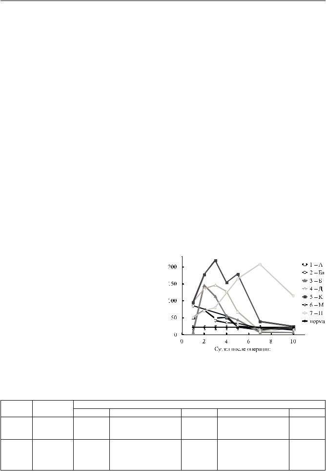
При исследовании концентрации NOx, MetHb и лактата в крови больных после ТЛ в динамике было обнаружено достоверное повышение уровня NOx в 3,6; 3,9; 4,3; 3,0 и 2,5 раза соответственно на 1; 2; 3; 4 и 5-е сутки после операции (табл. 1). Графически динамикауровняNOx послеТЛотдельноукаждого больного представлена на рис. 1. Из рис. 1 видно, что на протяжении 5 суток после операции у всех больных уровень NOx в сыворотке крови был выше нормы, далее у 6 пациентов уровень NOx постепенно снижался и к 10-м суткам практически достигал нормальных значений. Концентрация MetHb оставалась в пределах нормы, уровень лактата достоверно повышался в 3,4; 2,3; 2,2; 1,4 и 2,0 раза соответственно на 1; 2; 3; 4 и 5-е сутки исследования
(табл. 1).
Наши данные согласуются с данными Steudel W. et al., где показано, что значительная метгемоглобинемия или образование NO2 редко встречается у пациентов, вдыхающих NO в дозах ≤ 80 ppm [10]. Проходя через альвеолярно-капиллярную мембрану, иNO может взаимодействовать с кислородом,
Рис. 1. Динамика уровня NOx у больных после ТЛ |
|
Динамика NOx, MetHb и лактата в группе больных (n = 4) после ТЛ |
Таблица 1 |
|||||||
|
|
||||||||
|
(данные представлены в виде медианы и интерквартильного размаха) |
|
|||||||
Показа- |
Норма |
|
|
|
Сутки |
|
|
|
|
тель |
1 |
2 |
3 |
4 |
5 |
7 |
10 |
||
|
|||||||||
NOx, |
18,61 |
67,73* |
72,05 * |
79,49* |
54,98* |
45,77* |
13,37 |
17,38 |
|
(50,48– |
(71,02– |
(63,87– |
(52,78– |
(23,49– |
(11,69– |
(15,11– |
|||
мкмоль/л |
(17,70–23,62) |
||||||||
87,30) |
103,63) |
112,80) |
91,09) |
91,75) |
110,29) |
44,94) |
|||
|
1,3 |
||||||||
MetHb, |
1,5 |
1,5 |
1,0 |
1,1 |
1,0 |
0,7 |
0,8 |
||
% |
(1,0–1,4) |
(1,3–1,5) |
(1,4–1,7) |
(1,0–1,2) |
(0,9–1,3) |
(0,8–1,1) |
(0,7–0,9) |
(0,7–1,0) |
|
Лактат, |
1,1 |
3,7 (1,9– |
2,5 (2,4– |
2,4 |
1,5 |
2,2 |
1,3 (1,2–1,4) 1,4 (1,2–1,8) |
||
ммоль/л |
(0,9–1,2) |
5,7)* |
2,7)* |
(2,1–2,8)* |
(1,5–2,1)* |
(1,9–2,3)* |
|||
Примечание. * p < 0,05 по отношению к норме.
40

ТРАНСПЛАНТАЦИЯ ОРГАНОВ
образуя NOx, или вступать в связь с оксигемоглобином, образуя не только NOx, но и MetHb. Учитывая метаболизм иNO в организме, мы изучали взаимосвязь иNO с концентрацией MetHb в цельной крови
ис NOx в сыворотке крови у больных после ТЛ. В нашем исследовании в общей группе больных
после ТЛ наблюдались следующие достоверные положительные корреляции: между иNO и концентрацией NOх, иNO и концентрацией MetHb, иNO
иконцентрацией лактата, концентрацией NOx и MetHb, концентрацией NOx и лактата, концентрацией MetHb и лактата (табл. 2). Эти данные указывают на метаболизм иNO в организме с образованием NOх и MetHb, который в свою очередь может приводить к увеличению концентрации лактата в крови за счет тканевой гипоксии.
Более того, в наших исследованиях обнаружены достоверные положительные корреляции отдельно у каждого больного после ТЛ:
−между иNO и концентрацией NOx у больных 2, 3, 4 (табл. 3);
−между иNO и концентрацией MetHb у больной 4 (табл. 3);
−между иNO и концентрацией лактата у больных
1, 4, 7 (табл. 3);
−между концентрацией NOx и MetHb у больной 1
и 4 (табл. 3);
−между концентрацией MetHb и лактата у больной 4 (табл. 3).
Полученные нами корреляции по конкретным больным также подтверждают тот факт, что иNO метаболизируетсяворганизмесобразованиемглавным образом NOх.
Несмотря на увеличение количества ТЛ, ежегодно проводимых во всем мире, необходимо проводить углубленные исследования метаболизма NO в сыворотке крови пациентов с успешными ТЛ и пациентов с острым или хроническим отторжением трансплантата. Изучению концентрации стабильных метаболитов NO у больных с ТЛ посвящено всего несколько работ. Так, De Andrade et al. [11] отмечали значительное увеличение концентрации общего NOx в бронхоальвеолярной жидкости, полученной при лаваже (БАЛ), и в сыворотке крови больных после ТЛ по сравнению с контролем. Эти же ученые показали, что концентрация NOx в сыворотке крови больных коррелирует со степенью воспаления дыхательных путей
ифиброза вследствие отторжения трансплантата [12]. Reid et al. [13] подтвердили данные об увеличении уровня нитритов у больных после ТЛ, однако при выделении группы стабильных пациентов
ипациентов с облитерирующим бронхиолитом только у последних наблюдалось значительное увеличение уровня нитритов в БАЛ. Повышение уровня циркулирующих нитратов наблюдалось в экспериментальных исследованиях на животных
сострым клеточным отторжением трансплантата
[14, 15].
Таблица 2
Взаимосвязь иNO, уровня NOx, MetHb и лактата в группе больных после ТЛ
|
иNO |
NOx |
MetHb |
NOx |
R = 0,866; p = 0,012; (n = 7) |
− |
R = 0,870; p = 0,002; (n = 4) |
MetHb |
R = 0,840; p = 0,036; (n = 4) |
R = 0,870; p = 0,002; (n = 4) |
− |
Лактат |
R = 0,828; p = 0,042; (n = 4) |
R = 0,767; p = 0,016; (n = 4) |
R = 0,854; p = 0,003; (n = 4) |
|
|
|
|
|
|
|
|
Таблица 3 |
|
|
|
Взаимосвязь иNO, уровня NOx, MetHb и лактата отдельно у каждого пациента после ТЛ |
|||||||
Пациенты |
|
|
Корреляции |
|
|
||||
иNO и NOx |
иNO и MetHb |
иNO и лактат |
NOx и MetHb |
NOx и лактат |
MetHb и лактат |
||||
|
|
|
|||||||
1 |
– А |
|
R = 0,545, |
R = 0,521, |
R = 0,698, |
R = 0,720, |
R = 0,550, |
R = 0,418, |
|
|
p = 0,129 |
p = 0,150 |
p = 0,037 |
p = 0,029 |
p = 0,125 |
p = 0,262 |
|||
|
|
|
|||||||
2 |
– Бл |
|
R = 0,828, |
R = 0,105, |
R = 0,0, |
R = 0,174, |
R = −0,429, |
R = −0,261, |
|
|
p = 0,042 |
p = 0,843 |
p = 1,0 |
p = 0,742 |
p = 0,397 |
p = 0,618 |
|||
|
|
|
|||||||
3 |
– Б |
|
R = 0,779, |
|
|
|
|
|
|
|
p = 0,013 |
|
|
|
|
|
|||
|
|
|
|
|
|
|
|
||
4 |
– Д |
|
R = 0,853, |
R = 0,880, |
R = 0,717, |
R = 0,900, |
R = 0,581, |
R = 0,732, |
|
|
p = 0,002 |
p = 0,0008 |
p = 0,020 |
p = 0,0004 |
p = 0,078 |
p = 0,016 |
|||
|
|
|
|||||||
5 |
– К |
|
R = 0,293, |
|
|
|
|
|
|
|
p = 0,573 |
|
|
|
|
|
|||
|
|
|
|
|
|
|
|
||
6 |
– М |
|
R = 0,393, |
|
|
|
|
|
|
|
p = 0,441 |
|
|
|
|
|
|||
|
|
|
|
|
|
|
|
||
7 |
– П |
|
R = −0,693, |
R = 0,044, |
R = 0,830, |
R = −0,328, |
R = −0,487, |
R = 0,064, |
|
|
p = 0,039 |
p = 0,911 |
p = 0,006 |
p = 0,389 |
p = 0,183 |
p = 0,871 |
|||
|
|
|
|||||||
41

ВЕСТНИК ТРАНСПЛАНТОЛОГИИ И ИСКУССТВЕННЫХ ОРГАНОВ |
том XV № 3–2013 |
В наших исследованиях у больных с ТЛ обнаружено достоверное повышение уровня NOx с 1-х по 5-е сутки после операции примерно в 3–4 раза и выявлена достоверная положительная корреляция между иNO и концентрацией NOх. Известно, что в синтезе эндогенного NO принимают участие нейрональная (nNOS), индуцибельная (iNOS) и эндотелиальная (eNOS) изоформы синтазы оксида азота [16]. nNOS и eNOS поддерживают низкий стационарный уровеньNO, которыйнепревышаетнесколькихмикромолей и необходим для поддержания биологических функций организма. iNOS экспрессируется во многихклетках(моноциты/макрофаги, нейтрофилы, гладкомышечные клетки, сосудистый эндотелий) после иммунологических стимулов и при воспалении. При участии iNOS высвобождается большое количество NO, стационарный уровень NO при этом достигает сотни микромолей и поддерживается в течение от нескольких часов до нескольких дней, что зависит от длительности воздействия [17]. Заметное увеличение концентрации NOx в сыворотке крови больныхна1–5-есуткипослеТЛ, возможно, связано нетолькосметаболизмомиNO доNOх, атакжесактивацией iNOS, что требует дальнейшего изучения.
ЗАКЛЮЧЕНИЕ
Полученные данные свидетельствуют о том, что при использовании ингаляционного оксида азота у больных после трансплантации легких необходимо контролировать как концентрацию вводимого ингаляционного оксида азота и метгемоглобина в крови, так и уровень нитрита/нитрата. Обнаруженная положительная корреляция между концентрациями нитрит/нитрата и лактата в течение первых пяти суток после трансплантации легких требует дальнейшего изучения.
СПИСОК ЛИТЕРАТУРЫ
1.Kuga T., Mohri M. Bradykinin-induced vasodilatation of human coronary arteries in vivo: role of nitric oxide and angiotensin-converting enzyme. J. Am. Coll. Cardiol. 1997; 30: 108–112.
2.Cooke J.P., Dzau V.J. Nitric oxide synthase: Role in the genesis of vascular disease. Annu. Rev. Med. 1997; 48: 489–509.
3.Rossaint R., Falke K.J., Lopez F., Slama K., Pison U., Zapol W.M. Inhaled nitric oxide for the adult respiratory distress syndrome. N. Engl. J. Med. 1993; 328: 399–405.
4.Ichinose F., Roberts J.D. Jr., Zapol W.M. Inhaled nitric oxide: a selective pulmonary vasodilator: current uses and therapeutic potential. Circulation. 2004; 109: 3106–3111.
5.Young J.D., Sear J.W., Valvini E.M. Kinetics of methaemoglobin and serum nitrogen oxide production during inhalation of nitric oxide in volunteers. Br. J. Anaesth. 1996; 76: 652–656.
6.Weinberger B., Laskin D.L., Heck D.E., Laskin J.D. The toxicology of inhaled nitric oxide. Toxicol. Sci. 2001; 59 (1): 5–16.
7.Sokol G.M., Van Meurs K.P., Wright L.L., Rivera O., Thorn W.J., Chu P.M., Sams R.L. Nitrogen dioxide formation during inhaled nitric oxide therapy. Clin. Chem. 1999; 45 (3): 382–387.
8.Kirmse M., Hess D., Fujino Y., Kacmarek R.M., Hurford W.E. Delivery of inhaled nitric oxide using the Ohmeda INOvent Delivery System. Chest. 1998; 113 (6): 1650–1657.
9. Голиков П.П., Николаева Н.Ю. Метод определения нитрита/нитрата (NOx) в сыворотке крови. Вопр.
биомед. химии. 2004; 1: 79–85.
10.Steudel W., Hurford W.E., Zapol W.M. Inhaled nitric oxide: basic biology and clinical applications. Anesthesiology. 1999; 91: 1090–1121.
11.De Andrade J.A., Crow J.P., Viera L., Bruce Alexander C., Randall Young K., McGiffin D.C., Zorn G.L., Zhu S., Matalon S., Jackson R.M. Protein nitration, metabolites of reactive nitrogen species, and inflammation in lung allografts. Am. J. Respir. Crit. Care Med. 2000; 161 (6): 2035–2042.
12.De Andrade J.A., Crow J., Viera L., Bruce Alexander C., Randall Young K., McGiffin D.C., Zorn G.L., Matalon S., Jackson R.M. Reactive nitrogen species, airway inflammation, and fibrosis in lung transplant. Chest. 2001; 120: 58–59.
13.Reid D., Snell G., Ward C., Krishnaswamy R., Ward R.,
Zheng L., Williams T., Walters H. Iron overload and nitric oxide-derived oxidative stress following lung transplantation. J. Heart Lung Transplant. 2001; 20: 840–849.
14. Wiklund L., Lewis D.H., Sjöquist P.O., Nilsson F., Tazelaar H., Miller V.M., McGregor C.G. Increased levels of circulating nitrates and impaired endothelium-mediated vasodilation suggest multiple roles of nitric oxide during acute rejection of pulmonary allografts. J. Heart Lung Transplant. 1997; 16 (5): 517–523.
15. Worrall N.K., Boasquevisque C.H., Misko T.P., Sullivan P.M., Ferguson T.B. Jr., Patterson G.A. Inducible nitric oxide synthase is expressed during experimental acute lung allograft rejection. J. Heart Lung Transplant. 1997; 16: 334–339.
16.Johnson M.L., Billiar T.R. Roles of nitric oxide in surgical infection and sepsis. World J. Surg. 1998; 22: 187– 196.
17.Воспельникова Н.Д. Оксид азота как регулятор клеточных функций. Биохимические основы патологических процессов. Под ред. Е.С. Северина. М. Меди-
цина. 2000; 10: 266–291.
REFERENCES
1.Kuga T., Mohri M. Bradykinin-induced vasodilatation of human coronary arteries in vivo: role of nitric oxide and angiotensin-converting enzyme. J. Am. Coll. Cardiol. 1997; 30: 108–112.
2.Cooke J.P., Dzau V.J. Nitric oxide synthase: Role in the genesis of vascular disease. Annu. Rev. Med. 1997; 48: 489–509.
42

ТРАНСПЛАНТАЦИЯ ОРГАНОВ
3.Rossaint R., Falke K.J., Lopez F., Slama K., Pison U., Zapol W.M. Inhaled nitric oxide for the adult respiratory distress syndrome. N. Engl. J. Med. 1993; 328: 399–405.
4.Ichinose F., Roberts J.D. Jr., Zapol W.M. Inhaled nitric oxide: a selective pulmonary vasodilator: current uses and therapeutic potential. Circulation. 2004; 109: 3106–3111.
5.Young J.D., Sear J.W., Valvini E.M. Kinetics of methaemoglobin and serum nitrogen oxide production during inhalation of nitric oxide in volunteers. Br. J. Anaesth. 1996; 76: 652–656.
6.Weinberger B., Laskin D.L., Heck D.E., Laskin J.D. The toxicology of inhaled nitric oxide. Toxicol. Sci. 2001; 59 (1): 5–16.
7.Sokol G.M., Van Meurs K.P., Wright L.L., Rivera O., Thorn W.J., Chu P.M., Sams R.L. Nitrogen dioxide formation during inhaled nitric oxide therapy. Clin. Chem. 1999; 45 (3): 382–387.
8.Kirmse M., Hess D., Fujino Y., Kacmarek R.M., Hurford W.E. Delivery of inhaled nitric oxide using the Ohmeda INOvent Delivery System. Chest. 1998; 113 (6): 1650–1657.
9.Golikov P.P., Nikolaeva N.Yu. Method for determination of nitrite/nitrate (NOx) in serum. Vopr. biomed. himii. 2004; 1: 79–85 (in rus).
10.Steudel W., Hurford W.E., Zapol W.M. Inhaled nitric oxide: basic biology and clinical applications. Anesthesiology. 1999; 91: 1090–1121.
11.De Andrade J.A., Crow J.P., Viera L., Bruce Alexander C., Randall Young K., McGiffin D.C., Zorn G.L., Zhu S., Matalon S., Jackson R.M. Protein nitration, me-
tabolites of reactive nitrogen species, and inflammation in lung allografts. Am. J. Respir. Crit. Care Med. 2000; 161 (6): 2035–2042.
12.De Andrade J.A., Crow J., Viera L., Bruce Alexander C., Randall Young K., McGiffin D.C., Zorn G.L., Matalon S., Jackson R.M. Reactive nitrogen species, airway inflammation, and fibrosis in lung transplant. Chest. 2001; 120: 58–59.
13.Reid D., Snell G., Ward C., Krishnaswamy R., Ward R., Zheng L., Williams T., Walters H. Iron overload and nitric oxide-derived oxidative stress following lung transplantation. J. Heart Lung Transplant. 2001; 20: 840–849.
14. Wiklund L., Lewis D.H., Sjöquist P.O., Nilsson F., Tazelaar H., Miller V.M., McGregor C.G. Increased levels of circulating nitrates and impaired endothelium-mediated vasodilation suggest multiple roles of nitric oxide during acute rejection of pulmonary allografts. J. Heart Lung Transplant. 1997; 16 (5): 517–523.
15. Worrall N.K., Boasquevisque C.H., Misko T.P., Sullivan P.M., Ferguson T.B. Jr., Patterson G.A. Inducible nitric oxide synthase is expressed during experimental acute lung allograft rejection. J. Heart Lung Transplant. 1997; 16: 334–339.
16.Johnson M.L., Billiar T.R. Roles of nitric oxide in surgical infection and sepsis. World J. Surg. 1998; 22: 187– 196.
17.Vospel’nikova N.D. Nitric oxide as a regulator of cellular functions. Biohimicheskie osnovy patologicheskih processov. Pod red. E.S. Severina. M. Medicina. 2000; 10: 266–291 (in rus).
43

ВЕСТНИК ТРАНСПЛАНТОЛОГИИ И ИСКУССТВЕННЫХ ОРГАНОВ |
том XV № 3–2013 |
INFECTIONS IN THE INTENSIVE CARE UNIT FOLLOWING LIVER TRANSPLANTATION: PROFILE OF A SINGLE CENTER
Otan E., Usta S., Aydin C., Karakas S., Unal B., Mamedov R., Kayaalp C., Yilmaz S.
Inonu University, School of Medicine Dep of General Surgery and Institute for Liver Transplantation, Malatya, Turkey
Introduction. Despite the advances in antibiotherapy and critical care management, infectious complications remain among the leading complications after liver transplantation related with mortality and morbidity. This study analysis the incidence and pattern of infections and possible prognostic factors of infectious complications retrospectively in a single center. Patients and Methods. Results of 30 consecutive patients with a primary liver transplantation history in a single center between August 2011 and August 2012 and a positive culture result in the first month in the ICU were analysed retrospectively. Results. During the first 1 month stay in the ICU postoperatively 30 (13,63%) patients had at least 1 infection. Total number of infections were 68. Mortality rate of the infected patients was 53,3% (n = 16). Among these infections, 25 (36,76%) of them were in deep surgical sites. Eighteen of the 30 patients (60%) were infected with a single microorganism. Eleven patients (36,66%) had a single infection episode. Microorganism were gram negative in 52 (76,47%) of the infections, gram positive in 14 (20,58%) of the infections, rest of the 2 (2,94%) infections were due to Candidiasis. Among the possible risk factors contributing to mortality, there was a statistically significant difference (p < 0,001) between the platelet counts of the mortality and surviving groups of the patients. Conclusion. Infections are among the preventable risk factors for mortality and morbidity after liver transplantation. Our data reveals a significant relation between trombocytopenia and mortality among the infected patients. Further studies focusing on this relation would expose the mechanisms and any possible contribution in clinical management of the patients.
Key words: liver transplantation, infection, immunosuppression, intensive care.
INTRODUCTION
Liver transplantation (LT) is the curative treatment option for end stage liver disease. Despite the advances in immunosuppressive treatment and antibioprophylaxy regimes, infectious complications remain to be the most frequent complication associated with significant increase in mortality and morbidity [1]. Vulnerability of the liver transplant patients to infections is related with technical difficulty of the procedure, preoperative abdominal contamination and underlying disease [1]. Most of the bacteremia episodes occur in first 3 months following transplantation with a rate of 33–68% [1, 2].
Following LT, overall mortality in relation with infection is reported from 30 to 53 % [3, 4].
The aim of this study was to examine the incidence and pattern of infections and prognostic factors associated with mortality following LT.
PATIENTS AND METHODS
We retrospectively analyzed 220 patients who had undergone liver transplantation between August 2011 and August 2012. Results of 30 consecutive patients with a primary liver transplantation history between August 2011 and August 2012 and a positive culture result in the first month in the ICU were included to the study.
Patients already under antibiotherapy with a history of preoperative infection and patients postoperatively hospitalised out of ICU were excluded from the study.
Data of age, gender, aetiology of the liver disease, type of transplantation (deceased, cadaveric), timing of surgery (emergent or elective), immunosuppressive regime, WBC, PLT, liver function rest results on day of positive culture result, model for end stage liver disease (MELD) score, history of diabetes mellitus (DM), preoperative hospitalisation history, isolated microorganisms and infection sites were analysed retrospectively.
Immunosuppressive and Antibioprophylaxy Protocol
Without a history of ongoing infection therapy, periopertaive antibioprophylactic regimen consisted of 1000 mg Ampicillin/sulbactam administered intraveously every 4 hours. Same regimen is continued postoperatively twice daily for 48 hours and discontinued in cases with no clinical signs for infection.
Immunosuppressive methylprednisolone treatment is initialised in the ICU at a dose of 100 mg/day is tapered according to the greft function status. On postoperative day 3, without any contraindication, tacrolimus 2 mg/day and cyclosporin 200 mg/day are initiated.
44

ТРАНСПЛАНТАЦИЯ ОРГАНОВ
Microbiological Analysis
Blood culture testing was carried out with an automated microbiology growth and detection system (BACTEC™ Instrumented blood culture systems Sparks, MD. USA). Conventional methods were used for rest of the specimens. Antibiotic susceptibility was determined by minimum inhibitory concentration test.
RESULTS
Among 220 patients, 121 of them male (55%) and 99 were female (45%) with a mean age of 41,07 ± 17,89. Number of patients underwent living donor liver transplantation (LDLT) was 166 (75,45%) and cadaveric liver transplantation was 54 (24,54%). Among these liver transplantation operations, indication was acute liver failure in 132 (60%) patients and rest of the 88 patients (40%) underwent surgery as a result of elective conditions. During the first 1 month stay in the ICU postoperatively 30 (13,63%) patients had at least 1 infection. Total number of infections were 68. Seventeen (56,7%) of these patients were male and 13 (43,3%) were female with a mean age of 37,77 ± 21,08. Mortality rate was 53,3% (n = 16) among the infected patients. Among these infections, 25 (36,76%) of them were in deep surgical sites. Eighteen of the 30 patients (60%) were infected with a single microorganism. Eleven patients (36,66%) had a single infection episode. Microorganism were gram negative in 52 (76,47%) of the infections, gram positive in 14 (20,58%) of the infections, rest of the 2 (2,94%) infections were due to Candidiasis. Infection site disturbance and microbiological disturbance details of the infections are given in table 1 and 2.
Infection site disturbance |
Table 1 |
|
|
||
Site of Infection |
n |
% |
Deep surgical site |
25 |
36,76 |
Superficial surgical site |
3 |
4,41 |
Bloodstream |
24 |
35,29 |
Urinary |
7 |
10,29 |
Sputum |
4 |
5,88 |
Catheter |
5 |
7,35 |
Total |
68 |
100 |
Among the pre-, intraand post-operative conditions contributing to mortality; microbiological disturbance details are given in table 3–9.
Discussion
Bacterial infections following liver transplantation remain among the leading preventable factors contributing to morbidity. More than half of the bacterial infections occur within the 1 st month after transplantation [5, 6]. In our study, rate of infection in the 1 st month among patients in the ICU was 13,63%. This relatively lower rate is thought to be related with the study design, where patients already under antibiotherapy with a history of preoperative infection and patients postoperatively hospitalised out of ICU were excluded.
Although gram-positive bacteria have a predominance, there is a notable increase in incidence of blood-stream infections due to gram-negative organisms following liver transplantation [7–9]. Our study results reveal a significant gram-negative infections dominancy with a rate of 76,47%. Among blood-stream
Table 2
Microbiological disturbance
Microorganism |
Sputum |
Bloodstream |
Urine |
|
Surgical Site Infection |
Catheter |
|
Total |
||
(gram stain + or –) |
n (%) |
n (%) |
n (%) |
Deep n (%) |
Superficial n (%) |
n (%) |
n (%) |
|||
Pseudomonas (–) |
1 (1,47) |
3 |
(4,41) |
– |
|
– |
– |
– |
4 |
(5,88) |
E. coli (–) |
– |
3 |
(4,41) |
1 (1,47) |
6 |
(8,82) |
– |
2 (3,03) |
12 |
(17,64) |
Acinetobacter (–) |
3 (4,41) |
8 (11,76) |
1 (1,47) |
8 (11,76) |
1 (1,47) |
1 (1,47) |
22 |
(32,35) |
||
Clebsiella (–) |
1 (1,47) |
3 |
(4,41) |
2 (3,03) |
4 |
(5,88) |
1 (1,47) |
1 (1,47) |
12 |
(17,64) |
Enterococ (+) |
– |
4 |
(5,88) |
1(1,47) |
3 |
(4,41) |
– |
1 (1,47) |
9 (13,23) |
|
Candida |
– |
|
– |
1(1,47) |
1 |
(1,47) |
– |
– |
2 |
(3,03) |
Strep (+) |
– |
1 |
(1,47) |
– |
|
– |
– |
– |
1 |
(1,47) |
S. aureus (+) |
– |
1 |
(1,47) |
– |
|
– |
1 (1,47) |
– |
2 |
(3,03) |
Proteus (–) |
– |
|
– |
1(1,47) |
|
– |
– |
– |
1 |
(1,47) |
Citrobacter (–) |
– |
1 |
(1,47) |
– |
|
– |
– |
– |
1 |
(1,47) |
Coag. (–) staph (+) |
– |
|
– |
– |
2 |
(3,03) |
– |
– |
2 |
(3,03) |
Total (n) |
5 (7,35) |
24 |
(35,29) |
7 (10,29) |
24 |
(35,29) |
3 (4,41) |
5 (7,35) |
|
68 |
For correspondence: Mamedov Ruslan. Address: Inonu Universitesi Tip Fakultesi, Turgut Ozal Tip Merkezi, Genel Cerrahi AD, Elazig Yolu 15. Km, 44280 Malatya, TURKEY
Tel: +905370466537. E-mail: rmamedov2001@yahoo.com
45

ВЕСТНИК ТРАНСПЛАНТОЛОГИИ И ИСКУССТВЕННЫХ ОРГАНОВ |
том XV № 3–2013 |
infections of our study, gram-negative microorganisms acount for 75% (n = 18) of the cases.
Previous studies high-lighted the tendency and vulnerability to infections of LT patients as a result of technical complexity and immunosuppression [1, 10]. In our study, these conditions led to infections in 30 patients where Acinetobacter was the most frequent pathogen microorganism (Table 2).
Catheter related infections are reported to be the most frequent sites of bacteremia with an incidence of 23 to 30% followed by intra-abdominal/biliary infections with an incidence of 7 to 10% [2, 11]. In our study, catheters were the site for 7,35% of the infections, whereas intra-abdominal (deep-surgical) infections had
a relatively high incidence of 37,76%. This relatively high incidence is supposed to be associated with latent contamination in the abdominal cavity rather than failure in intensive care, as similarly stated in previous studies [12]. Although there have been several studies concerning the surveillance and risk factors of infections following liver transplantation, none of them stated a significant risk factor, which can be monitored during follow-up, related with mortality when liver transplant donors are infected.
In a study of Saner F.H., pulmonary infections following liver transplantation are stated to significantly detoriate the outcome [13]. Our study based on patients’ positive culture results were mostly from deep
Table 3
Microbiological Disturbance in Patients without Preoperative Hospitalisation History
Microorganism |
Bloodstream |
Urine |
Cath |
Total |
E. coli |
1 |
1 |
1 |
3 |
Total (n) |
1 |
1 |
1 |
3 |
|
Microbiological Disturbance in Diabetic Patients |
|
Table 4 |
|||
|
|
|
||||
Microorganism |
Bloodstream |
Urine |
Surgical Site Infection |
Cath |
Total |
|
|
|
|
Deep |
Superficial |
|
|
Pseudomonas |
– |
– |
1 |
– |
– |
1 |
E. coli |
1 |
1 |
– |
– |
– |
2 |
Acinetobacter |
– |
– |
2 |
– |
– |
2 |
Clebsiella |
– |
– |
|
– |
1 |
1 |
Enterococ |
1 |
– |
– |
– |
– |
1 |
Candida |
1 |
– |
– |
– |
– |
1 |
Strep |
1 |
– |
– |
– |
– |
1 |
S. aureus |
– |
– |
1 |
– |
– |
1 |
Total (n) |
4 |
1 |
4 |
|
1 |
10 |
Table 5
Microbiological Disturbance in Patients with Preoperative Hospitalisation History
Microorganism |
Sputum |
Bloodstream |
Surgical Site Infection |
Cath |
Total |
|
|
|
|
Deep |
Superficial |
|
|
Pseudomonas |
– |
1 |
– |
– |
– |
1 |
E. coli |
– |
1 |
1 |
– |
1 |
3 |
Acinetobacter |
– |
3 |
5 |
– |
– |
8 |
Clebsiella |
1 |
1 |
1 |
– |
1 |
4 |
Enterococ |
– |
2 |
– |
– |
1 |
3 |
Candida |
– |
2 |
– |
– |
– |
2 |
Strep |
– |
1 |
– |
– |
– |
1 |
S. aureus |
– |
– |
1 |
– |
– |
1 |
Proteus |
– |
1 |
– |
– |
– |
1 |
Citrobacter |
– |
1 |
1 |
– |
– |
2 |
Coag. (–) Staph |
– |
– |
1 |
– |
– |
1 |
Total (n) |
1 |
13 |
10 |
– |
3 |
27 |
46

ТРАНСПЛАНТАЦИЯ ОРГАНОВ
Table 6
Microbiological Disturbance in Patients with Living Donor Liver Transplantation
Microorganism |
Sputum |
Bloodstream |
Urine |
Surgical Site Infection |
Cath |
Total |
|
|
|
|
|
Deep |
Superficial |
|
|
E. coli |
– |
2 |
1 |
1 |
– |
1 |
5 |
Acinetobacter |
– |
3 |
– |
3 |
– |
– |
6 |
Clebsiella |
1 |
1 |
– |
1 |
– |
1 |
4 |
Enterococ |
– |
2 |
– |
– |
– |
– |
2 |
Candida |
– |
2 |
– |
– |
– |
– |
2 |
Strep |
– |
1 |
– |
– |
– |
– |
1 |
Proteus |
– |
1 |
– |
– |
– |
– |
1 |
Citrobacter |
– |
1 |
– |
1 |
– |
– |
2 |
Coag. (–) Staph |
– |
– |
– |
2 |
– |
– |
2 |
Total (n) |
1 |
13 |
1 |
8 |
|
2 |
25 |
Table 7
Microbiological Disturbance in Patients with Cadaveric Liver Transplantation
Microorganism |
Bloodstream |
Surgical Site Infection |
Cath |
Total |
|
|
|
Deep |
Superficial |
|
|
Pseudomonas |
1 |
– |
– |
– |
1 |
E. coli |
– |
– |
– |
1 |
1 |
Acinetobacter |
– |
2 |
– |
– |
2 |
Enterococ |
– |
– |
– |
1 |
1 |
Total (n) |
1 |
2 |
– |
2 |
5 |
Table 8
Comparrison of preand post-operative conditions among intected patients contributing to mortality
Condition |
Mortality group (n, %) |
Surviving group (n, %) |
p value |
Diabetes mellitus history |
5 (31,3%) |
6 (42,9%) |
0,51 |
Preoperative hospitalisation history |
15 (93,8%) |
12 (85,7%) |
0,46 |
Table 9
Comparrison of possible risk factors contributing to mortality among infected patients
Condition |
Mortality group |
Surviving group |
p value |
||
WBC |
11 230 |
± 10 690 |
8900 |
± 4810 |
0,45 |
Plt |
51 750 |
± 46 393 |
108 000 |
± 92 530 |
0,04 * |
AST |
314,06 ± 52,5 |
290,79 |
± 57,00 |
0,92 |
|
ALT |
230,63 ± 78,0 |
200,64 |
± 103,0 |
0,83 |
|
T. bil. |
11,02 ± 9,64 |
5,56 |
± 4,69 |
0,064 |
|
MELD score |
28,44 |
± 13,74 |
25,07 |
± 10,16 |
0,45 |
surgical site and blood samples collected in case of suspicion for infection. On the other hand, the mentioned study defines pneumonia based on pulmonary infiltrates with clinical symptoms of lower respiratory infection, without positive culture result. Bloodstream infections were the second most frequent site for infection (Table 1), some of which are a result of respiratory tract infection related bacteremia.
In our study, overall Acinetobacter species infections of any site had the highest relation with mortality. Study about bloodstream infections among transplant recipients by Moreno A et al emphasize that bloodstream infections caused by multiple an- tibiotic-resistant Pseudomonas aeruginosa or Acinetobacter baumanii are associated with significantly higher mortality and increase of stated nosocomial
47

ВЕСТНИК ТРАНСПЛАНТОЛОГИИ И ИСКУССТВЕННЫХ ОРГАНОВ |
том XV № 3–2013 |
infections is a worldwide problem resulting in higher mortality and morbidity rates following liver transplantation [11].
Our study reveals a significant relation between thrombocytopenia and mortality (Table 9). We believe that low platelet count would be a remarkable predictor for mortality among infected patients following liver transplantation.
Statement for Conflict of Interest:
Hereby we certify that there is no conflict of interest with any financial organization regarding the material discussed in the manuscript.
REFERENCES
1.Kim Y.J., Kim S.I., Wie S.H. Infectious complications in living-donor liver transplant recipients: a 9-year singlecenter experience. Transplant Infectious Disease. 2008; 10: 316–324.
2.Linares L., Garc í a-Goz J.F., Cervera C. Early bacteremia after solid organ transplantation. Transplantation proceedings. 2009; 41: 2262–2264.
3.Patel R., Paya C.V. Infections in solid-organ transplant recipients. Clin. Microbiol. Rev. 1997; 10: 86–124.
4.Syndman D.R. Infection in solid organ transplantation.
Transpl. Infect. Dis. 1999; 1: 21–28.
5.Rhee K.W., Oh S.H., Kim K.M. Early bloodstream infection after pediatric living donor liver transplantation.
Transplantation proceedings. 2012; 44: 794–796.
6.George D.L., Arnow P.M., Fox A.S. Bacterial infection as a complication of liver transplantation: epidemiology and risk factors. Rev. Infect. Dis. 1991 May–Jun; 13 (3): 387–396.
7.Sganga G., Spanu T., Bianco G. Bacterial bloodstream infections in liver transplantation: etiologic agents and antimicrobial susceptibility profiles. Transplantataion Proceedings. 2012; 44: 1973–1976.
8.Wade J.J., Rolando N., Hayllar K. Bacterial and fungal infections after liver transplantation: an analysis of 284 patients. Hepatology. 1995; 21: 1328–1336.
9.Soong R.S., Chan K.M., Chou H.S. The risk factors for early infection in adult living donor liver transplantation recipients. Transplantation proceedings. 2012; 44: 784–786.
10.Patel G., Huprikar S. Infectious complications after orthotopic liver transplantation. Semin Respir Crit Care Med. 2012; 33: 111–124.
11.Moreno A., Cervera C., Gavald á J. Bloodstream infections among transplant recipients: results of a nationwide surveillance in Spain. Am. J. Trans. 2007; 7: 2579–2586.
12.Winston D.J., Emmanouilides C., Busuttil R.W. Infections in liver transplant recipients. Clin. Infect. Dis. 1995; 21: 1077–1189.
13.Saner F.H., Damink S., Pavlakovic G. Pulmonary and blood stream infections in adult living donor and cadaveric liver transplant patients. Transplantation. 2008; 85 (11): 1564–1568.
48