

трансплантация / Transplantologia_2_2013_web_статины_после_ТС
.pdfРЕГЕНЕРАТИВНАЯ МЕДИЦИНА И КЛЕТОЧНЫЕ ТЕХНОЛОГИИ
Рис. 7. Доминирование дискоцитов среди немногочисленных эритроцитов и тромбоцитов на поверхности
мембраны плазмофильтра. Плазмаферез с лазерным облучением. СЭМ, 1000
Последние благодаря их форме и размерам отчетливо различимы на поверхности мембран плазмофильтров при больших увеличениях (рис. 8).
На поверхности сеток плазмофильтров, подвергнутых лазерному облучению во время плазмафереза, также реже выявляются эритроциты и другие дискретные структуры.
Проведенный подсчет соотношения дискоцитов и патологических форм эритроцитов на поверхности мембран плазмофильтров показывает, что у па-
циентов с миастенией доля эхиноцитов составляет
52 ± 1,5%, стоматоцитов – 14 ± 1%.
ОБСУЖДЕНИЕ
Эритроциты, как самые дифференцированные клетки организма человека и млекопитающих животных, лишенные всех органелл и ядра, являются наиболее чувствительными клетками к изменению
различных параметров крови и другим воздействиям [11, 13, 14]. Морфологически это проявляется,
в первую очередь, изменениями формы эритроцитов, обусловленными соответствующими структур- но-функциональными сдвигами плазматических мембран. В норме эритроцит имеет форму двояковогнутого диска – дискоцита. Однако и в норме встречаются некоторые патологические формы эритроцитов, не являющиеся патогномоничными для какой-либо патологии крови. Это стоматоциты: эритроциты с углублениями – эхиноциты, эритроциты с выростами, эритроциты с гребнем и другие. Определенную долю составляют так называемые
необратимые патологические формы эритроцитов
[11, 13, 14].
Все методы современной медицины, связанные с использованием различных аппаратов, с которыми контактирует кровь, неизбежно связаны с механическими и другими воздействиями на ее клетки.
Рис. 8. Тромбоциты на поверхности мембраны плазмо-
фильтра. Плазмаферез с лазерным облучением. СЭМ,
4000
В первую очередь – с воздействиями на самые мно-
гочисленные клетки, составляющие ее основу – эритроциты [1, 8–11, 13, 14].
Лазерные воздействия на кровь, как вне организма, так и при внутрисосудистом лазерном облучении крови, способствуют редукции патологических
форм эритроцитов и возрастанию доли дискоцитов
[1, 9–12, 15, 16].
Проведенные нами исследования показали возрастание патологических форм эритроцитов в периферической крови при миастении. В комплексе лечебных мероприятий последней для элиминации антител, блокирующих ацетилхолиновые рецепторы и затрудняющие передачу нервных импульсов к мышцам, что является важнейшей патогенетической составляющей при этой патологии, широко используется плазмаферез.
СЭМ-исследования показали существенное возрастание числа эритроцитов, особенно их патологических форм, на мембранах плазмофильтров. Лазерное облучение контуров плазмофильтров приводит к уменьшению количества эритроцитов на их поверхности и к редукции доли патологических форм эритроцитов.
ВЫВОДЫ
1.При миастении в периферической крови возрастает доля патологических форм эритроцитов.
2.Плазмаферез способствует увеличению доли патологических форм эритроцитов, как в периферическойкрови, такинакомпонентахплазмофильтров.
3.Облучение лазером с длиной волны 0,63 мкм элементов плазмофильтров во время плазмафереза
значительно уменьшает число эритроцитов на их поверхности, особенно их патологических форм.
4.Лазерное облучение плазмофильтров во время плазмафереза является эффективным меропри-
79

ВЕСТНИК ТРАНСПЛАНТОЛОГИИ И ИСКУССТВЕННЫХ ОРГАНОВ |
том XV № 2–2013 |
ятием, редуцирующим долю патологических форм эритроцитов и способствующим нормализации соотношения дискоцитов и патологических форм эритроцитов.
СПИСОК ЛИТЕРАТУРЫ
1.Бутаев А.Х. Изменение формы эритроцитов при ми-
астении и возможности лазерного облучения крови в ее коррекции // Лазерная медицина. 2011; 15 (4): 22–25.
Butaev A.Kh. Alterations of erythrocytes’ shape in myasthenia and possibilities of laser irradiation of blood in their correction // Laser medicine. 2011; 15 (4): 22–25.
2.Гехт Б.М., Санадзе А.Г., Касаткина Л.Ф., Сид-
нев Д.В., Щербакова Н.И. Диагностика и лечение миастении // Информационное письмо МЗ РФ. М., 2003: 24.
Geht B.M., Sanadze A.G., Kasatkina L.F., Sidnev D.V., Shherbakova N.I. Diagnosis and treatment of myasthenia gravis // Information Letter Mh RF. M., 2003: 24.
3.Исмаилов Д.А., Бутаев А.Х., Байбеков И.М. Форма эритроцитов периферической крови, до и после хирургического лечения миастении // Пластическая и реконструктивная хирургия. Украина, 2007; 2 (VIII): 142–144.
Ismailov D.A., Butaev A.Kh., Baybekov I.M. Form of red blood cells of peripheral blood before and after surgical treatment of myasthenia gravis // Plastic and Reconstructive Surgery. Ukraine, 2007; 2 (VIII): 142–144.
4.Cole R.N., Reddel S.W., Gervasio O.L., Phillips W.D.
Anti-MuSK patient antibodies disrupt the mouse neuromuscular junction // Ann. Neurol. 2008; 63 (6): 782–789.
5.Gajdos P., Chevret S., Toyka K. Plazma exchange for generalized myasthenia gravis. Cochrane Database of Systematic Reviews. 2011, Issue 4.
6.Jensen P., Bril V. A comparison of the effectiveness of intravenous immunoglobulin and plasma exchange as preoperative therapy of myasthenia gravis // Clinical Neuromuscular Disease. 2008; 9 (3): 352–355.
7. Perez-Nellar J., Dominguez |
A.M., Llorens-Figue- |
roa J.A., Ferra-Betancourt A., |
Pardo A., Quiala M. |
Z Gali. A comparative study of intravenous immunoglobulin and Plasmapheresis preoperatively in myasthenia [Estudio comparative entre immunoglobulina intravenosa y plasmaferesis en el perioperatorio de la myasthenia gravis]. Revista de Neurologia. 2001; 33 (5): 413–416.
8.Yeh J.H., Chen W.H., Huang K.M., Chiu H.C. Prethymecthomy plasmapheresis in myasthenia gravis // Clinical Apheresis. 2005; 20 (4): 217–221.
9.Байбеков И.М., Бутаев А.Х. Влияние экстракорпо-
ральной детоксикации и лазерного облучения кро-
ви на форму эритроцитов и микроциркуляцию // VIII международная конференция «Актуальные ас-
пекты экстракорпорального очищения крови в интенсивной терапии». М., 31 мая–1 июня 2012: 60.
Baybekov I.M., Butaev A.Kh. Effect of extracorporeal detoxication and of laser irradiation of blood on the form of red blood cells and on microcirculation // VIII International Conference “Urgent aspects of extracorporeal blood detoxication in intensive therapy”. M., May 31– June 1 2012: 60.
10.Бутаев А.Х., Стрижков Н.А., Саидханов Б.А., Бай-
беков И.М. Изменение формы эритроцитов при ис-
кусственном кровообращении и экстракорпоральной детоксикации и их коррекция с помощью лазерного воздействия // VIII международная конференция «Актуальные аспекты экстракорпорального очищения крови в интенсивной терапии». М., 31 мая–
1 июня 2012: 31.
Butaev A.Kh, Strijkov N.A., Saidkhanov B.A., Baybekov I.M. Alterations of the forms of red blood cells during extracorporeal circulation and in extracorporeal detoxication and their correction with laser influence //
VIII International Conference “Urgent aspects of extracorporeal blood detoxication in intensive therapy”. M., 31 may–1 june 2012: 60.
11. Байбеков И.М., Мавлян-Ходжаев Р.Ш., Эрсте-
кис А.Г., Москвин С.В. Эритроциты в норме, пато-
логии и при лазерных воздействиях. Тверь: Триада, 2008: 255.
Baybekov I.M., Mavlyan-Khodjaev R.Sh., Erstekis A.G., Moskvin S.V. Erythrocytes in norm, pathology and under laser influence. Tver: Triada, 2008: 255.
12.Гейниц А.В., Москвин С.В., Азизов Г.А. Внутривенное лазерное облучение крови. М.–Тверь: Триада, 2006: 144.
Gejnic A.V., Moskvin S.V., Azizov G.A. Intravascular laser irradiation of blood. M.– Tver: Triada, 2006: 144.
13.Новодержкина Ю.К. Шишканова З.Г., Козинец Г.И.
Конфигурация и поверхность клеток крови в норме и патологии. М.: Триада-фарм, 2004: 152.
Novoderzhkina Ju.K., Shishkanova Z.G., Kozinec G.I.
Configuration and the surface of blood cells in health and disease. М.: Triada-farm, 2004: 152.
14.Hoffman R., Benz E.J., Shattik S.J., Furie B., Cohen H.J., Silberstein L.E. (Eds). Hematology Basic Principles and Practice. N.Y., Churchill Livingstone. 2001: 1970.
15.Simunovic Z. (Ed-r) Lasers in medicine science and praxis in medicine, surgery dentistry and veterinary Trilogy updates with emphasis on LILT-photobiostimulation- photodynamic therapy and laser acupuncture. Locarno. 2009: 772.
16.Tuner J., Hode L. The New Laser Therapy Handbook. Stockholm: Prima Books, 2010: 847.
80

РЕГЕНЕРАТИВНАЯ МЕДИЦИНА И КЛЕТОЧНЫЕ ТЕХНОЛОГИИ
IN VIVO CONFOCAL MICROENDOSCOPY: FROM
THE PROXIMAL BRONCHUS DOWN TO THE PULMONARY ACINUS
Thiberville L.1, 2, Salaün M.1, 2, Bourg-Heckly G.3
1Rouen University Hospital
2LITIS EA 4108 (groupe Quant-IF), Faculte´ de Me´decine-Pharmacie, Rouen
3UMR CNRS 7033, BioMoCeTi, Universite´ Pierre et Marie Curie – Paris 6, France
In vivo endoscopic microscopy aims to provide the clinician with a tool to assess architecture and morphology of a living tissue in real time, with an optical resolution similar to standard histopathology. To date, available microendoscopic devices use the principle of fluorescence confocal microscopy, and thereby mainly analyse the spatial distribution of specific endogenous or exogenous fluorophores. Fluorescence microendoscopes devoted to respiratory system exploration use a bundle of optical fibres, introduced into the working channel of the bronchoscope. This miniprobe can be applied in vivo onto the bronchial inner surface or advanced into a distal bronchiole down to the acinus, to produce in situ, in vivo microscopic imaging of the respiratory tract in real time.
Fluorescence confocal microendoscopy has the capability to image the epithelial and subepithelial layers of the proximal bronchial tree, as well as the more distal parts of the lungs, from the terminal bronchioles down to the alveolar ducts and sacs. Potential applications include in vivo microscopic assessment of early bronchial cancers, bronchial wall remodelling evaluation and diffuse peripheral lung disease exploration, as well as in vivo diagnosis of peripheral lung nodules. The technique has also the potential to be coupled with fluorescence molecular imaging. This chapter describes the capabilities and possible limitations of confocal microendoscopy for proximal and distal lung exploration.
Key words: bronchoscopy, confocal microscopy, in vivo microimaging, precancerous conditions, probe-based confocal laser endomicroscopy, pulmonary alveoli.
With recent advances in computer and optics engineering, diagnostic endoscopy of the respiratory tract has now entered the era of microscopic imaging [1, 2]. Currently available microendoscopic devices use the principle of confocal fluorescence microscopy, where the microscope objective is replaced by optical fibres, which conduct both the excitation light to the tissue and the tissue fluorescence back from the fibre tip. Such systems analyse the spatial distribution of specific tissue fluorophores, or alternatively exogenous fluorophores, at the interface with the distal part of the optics. Over the last 5 yrs, fluorescence confocal microendoscopy has been successfully applied to the in vivo explorations of the gastric and colonic mucosae, the biliary tract [3– 6] and, more recently, to the microscopic imaging of the proximal and distal respiratory systems [7, 8].
Translating confocal microscopy into the clinic for endomicroscopic explorations is the subject of significant scientific efforts [9, 10], which recently ended in the availability of commercial systems for both animal [11–13] and human in vivo explorations [5, 7, 8, 14]. To take into account the problem of both the small size and relative inaccessibility of the human respiratory system, respiratory endomicroscopic systems use the principle of fibred confocal fluorescence microscopy (FCFM) or catheter-based confocal microscopy.
PRINCIPLES OF FCFM
Confocal microscopy allows in vivo optical sectioning of cells and tissue with enhanced lateral and axial
resolutions [9, 10], resulting in the imaging of a thin slice of a sample. The principle of confocal microscopy relies on both the use of a narrow point source on the illumination path and of a small aperture or pinhole on the light detection path. According to this principle, a laser source (the point source) focuses on a single spot in the sample and the light emitted from this focal point is imaged through the pinhole onto a detector. This results in the rejection of out-of-focus information from the material above and below a very thin plane of focus. The illumination and detection systems being conjugated on the same focal plane are termed “confocal”. In order to obtain a two-dimensional image within the tissue, confocal microscopes must scan the sample in both lateral dimensions.
Respiratory confocal microendoscopes differ from the first confocal fluorescence endoscopes used for gas- tro-intestinal (GI) imaging by the technical approach used to scan the tissue. The Optiscan®/Pentax endomicroscope used for GI system exploration [15] uses a small scanhead (4.5 cm long, 3.5 mm diameter) that is included in the distal end of the endoscope. Optiscan® endomicroscopic images from the GI tract appear very close to conventional histology, with a lateral resolution below 1 μm and optical slices of 7 μm for a field of view of 475 475 μm. Two drawbacks explain why this system is not yet available for respiratory tract imaging. First, because of the added sizes of the distal scanhead, working channel, conventional light guide and CCD camera, the diameter of the distal tip of the endoscope is currently larger than 12 mm, a size barely compatib-
81

ВЕСТНИК ТРАНСПЛАНТОЛОГИИ И ИСКУССТВЕННЫХ ОРГАНОВ |
том XV № 2–2013 |
le with the exploration of the human trachea and large main bronchi. Secondly, the miniaturisation of the distal scanhead results in scanning rates of 1 frame·s–1, which needs a very efficient stabilisation system of the distal tip of the endoscope onto the mucosae, in order to produce crisp microscopic images of the epithelium.
To date, the only commercially available confocal endomicroscope for respiratory explorations (Cellvizio®, Mauna Kea Technologies, Paris, France), which also allows GI explorations, uses the principle of proximal scanning, in which the illumination light scans the proximal part of a coherent fibre bundle or miniprobe. This bundle conducts the light back and forth from the imaged area at the tip of the miniprobe. The light delivery, scanning, spectral filtering and imaging systems are located at the proximal part of the device, the distal part being a separate miniprobe, including both the fibre bundle and its connector to the laser scanning unit (fig. 1).
This fibre bundle based system, also described as FCFM, uses very thin and flexible miniprobes (300 μm–2 mm in diameter) that can contain up to
30,000 compacted microfibres. Similar to conventional confocal microscopes, FCFM uses two rapidly moving mirrors to scan the microfibres across the coherent fibre bundle in a raster fashion. Each microfibre, scanned one at a time by the laser light, acts as a light delivery and collection system and is, in essence, its own pinhole.
The main advantages of this design is the very small size of the probe that can reach the more distal part of the lungs, as well as the fast image collection speed that helps to avoid artefacts due to tissue movement.
Specific miniprobes for bronchial and alveolar imaging have a diameter of 1 mm, twice the size of an alveolar duct (fig. 1). These miniprobes can enter the 2-mm working channel of adult bronchoscopes and reach any part of the lungs except the apical and posterior segments of both upper lobes, due to the relative stiffness of the probe's distal part (fig. 2) [8]. Respiratory miniprobes are devoid of distal optics and have a depth of focus of 0–50 μm, a lateral resolution of 3 μm for a field of view of 600 600 μm. The system produces endomicroscopic imaging in real time at 9–12 frames·s–1.
а |
Laser source |
|
Photodetector
Tissue sample
Dichroic filter
|
Х |
|
|
Image bundler |
|
|
|
|
|
|
Beam scanning |
|
|
|
|
|
Y |
||
|
|
|
|
|
b |
|
c |
||
Fig. 1. – a) Schematic diagram of a fibred confocal fluorescence microscope (Cellvizio®, Mauna Kea Technologies, Paris, France), and relative sizes of the 1.4-mm Alveoflex® miniprobe tip and the b) bronchiolar and c) alveolar structures. The white bar corresponds to the diameter of the fibre bundle and field of view (600 m). Reproduced from [8]
Luc Thiberville – Principal Investigator, Chef du Service Pneumologie, Rouen University Hospital, CHU de Rouen, 1 rue de Germont, 76031 Rouen Cedex, France. Dr. Mathieu Salaun – Material Sciences researcher CHU de Rouen Pneumologie. Dr. Grenevieve BourgHeckly – Clinical Research Universite Pierre et Marie Curie
For correspondence: Luc Thiberville. Clinique Pneumologique, Hospital Charles Nicolle, CHU de Rouen, 1 rue de Germont, 76031 Rouen Cedex, France. Phone +33 2 32 88 82 47. E-mail: luc.thiberville@univ-rouen.fr.
82

РЕГЕНЕРАТИВНАЯ МЕДИЦИНА И КЛЕТОЧНЫЕ ТЕХНОЛОГИИ
|
|
|
а |
b |
|
|
|
c |
d |
Fig. 2. – Cellvizio® systems and specifically designed miniprobes for lung exploration (Mauna Kea Technologies, Paris, France). a) Cellvizio® device, b) Alveoflex® miniprobe entering the bronchoscope working channel, c) 660 nm red light produced by the Cellvizio® 660 device, and d) 488 nm blue light produced by the Cellvizio® lung device
Two different wavelengths are available (fig. 2). The Cellvizio® 488 nm is used for autofluorescence imaging of the respiratory tract as well as for fluorescein-induced imaging of the GI tract [6–8]. Another device at 660 nm excitation can be used for epithelial cell imaging after topical application of exogenous fluorophores such as methylene blue [16–18].
The main limitations of the system are related to its maximal imaging capabilities (30,000 pixels), which restrict the lateral resolution to the fibre intercore distance (3 μm), and the fact that the focus point of the system cannot be adjusted. Interpretation of the data also relies on the fluorescence properties of the imaged tissue.
HUMAN IN VIVO CONFOCAL MICROIMAGING OF THE NORMAL LUNG USING FCFM
Early ex vivo experiments using fresh adult human lung samples have demonstrated that, at 488 nm excitation wavelength, FCFM produces a thin fibre network imaging, corresponding to the connective tissue autofluorescence of the lungs (fig. 3). This made it possib-
le to conduct in vivo microscopic explorations during bronchoscopy in humans, from the proximal bronchial tree down to the most distal acinar structures of the lung.
Nature of the autofluorescence bronchial and alveolar signals
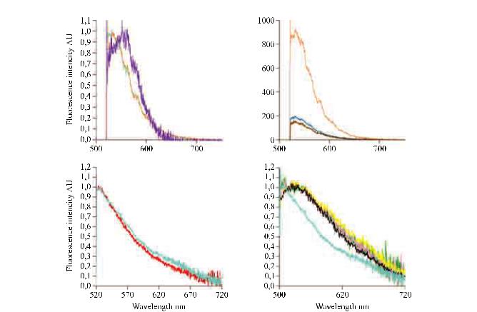
Fluorescence properties of the bronchial mucosae at 488 nm excitation are determined by the concentration of various cellular and extracellular fluorophores, including the intracellular flavins, that could originate from the epithelial cells, and specific crosslinks of collagens and elastin present in the subepithelial areas [10, 19, 20]. Microspectrometer experiments coupled with FCFM imaging have clearly demonstrated that the main fluorescence signal emitted after 488 nm excitation from both the bronchial and alveolar human system originates from the elastin component of the tissue (fig. 4) [7, 8, 21]. In contrast, the collagen fluorescence does not seem to significantly affect the FCFM image produced at 488 nm, the fluorescence yield of collagen at this wavelength being at least one order of magnitude smaller than that of elastin. Along the same lines, flavin cellular autofluorescence appears too weak
83

ВЕСТНИК ТРАНСПЛАНТОЛОГИИ И ИСКУССТВЕННЫХ ОРГАНОВ |
том XV № 2–2013 |
|
|
|
а |
b |
|
|
|
c |
d |
|
|
|
e |
f |
Fig. 3. – Ex vivo confocal imaging of bronchial and lung biopsies. a and b) Confocal fluorescence and reflectance imaging of fresh bronchial tissue. a) Autofluorescence images of submucosa ~25–30 m below surface, 437 nm illumination. b) Cell nuclei reflectance and fluorescence images superimposed. c and d) Extra-alveolar vessel and adjacent alveolar walls, fixed lung section. c) Conventional histology and d) corresponding fibred confocal fluorescence microscopy (FCFM) imaging (autofluorescence, 488 nm illumination). e and f) Ex vivo FCFM imaging offresh parenchymal lung tissue section (488 nm illumination). e) Autofluorescence, lung elastic network and alveolar mouth imaging. f) FCFM imaging after topical application of acriflavin onto the lung section. FCFM shows fluorescent acriflavin-stained nuclei from pneumocytes, in addition to the elastin framework of the alveolar duct. Scale bars: a) 10 m; d–f) 50 m. a and b) Courtesy of C. MacAuley, British Columbia Cancer Research Centre, Vancouver, BC, Canada; c and d) reproduced from [8]; e and f) L. Thiberville, Rouen University Hospital, Rouen, France
84
РЕГЕНЕРАТИВНАЯ МЕДИЦИНА И КЛЕТОЧНЫЕ ТЕХНОЛОГИИ
а |
|
b |
c |
|
d |
Fig. 4. – Nature of the fibred confocal fluorescence microscopy signal (autofluorescence, 488 nm illumination) in proximal bronchi and alveoli, in both smokers and nonsmoking subjects, as deduced from in vivo spectrometry experiments. a) Typical normalised autofluorescence spectra of healthy bronchial mucosa (orange), elastin powder (pale blue) and collagen (purple) excited at 488 nm. b) Autofluorescence spectra of healthy bronchial mucosa (orange), carcinoma in situ (dark blue) and Mounier Kühn syndrome (brown) at 488 nm excitation. Autofluorescence emission spectra of alveolar systems from c) nonsmokers (red) and d) healthy smokers (green, pink, yellow), during in vivo alveoscopy. c and d) Pale blue: elastin powder. d) Black: smoked cigarette. AU: arbitrary units. a and b) Reproduced from [7], with permission from the publisher; c and d) reproduced from [8]
to allow imaging of the epithelial layer using 488 nm FCFM [22].
FCFM devices using shorter wavelength may produce slightly different imaging of the bronchial wall connective tissue (fig. 3). However, imaging the epithelial layer on top of the basement membrane network needs another approach, the accessible way being currently the use of an exogenous fluorescent dye [18, 23]. In the future, devices based on multiple wavelengths [24], the adjunction of a reflectance device [10] or a multiphoton approach [9] may enable imaging of collagen, elastin and flavins simultaneously.
As a result, 488 nm excitation FCFM specifically images the elastin respiratory network that is contained in the basement membrane of the proximal airways and participates in the axial backbone of the peripheral interstitial respiratory system.
In vivo autofluorescence microimaging of the proximal bronchial wall
FCFM can easily be performed during a fibreoptic bronchoscopy under local anaesthesia [7, 8]. The tech-
nique of in vivo bronchial FCFM imaging is simple: the miniprobe is introduced into the 2-mm working channel of the bronchoscope and the probe tip applied onto the bronchial mucosae under sight control. The depth of focus being 50 μm below the contact surface, the system can image the first layers of the bronchial subepithelial connective tissue, presumably the lamina densa and the lamina reticularis [7].
At 488 nm excitation, FCFM produces very precise microscopic fluorescent images of the bronchial basement membrane zone. As seen in figure 5, in vivo FCFM bronchial microimaging reveals a mat of large fibres mainly oriented along the longitudinal axis of the airways with crosslinked smaller fibres, as well as larger openings (100–200 μm) corresponding to the bronchial gland origins. In vivo, the technique also makes it possible to record high-resolution images of small airways such as terminal bronchioles, which are recognisable by the presence of the helicoidal imprint of the smooth muscle on the inner part of the bronchiole (fig. 5) [7].
Application of the FCFM imaging system for the exploration of proximal bronchial diseases is still at its
85

ВЕСТНИК ТРАНСПЛАНТОЛОГИИ И ИСКУССТВЕННЫХ ОРГАНОВ |
том XV № 2–2013 |
|
|
|
а |
b |
|
|
|
c |
d |
Fig. 5. – In vivo fibred confocal fluorescence microscopy imaging of human bronchi and bronchioles. a) Proximal bronchus, opening of a bronchial gland (#). b) Main bronchus, elastic fibred network oriented along the longitudinal axis of the airways. c) Distal bronchiole showing helicoidal imprints of smooth muscles (arrow). d) Transitional bronchiole showing an alveolar bud (arrowhead). Scale bars: 50 m
beginning, with early results being published on bronchial wall remodelling in benign [7] and preinvasive bronchial lesions [7, 18].
In vivo assessment of bronchial wall remodelling
FCFM imaging of the bronchial wall microstructure underlying premalignant epithelia is significantly modified [7]. In these precancerous conditions, the elastic fibred pattern of the lamina reticularis disappears in most cases, while appearing disorganised in about onethird of the lesions, supporting the hypothesis of an early degradation of the basement membrane components in preinvasive bronchial lesions (fig. 6) [7]. Whether early remodelling of the lamina reticularis is associated with specific outcomes of the lesions should be further explored.
However, while this observation shed some light on the origin of the autofluorescence defect in precancerous bronchial lesions, the absence of epithelial cell
visualisation in this study did not allow the technique to differentiate between the different grades of progression of the precancerous bronchial lesions such as metaplasia/dysplasia/ carcinoma in situ (see section on FCFM assessment of the bronchial epithelial cell layer, and figure 6).
Besides the study of the premalignant bronchial wall alterations, the application of FCFM could be extended to the field of nonmalignant bronchial diseases. In one study, a complete disappearance of the bronchial wall fibred connective network was observed in a tracheomegaly syndrome, a pathological condition related to a defect in elastic component of the bronchial wall [7]. The same study also observed a remarkable FCFM aspect in a case of sarcoidosis, corresponding to subepithelial granuloma upon bronchial sampling. Whereas still limited, these observations indicate that per endoscopic FCFM could be used to study specific basement membrane remodelling alterations such as in chronic bronchial inflammations, asthma and chronic obstructive pulmonary disease.
86

РЕГЕНЕРАТИВНАЯ МЕДИЦИНА И КЛЕТОЧНЫЕ ТЕХНОЛОГИИ
|
|
|
а |
b |
|
|
|
c |
d |
Fig. 6. – Epithelial and subepithelial confocal microendoscopy imaging of normal and precancerous lesions. a) Normal elastic fibred network of the basement membrane zone. b) Disorganised basement membrane zone elastic network at the vicinity of a bronchial carcinoma in situ (CIS). c) Regular normal bronchial epithelium and d) CIS. a and b) Images taken at 488 nm excitation without exogenous fluorophore (Cellvizio® 488). Under these conditions, only the elastin of the basement membrane is detectable and imaged. c and d) Taken at 660 nm excitation using topical methylene blue (0.1%), in order to image the epithelial layer (Cellvizio® 660). All images were recorded in vivo during bronchoscopy, but with two different laser scanning units (Cellvizio® 488 and 660; Mauna Kea Technologies, Paris, France). Scale bars: a, c, d) 50 m; b) 40 m. Source: L. Thiberville, Rouen University Hospital, Rouen, France
FCFM ASSESSMENT OF THE BRONCHIAL EPITHELIAL CELL LAYER
In order to be successfully applied to the exploration of precancerous/cancerous bronchial epithelium, the FCFM technique would need to be coupled with the use of an exogenous nontoxic fluorophore. Ex vivo studies have shown that the resolution of the system is not a limitation for nuclear or cellular imaging [7, 8]. Exogenous fluorophores that could be activated at 488 nm, such as acriflavin (a putative mutagen agent) or fluorescein solution, which does not stain the nuclei [25], are not approved for intrabronchial use. Recently, Lane et al. [23] have used a confocal microendoscope prototype at 488 nm excitation and topical physiological pH cresyl violet to provide cellular contrast in the bronchial epithelium both in vitro and in vivo.
Methylene blue is a nontoxic agent which is commonly used during bronchoscopy for the diagnostic of bronchopleural fistulae. It is also used in gastroenterology for chro- mo-endoscopic detection of precancerous lesions [26–28], as well as for in vivo microscopic examination of the GI tract and bronchus using a novel endocytoscopic system [29, 30]. Methylene blue is a potent fluorophore that enters the nuclei and reversibly binds to the DNA, before being reabsorbed by the lymphatics. In order to give a fluorescent signal, methylene blue needs to be excited around 660 nm, and is therefore accessible to FCFM intravital imaging using this excitation wavelength.
Preliminary study has demonstrated that Cellvizio® 660/topical methylene blue makes it possible to reproducibly image the epithelial layer of the main bronchi (fig. 6) [18]. Future studies using this technique could make it possible to differentiate normal, premalignant
87

ВЕСТНИК ТРАНСПЛАНТОЛОГИИ И ИСКУССТВЕННЫХ ОРГАНОВ |
том XV № 2–2013 |
and malignant alterations at the microscopic level in vivo. If this strategy is successful, FCFM may become a very powerful technique for in vivo diagnosis of early malignant and premalignant conditions of the bronchial tree, allowing the analysis of both the epithelial and subepithelial layers during the same procedure.
DISTAL AND TRANSITIONAL BRONCHIAL IMAGING
When progressing towards the more distal parts of the bronchial tree, small noncartilaginous bronchioles are easily recognisable because of the helicoidal imprint of the smooth muscles on the bronchiolar walls (fig. 5). However, the orthogonal branching and the small calibre of the terminal and respiratory bronchioles in humans compared with the probe size implies that the progression of the probe towards the acinus regularly bypasses the transitional respiratory bronchioles. While FCFM images of alveolar buds in respiratory bronchio-
les could be observed casually (fig. 5) [8], FCFM study of the distal membranous and respiratory bronchioles appears difficult, unless thinner probes, currently devoted to experimental animal imaging, become clinically available in the future [13].
FCFM IMAGING OF THE ACINUS AND PERIPHERAL CONNECTIVE TISSUE NETWORK
Earlier work has demonstrated that elastin represents up to 50% of the peripheral lung connective tissue fibres [31]. Following the first work on the proximal bronchi, FCFM rapidly appeared to be able to image the elastic framework of the distal lung as well [8]. In the acinus, elastin is present in the axial backbone of the alveolar ducts and alveolar entrances, as well as in the external sheath of the extra-alveolar microvessels [32, 33], explaining the nature of the intra-acinar FCFM imaging (figs 3 and 7).
|
|
|
а |
b |
|
|
|
c |
d |
Fig. 7. – In vivo fibred confocal fluorescence microscopy (FCFM) imaging during alveoscopy, 488 nm illumination. a and c) Nonsmoking subject. Elastin framework of a) an alveolar mouth (diameter 267.7 m (double arrow)) and c) extra-alveolar microvessel (arrow). b and d) FCFM imaging of smoker alveoli, showing alveolar walls, edge of an alveolar duct (arrows) and alveolar macrophages. Scale bars: 50 m
88