

- •Dedication
- •Preface
- •Acknowledgments
- •Figure Credits
- •Expert Consultants and Reviewers
- •Contents
- •Descriptive Terms for Normal Cells
- •Descriptive Terms for Abnormal Cells and Tissues
- •Epithelium
- •Glands
- •Introduction and Key Concepts for Connective Tissue
- •Cartilage
- •Bone
- •Introduction and Key Concepts for the Nervous System
- •Peripheral Blood Cells
- •Hemopoiesis
- •Introduction and Key Concepts for the Circulatory System
- •The Cardiovascular System
- •Introduction and Key Concepts for the Lymphoid System
- •Cells in the Lymphoid System
- •Introduction and Key Concepts for the Respiratory System
- •Conducting Portion
- •Respiratory Portion
- •Introduction and Key Concepts for the Urinary System
- •Introduction and Key Concepts for the Integumentary System
- •Oral Mucosa
- •Teeth
- •Introduction and Key Concepts for the Digestive Tract
- •Introduction and Key Concepts for the Endocrine System
- •Introduction and Key Concepts for the Male Reproductive System
- •Introduction and Key Concepts for the Female Reproductive System
- •Introduction and Key Concepts for the Eye
- •Introduction and Key Concepts for the Ear
- •Introduction
- •Preservation versus Fixation
- •Fixatives and Methods of Fixation
- •Sectioning and Mounting
- •Staining
- •Index

280 UNIT 3 ■ Organ Systems
Figure 15-11B |
Villi of the Small Intestine |
Figure 15-12A |
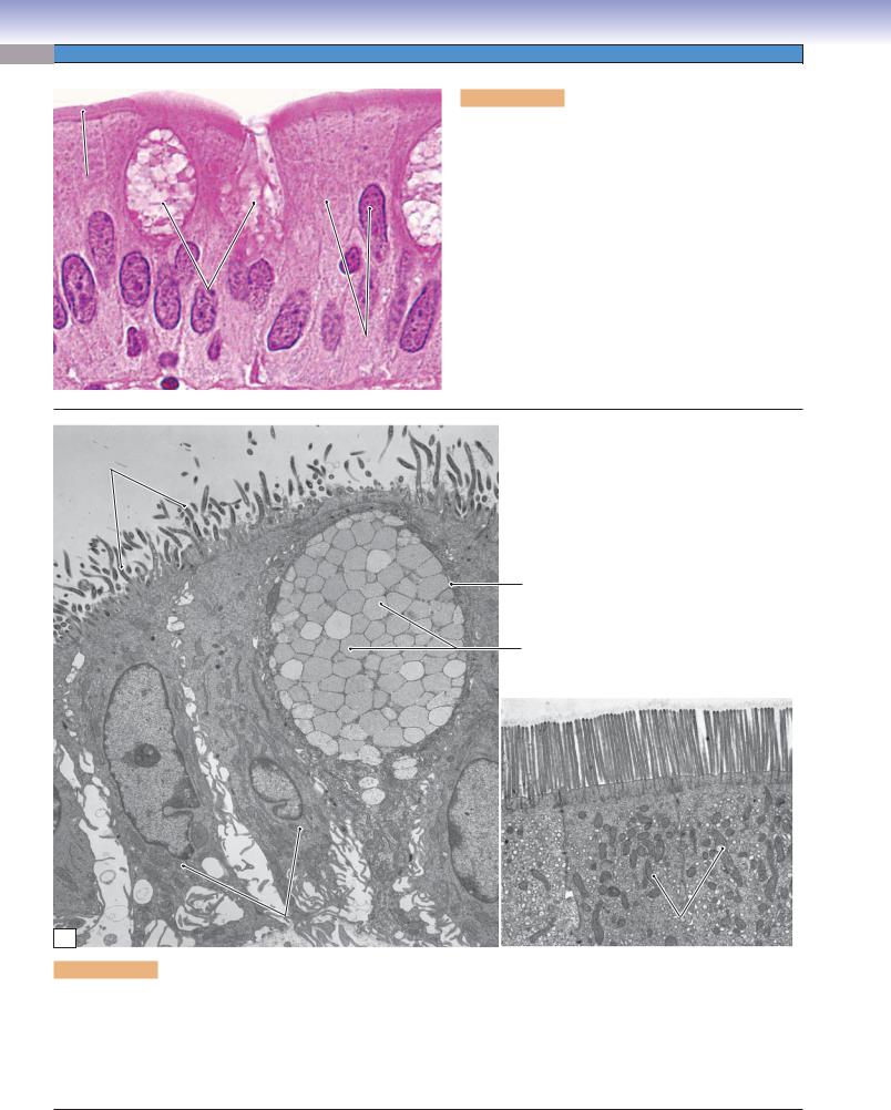
Columnar Absorptive and Goblet Cells of the Small Intestine |
Figure 15-12B |
Goblet, Columnar Absorptive Cells, and Microvilli |
Figure 15-13A |
Paneth Cells, Small Intestine |
Figure 15-13B |
Enteroendocrine Cells, Small Intestine |
Figure 15-14A |
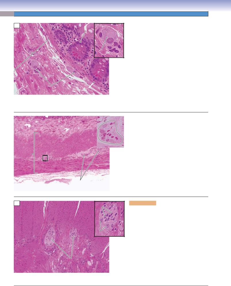
Submucosal/Meissner Plexus, Small Intestine |
Figure 15-14B |
Muscularis Externa, Small Intestine |
Figure 15-14C |
Myenteric/Auerbach Plexus, Muscularis Externa of the Small Intestine |
Figure 15-15A |
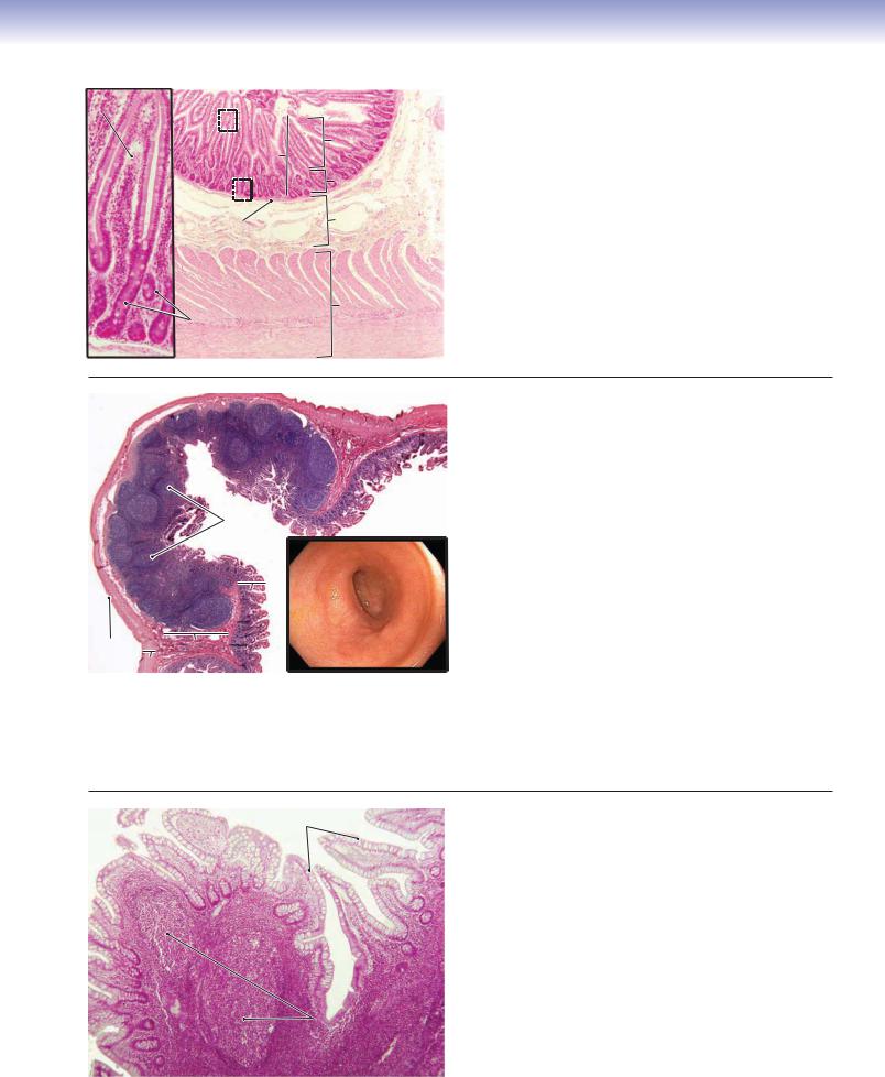
Jejunum, Small Intestine |
Figure 15-15B |
Ileum with Peyer Patches, Small Intestine |
Figure 15-15C |
Mucosa of the Ileum, Small Intestine |
Large Intestine |
|
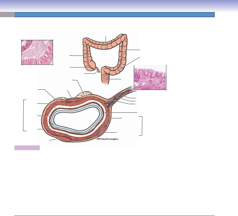
Figure 15-16 |
Overview of the Large Intestine |
Figure 15-17A,B |
Colon, Large Intestine |
Figure 15-17C |
Mucosa of the Colon, Large Intestine |
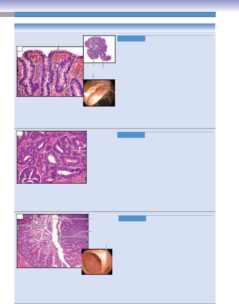
Figure 15-18A |
Clinical Correlation: Colon Polyps |
Figure 15-18B |
Clinical Correlation: Colorectal Cancer |
Figure 15-18C |
Clinical Correlation: Meckel Diverticulum |
Figure 15-19A |
Appendix and Cecum |
Figure 15-19B |
Anorectal Junction |
Figure 15-19C |
Clinical Correlation: Hemorrhoids |
Figure 15-20A |
Clinical Correlation: Ulcerative Colitis |
Figure 15-20B |
Clinical Correlation: Crohn Disease |
Synopsis 15-1 |
Pathological and Clinical Terms for the Digestive Tract |
Table 15-1 |
Digestive Tract |
Introduction and Key Concepts for the Digestive Tract
The digestive system is composed of the oral cavity, digestive tract, and digestive glands with associated organs. The “Oral Cavity” is discussed in Chapter 14; the “Digestive Glands and Associated Organs” are discussed in Chapter 16. The digestive tract is discussed in this chapter. It includes the esophagus, stomach, small intestine, and large intestine. The digestive tract is a continuation of the oral cavity, and its main functions are to ingest food and to digest the food as it passes along the tract. In this process, nutrients and water are absorbed, and waste materials are prepared for elimination from the body. Each section of the digestive tract has its unique histological features, which are closely associated with the function of that part of the tract, although there are some common characteristics: (1) Organs of the digestive tract are all hollow; (2) they are composed of four general tunic layers: mucosa, submucosa, muscularis externa, and adventitia or serosa; (3) they are innervated by the enteric portion of the autonomic nervous system, also known as the enteric nervous system (or the “second brain”); (4) they include epithelium, connective tissue, muscle, blood and lymphatic vessels, lymphatic nodules, and nerve fibers; and (5) they contain glands in the lamina propria or submucosa.
General Structure of the
Digestive Tract
Based on its histological organization, the wall of the digestive tract can be divided into four tunics (Fig. 15-3).
1.Mucosa is the innermost layer of the digestive wall. It includes epithelium, lamina propria, and muscularis mucosae. The epithelium consists of simple columnar epithelium lining most of the tract and stratified squamous epithelium lining the two ends, the esophagus and anal canal. The lamina propria is a loose connective tissue that contains abundant ground substance, many fibers, and numerous connective tissue cells such as fibroblasts, macrophages, mast cells, plasma cells, and leukocytes (see Chapter 4, “Connective Tissue”). Various types of glands are found in the lamina propria depending on the region of the digestive tract. The muscularis mucosae is a very thin layer of smooth muscle, which is the boundary between the mucosa and the submucosa. It is usually arranged in an inner circular and outer longitudinal layer. However, the muscularis mucosae varies in different regions, and it is often difficult to distinguish between the muscle layers.
2.Submucosa is a thick layer of dense irregular connective tissue. This layer contains blood vessels, lymphatic vessels,

CHAPTER 15 ■ Digestive Tract |
281 |
and submucosal (Meissner) plexuses, which contain nerve fibers and neurons of the enteric nervous system. In some regions of the digestive tract, this layer is characterized by mucous glands or lymphatic nodules.
3.Muscularis externa is composed of two or three oblique, circular, and longitudinal muscle layers, which vary from region to region. Most of the muscularis externa consists of smooth muscle fibers, but the upper and middle esophagi contain some skeletal muscle. The myenteric (Auerbach) plexuses (nerve fibers and neurons of the enteric nervous system) are located between the muscle layers. They innervate and control contraction of the muscularis externa.
4.Serosa and adventitia are coverings of the outermost wall of the digestive tract. Most parts of the digestive tract are covered by serosa, a thin layer of loose connective tissue lined by mesothelium. The mesothelium produces a lubricating fluid that reduces friction during movement of the organs against each other (see Chapter 3, “Epithelium and Glands,” Fig. 3-2A,B). The serosa is the visceral layer of the peritoneum and covers the wall of the digestive tract where it connects to the mesentery in the peritoneal cavity (intraperitoneal organs). The adventitia is a layer of loose connective tissue without mesothelium that covers the upper region of the esophagus, part of the duodenum, and the lower part of the digestive tract, such as the rectum and anal canal. Adventitia covers regions of the digestive tract where it is connected to other organs or to the body wall (e.g., retroperitoneal organs).
Esophagus
The esophagus is the upper part of the digestive tract, connecting the oral cavity to the stomach. The major function of the esophagus is to provide passage for food from the mouth to the stomach. The luminal surface of the esophagus is lined by nonkeratinized stratified squamous epithelium. Mucous glands called esophageal glands are located in the submucosa of the esophagus. The muscularis externa consists of two layers of muscle: inner circular and outer longitudinal layers. Both skeletal and smooth muscle fibers are found in the muscularis externa of the esophagus. The proportions of skeletal and smooth muscle fibers are different in different regions of the esophagus. The esophagus can be divided into three regions: the upper esophagus, middle esophagus, and lower esophagus
(Figs. 15-4A to 15-5A).
1.The upper esophagus connects the oropharynx to the middle esophagus. This segment contains numerous esophageal glands in the submucosa. These glands secrete mucus to lubricate the esophageal wall so that food will pass through easily. The upper esophagus contains only skeletal muscle fibers in the muscularis externa. These are voluntary muscle fibers and are innervated by the glossopharyngeal nerve (cranial nerve [CN] IX) (see Fig. 15-4B).
2.The middle esophagus has mucosa similar to that of the upper esophagus. The esophageal glands in the submucosa are less numerous than in the upper esophagus. The muscularis externa contains both skeletal and smooth muscles (Fig. 15-4C).
3.The lower esophagus connects the esophagus to the cardia of the stomach. This region contains large numbers of mucous glands in the lamina propria and submucosa. These are called esophageal cardiac glands and produce mucous
secretions to protect the lower esophagus from being damaged by reflux of acidic gastric juices from the stomach. The lower esophagus contains only smooth muscle fibers in the muscularis externa. These are controlled by the enteric branches of the vagus nerve (CN X) (see Fig. 15-5A).
Stomach
The stomach is a “J”-shaped sac (hollow) organ. It temporarily stores food, mixes food with gastric juice, and initiates the processing of food by breaking it down into simpler substances that are easier to digest. The stomach can be divided into the cardia, fundus, body, and pylorus. The inner surface of the stomach is lined by simple columnar epithelium composed mainly of surface mucous cells. The surface epithelium of the stomach is invaginated into the lamina propria to form gastric pits. These pits serve as ducts for the glands in the lamina propria, which vary from region to region in the stomach.
1.The cardiac region connects to the lower esophagus at the esophagogastric junction, which is characterized by a change from the nonkeratinized stratified squamous epithelium of the esophagus to the simple columnar epithelium of the stomach. A thickened smooth muscle ring called the gastroesophageal sphincter (lower esophageal sphincter) or cardiac sphincter surrounds the opening at the junction of the lower esophagus and cardiac region of the stomach. This smooth muscle contracts to prevent the acidic stomach contents from entering the esophagus. The glands in the lamina propria of the cardia are called cardiac glands and are branched tubular glands with coiled secretory portions. The cardiac gland contains mainly mucus-secreting cells and some stem cells, enteroendocrine cells, and, occasionally, parietal cells. The mucus-secreting cells mainly produce mucus and lysozymes. The mucus protects the stomach wall from acidic gastric juices; lysozymes destroy bacterial membranes, preventing bacterial infections (Fig. 15-7A).
2.The fundic and body regions form the largest portions of the stomach. Their mucosa has similar histological characteristics, including short gastric pits and long branched tubular glands in the lamina propria. The glands are called fundic or gastric glands in both the fundus and the body regions. The gastric glands contain mainly parietal cells and chief cells, along with some stem cells, mucous neck cells, and enteroendocrine cells. Parietal cells are more numerous in the superior regions of the glands; these cells produce large quantities of hydrochloric acid (HCl), creating an acidic environment to help digestion. Parietal cells also secrete intrinsic factor (IF), which is required for the absorption of vitamin B12. Chief cells are located in the more inferior regions of the glands; they secrete precursor enzymes such as pepsinogen, which is activated by HCl and becomes pepsin. Pepsin helps to break down proteins (particularly protein collagen) into simpler, more absorbable compounds. Chief cells also secrete precursors of lipases, which help in lipid digestion (Fig. 15-7B,C).
3.The pyloric region is the lower end of the stomach, which connects with the duodenum. Its mucosa is similar to that of the cardia, with long gastric pits and short, coiled secretory portions. A circular smooth muscle ring called the pylorus sphincter (pyloric valve) surrounds the end of the pylorus region. This valve controls the entry of stomach contents into the duodenum. The glands in the lamina propria of the pylorus are called pyloric glands and contain primarily

282 UNIT 3 ■ Organ Systems
mucus-secreting cells and two special types of enteroendocrine cells: gastrin-secreting cells (G cells) and somatostatinsecreting cells (D cells), as shown in Figure 15-8A. These enteroendocrine cells regulate gastric (HCl) secretion.
Small Intestine
The small intestine (Figs. 15-9 to 15-15C) is a hollow organ of small diameter that is typically 6 to 7 m long. It is the major site for the absorption of nutrients. Important features of the small intestine are villi and microvilli, which increase surface area for absorption. Intestinal glands called glands (crypts) of Lieberkühn are located in the lamina propria of the small intestine. Villi project into the lumen of the intestine; the glands of Lieberkühn open into the mucosa at the base of the villi (Fig. 15-11A,B). The small intestine can be divided into three parts: the duodenum, jejunum, and ileum.
1.The duodenum is the shortest segment of the small intestine, about 20 to 25 cm long. It has small openings called duodenal papillae (minor and major [Figs. 16-9A and 16-15A]), which allow pancreatic juice and bile to enter the digestive tract. It has a similar general structure to other parts of the small intestine (Fig. 15-9). However, the Brunner glands (mucussecreting gland) in the submucosa are a unique feature of the duodenum (Fig. 15-10A,B).
2.The jejunum is much longer than the duodenum, about 2.5 m long (two fifths of the rest of the small intestine). It has long villi and a somewhat increased number of goblet cells. It has neither Brunner glands nor Peyer patches (Fig. 15-15A).
3.The ileum is the longest segment, about 4 m long (three fifths of the rest of the small intestine). It has short villi with significantly increased numbers of goblet cells on the surface of the mucosa. There are clusters of lymphatic nodules in the lamina propria of the ileum; sometimes these lymphatic nodules extend into the submucosal layer. These clusters of lymphatic nodules are called Peyer patches and are unique to the ileum (Fig. 15-15B,C).
Large Intestine
The large intestine (Figs. 15-16 to 15-19B) is a hollow organ with a relatively large diameter compared to the small intestine and is about 1.5 m long. It is the last region of the digestive tract and is the major site for absorption of water and salts. It also forms, stores, and eliminates feces. Most of the regions of the large intestine have tunics that are similar to those of the small intestine, but there are no villi in the mucosa. There are large numbers of goblet cells in the large intestine. These cells produce
mucus, which helps in the formation of the feces and protects and lubricates the surface of the intestinal wall. The large intestine includes the cecum, appendix, colon, rectum, and anal canal.
1.The cecum is the most proximal region of the large intestine. It is a small, blind pouch of the large intestine where the ileum connects to the ascending colon. A sphincter muscle, a thickening of the muscularis mucosae, is called the ileocecal valve and is located at the junction of the ileum and cecum. It prevents the contents of the large intestine from backing up into the small intestine (Fig. 15-16).
2.The appendix is a small, blind tube that attaches to the posterior-medial wall of the cecum. It has the general tunic structure of the intestine and a small irregular lumen. There are many lymphatic nodules in the lamina propria (Fig. 15-19A).
3.The colon is the longest segment of the large intestine. It includes the ascending colon, transverse colon, descending colon, and sigmoid colon. The proximal half of the colon is responsible for the majority of the absorption of water and salt; the distal half of the colon has only a small absorptive function and is predominantly for processing and storing feces. The colon does not have villi; it has a smoother surface than the small intestine. Columnar absorptive cells and goblet cells line the mucosa. The large intestinal glands, the glands (crypts) of Lieberkühn, contain primarily goblet cells, columnar cells, enteroendocrine cells, and stem cells. Lymphatic nodules may also be found in the lamina propria. The muscularis externa consists of inner circular layers of muscle; the outer longitudinal smooth muscle layer becomes three teniae coli (Figs. 15-16 and 15-17A–C).
4.The rectum and anal canal are the last segments of the large intestine. The junction between the rectum and the anal canal is called the “anorectal junction.” The mucosa of the rectum is similar to that of the colon but has fewer glands of Lieberkühn. The main function of the rectum is the temporary storage of feces. The sensory receptors in the rectum send signals to the brain when feces need to be evacuated. The anal canal is the distal end of the large intestine. Most of the anal canal is lined by stratified squamous epithelium, although simple cuboidal epithelium may be present at the anorectal junction. Sebaceous glands and hair follicles may be found at or near the anal opening. There are many veins in the lamina propria and submucosa of the anal canal. The term hemorrhoids refers to the condition in which these veins become chronically swollen and inflamed in the rectal and anal regions (Fig. 15-19B).
CHAPTER 15 ■ Digestive Tract |
283 |
Esophagus |
|
Stomach |
|
|
|
|
|
|
Small intestine |
Transverse |
Large intestine |
|
colon |
|
|
|
Ascending colon
Descending Small  colon intestine
Cecum
 Sigmoid
 Rectum
Appendix
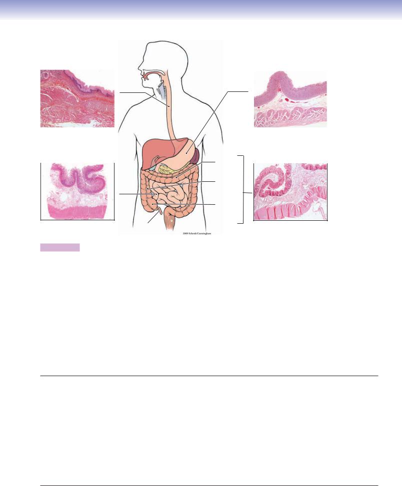
Figure 15-1. Overview of the digestive tract. H&E, 5 to 6
The digestive tract, also called the alimentary canal, includes the esophagus, stomach, small intestine, and large intestine. (1) The esophagus transports food to the stomach. Its luminal surface is lined by stratified squamous nonkeratinized epithelium and contains mucous glands in the lamina propria. It has both skeletal and smooth muscles in the muscularis externa. (2) The stomach temporarily stores and digests food. Its luminal surface is lined by simple columnar epithelium and contains gastric glands in the lamina propria. It has three layers of smooth muscle in the muscularis externa. (3) The small intestine digests and absorbs carbohydrates, proteins, and lipids. The small intestine includes the duodenum, jejunum, and ileum. The luminal surface is lined by simple columnar epithelium and contains the glands (crypts) of Lieberkühn in the lamina propria. There are two layers of smooth muscles (inner circular and outer longitudinal muscles) in the muscularis externa. (4) The large intestine includes the cecum, appendix, colon, rectum, and anal canal. The main functions of the large intestine are the absorption both of a large volume of the water that enters it (90%) and of electrolytes (e.g., Na+ and Cl−) and the formation of the feces. Most parts of the large intestine are lined by simple columnar epithelium, but stratified squamous epithelium lines the anal canal. The glands of Lieberkühn are located in the lamina propria. The muscularis externa contains an inner layer of circular smooth muscle and an outer layer of longitudinal smooth muscle, which forms three teniae coli (see Fig. 15-16). In general, the submucosa of the digestive tract is a thick layer of connective tissue containing blood vessels, lymphatic vessels, and submucosal (Meissner) plexuses consisting of nerve fibers and neuron cell bodies in the enteric nervous system. The myenteric (Auerbach) plexuses located in the muscularis externa are also components of the enteric nervous system.
Digestive Tract
I.Esophagus
A.Upper esophagus (skeletal muscle)
B.Middle esophagus (mixed skeletal and smooth muscles)
C.Lower esophagus (smooth muscle)
II.Stomach
A.Cardia
B.Fundus
C.Body
D.Pylorus
III.Small Intestine
A.Duodenum
B.Jejunum
C.Ileum
IV. Large Intestine
A.Cecum
B.Appendix
C.Colon (ascending, transverse, descending, and sigmoid)
D.Rectum
E.Anal canal
284 UNIT 3 ■ Organ Systems
Fig. 15-4BA
Fig. 15-4C
Fig. 15-5A
Fig. 15-7A
Fig. 15-7B
Fig. 15-10A,B |
Fig. 15-7C |

 Fig. 15-8A
Fig. 15-15A
Fig. 15-17A,B,C
Fig. 15-15B,C
Fig. 15-19A 
 Fig. 15-9B19B
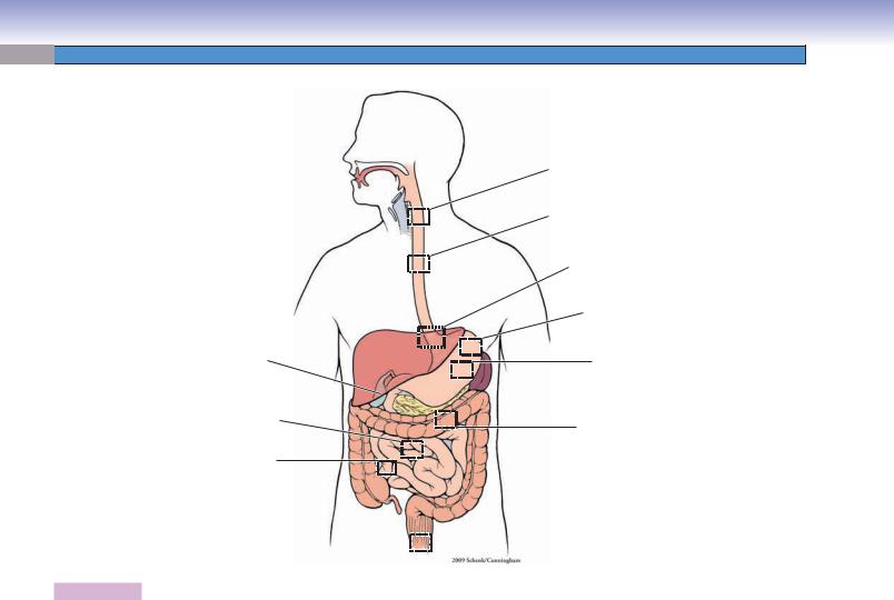
Figure 15-2. Orientation of detailed digestive tract illustrations.
Digestive Tract with Figure Numbers
Esophagus |
Figure 15-13A |
|
Figure 15-4A |
Figure 15-13B |
|
Figure 15-14A |
||
Figure 15-4B |
||
Figure 15-14B |
||
Figure 15-4C |
||
Figure 15-14C |
||
Figure 15-5A |
||
Figure 15-15A |
||
Figure 15-5B |
||
Figure 15-15B |
||
Figure 15-5C |
||
Figure 15-15C |
||
|
||
Stomach |
Large intestine |
|
Figure 15-6 |
||
Figure 15-7A |
Figure 15-16 |
|
Figure 15-7B |
Figure 15-17A |
|
Figure 15-7C |
Figure 15-17B |
|
Figure 15-8A |
Figure 17-17C |
|
Figure 15-8B |
Figure 15-18A |
|
Figure 15-8C |
Figure 15-18B |
|
Small intestine |
Figure 15-18C |
|
Figure 15-19A |
||
Figure 15-9 |
Figure 15-19B |
|
Figure 15-19C |
||
Figure 15-10A |
||
Figure 15-20A |
||
Figure 15-10B |
||
Figure 15-20B |
||
Figure 15-10C |
||
|
||
Figure 15-11A |
|
|
Figure 15-11B |
|
|
Figure 15-12A |
|
|
Figure 15-12B |
|
|
|
|
CHAPTER 15 ■ Digestive Tract |
285 |
Small intestine
|
|
|
4. Serosa / adventitia |
|
|
|
|
|
Outer longitudinal muscle 3. Muscularis |
||
|
|
|
Inner circular muscle |
|
externa |
|
|
|
|
|
|
|
|
Stomach |
2. Submucosa |
|
|
|
|
|
|
|
|
4. Serosa |
|
Muscularis mucosae |
|
|
|
|
|
|
Lamina propria |
1. Mucosa |
|
|
|
|
Epithelium |
||
|
Longitudinal |
|
|
|
|
|
|
|
|
|
|
|
muscle |
|
|
|
|
3. Muscularis |
Circular |
|
|
|
|
externa |
muscle |
|
|
|
|
|
Oblique |
|
|
|
|
|
muscle |
|
|
|
|
2. Submucosa |
|
Large intestine |
|
|
|
1. Mucosa |
|
Muscularis mucosae |
|
||
|
|
|
|
||
|
|
|
Lamina propria |
|
1. Mucosa |
|
|
|
Epithelium |
|
|
|
|
|
2. Submucosa |
||
|
|
|
Circular muscle |
|
3. Muscularis |
|
|
|
Longitudinal muscle |
||
|
|
|
band (teniae coli) |
|
externa |
|
|
|
|
|
|
4. Serosa/adventitia
Figure 15-3. General structure of the wall of the digestive tract.
The wall of the digestive tract can be divided into several basic tunics (layers) based on histologic organization. (1) Mucosa: epithelium, lamina propria, and muscularis mucosae. The epithelium is composed of simple columnar cells in most of the digestive tract, except for stratified squamous cells in the esophagus and anal canal. The lamina propria is a layer of loose connective tissue beneath the epithelium. The muscularis mucosae is a thin layer of smooth muscle; it marks the boundary between the mucosa and the submucosa.
(2) Submucosa: dense irregular connective tissue with blood vessels, lymphatic vessels, and submucosal plexuses (Meissner plexuses). Mucous glands may be present in this layer. (3) Muscularis externa: two or three layers of smooth muscle. It may also include skeletal muscle as in the esophagus. There are blood vessels and myenteric (Auerbach) plexuses that lie between the muscle layers. (4) Serosa/ adventitia: The outermost layer is called serosa if it is composed of loose connective tissue with blood vessels and nerves passing through and is covered by mesothelium. It is called adventitia if covered by a layer of connective tissue without mesothelium lining. The serosa covers organs within the abdominal or pelvic cavities (intraperitoneal), whereas the adventitia covers organs and serves as a capsule and attachment between the organs or between an organ and the body wall (retroperitoneal).
Tunics (Layers) of the Digestive Tract
1. |
Mucosa |
Outer longitudinal muscle layer |
|
Epithelium |
Oblique muscle layer (stomach) |
|
Lamina propria |
4. Serosa/Adventitia |
|
Muscularis mucosae |
Serosa: outermost layer composed of connective tissue |
2. |
Submucosa |
covered by mesothelium |
|
Mucous glands and lymphatic nodules may be present |
Adventitia: outermost layer composed of connective tissue |
3. |
Muscularis Externa |
without mesothelium covering |
|
Inner circular muscle layer |
|
|
|
|
286 UNIT 3 ■ Organ Systems
Esophagus
Muscularis
 externa
Submucosa
Mucosa
Lumen
Muscularis
mucosae Esophageal glands A
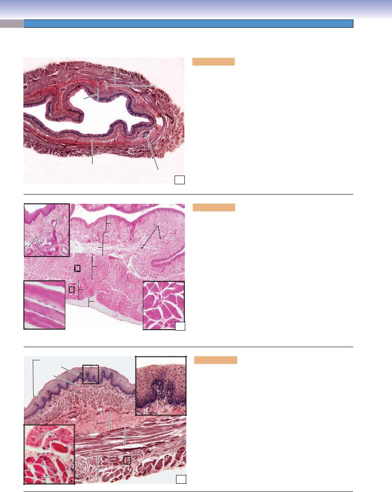
Figure 15-4A. Overview of the esophagus. H&E, 11
The esophagus can be divided into three regions: the upper, middle, and lower esophagus. The esophagus is a long tube that connects the oropharynx to the stomach. Like other parts of the digestive tract, the esophagus has mucosa, submucosa, muscularis externa, and adventitia/serosa. (1) The mucosa is composed of epithelium, lamina propria, and muscularis mucosae. The mucosa of the esophagus has folds extending into the lumen. Stratified squamous epithelium covers the inner surface of the esophagus. The muscularis mucosae is composed of a single layer of longitudinal smooth muscle. (2) The submucosa contains mucous glands called esophageal glands, which secrete mucus and provide lubrication that aids in swallowing. (3) The muscularis externa is composed of two layers of muscle organized into inner circular and outer longitudinal layers. (4) The outermost wall of the esophagus is commonly covered by adventitia (Fig. 15-4B), but the lower esophagus is covered by serosa.
Duct of |
|
|
glands |
|
Esophageal |
|
|
|
Esophageal |
Mucosa |
glands |
|
|
|
glands |
|
|
|
Submucosa |
|
|
Inner circular muscle |
|
Outer longitudinal |
Outer longitudinal muscle |
Inner circular |
(skeletal) muscle |
(skeletal) muscle |
|
|
Adventitia |
|
B
Figure 15-4B. Upper esophagus. H&E, 17; (upper inset)
40; (lower insets) 216
The upper esophagus, connecting to the oropharynx, is the first segment of the esophagus. The muscularis externa of the upper esophagus contains two layers of skeletal muscle. The skeletal muscle is a voluntary muscle that helps to initiate swallowing. This muscle is innervated by the glossopharyngeal nerve. The muscularis externa of the esophagus has distinguishable muscle components, which vary in different regions. The upper esophagus has skeletal muscle, the middle esophagus has mixed skeletal and smooth muscles, and the lower esophagus has only smooth muscle in the muscularis externa. The muscularis externa plays an important role in producing contractions of the esophagus that transport food from the oral cavity to the stomach. The major movement of the esophagus is peristalsis (waves of involuntary contraction), as in other parts of the digestive tract. The initiation of swallowing is voluntary because the upper esophagus contains only skeletal muscle fibers in the muscularis externa.
Mucosa
Epithelium
Epithelium
Lamina propria
Muscularis  mucosae
Submucosa |
|
Smooth muscle |
|
Inner circular muscle |
|
Outer longitudinal |
muscle |
Skeletal |
C |
muscle |
|
Figure 15-4C. Middle esophagus. H&E, 34; insets (left)
208, (right) 116
The mucosa of the esophagus is covered by a thick layer of nonkeratinized stratified squamous epithelium, which reflects its function in resisting abrasion and friction. The epithelium of the esophagus is continuously renewed by basal cells that migrate and differentiate from the basal layer. Most of the digestive tract is covered by simple columnar epithelium; only the two ends (esophagus and anal canal) are covered by stratified squamous epithelium. The muscularis externa of the middle esophagus contains mixed skeletal and smooth muscle fibers, which are organized in inner circular and outer longitudinal muscle bundles. Each skeletal muscle fiber is larger, has multiple nuclei positioned peripherally, and stains darker than the smooth muscle fiber. Each smooth muscle fiber has a single nucleus located in the center of the muscle fiber.

CHAPTER 15 ■ Digestive Tract |
287 |
A |
Simple |
columnar epithelium
 Mucosa
Stratified squamous epithelium
Submucosa
 Mucosa
 Submucosa
epithelium
Lamina Muscularis propria
externa
Esophageal |
cardiac glands |
Figure 15-5A. Lower esophagus, esophagogastric junction (esophagus on left; stomach on right of illustration). H&E, 11; insets (upper) 57; (lower) 45
The lower esophagus meets the stomach at the esophagogastric junction. The esophagus is lined by stratified squamous epithelium, and the cardiac region of the stomach is lined by simple columnar epithelium. The change in the lining epithelium reflects the change in function from a conduit for food transport to an organ of digestion. The muscularis externa of the lower esophagus is composed of two layers of smooth muscle fibers, which are controlled by the vagus nerve. Mucous glands are also found in the lamina propria of the lower esophagus (right inset). These glands are called esophageal cardiac glands. They produce mucus to protect the epithelial wall of the esophagus from the reflux of acidic gastric juices coming from the stomach.
In some patients, the epithelium of the lower esophagus (stratified squamous epithelium) changes to stomachlike epithelium (simple columnar epithelium). This pathologic change is called metaplasia. It is due to the long-term chemical irritation caused by gastroesophageal reflux.
CLINICAL CORRELATIONS
B |
Squamous |
epithelium |
|
|
Metaplastic |
|
Barrett |
|
epithelium with |
|
goblet cells |
|
Goblet cell |
Figure 15-5B. Barrett Esophagus. H&E, 48; inset 82 Barrett esophagus is a chronic complication of gastroesophageal reflux disease (GERD), characterized by metaplasia of the stratified squamous epithelium of the lower esophagus into a specialized glandular epithelium with goblet cells. Patients with Barrett esophagus have an increased risk of developing adenocarcinoma (cancer of the esophagus) of the distal esophagus. Common symptoms include heartburn, trouble swallowing, and weight loss. Endoscopically, Barrett esophagus appears as salmon-colored “tongues” of mucosa extending proximally from the gastroesophageal junction. This photomicrograph shows the metaplastic glandular epithelium with goblet cells that have replaced the normal squamous epithelium and the inflammatory cells (mainly lymphocytes and plasma cells) infiltrating the connective tissue.
 C
C
Squamous cell  carcinoma
Keratin pearl
Figure 15-5C. Esophageal Carcinoma. H&E, 97
Esophageal carcinoma is a malignant neoplasm that stems from the epithelial cells lining the inner surface of the esophagus. Worldwide, squamous cell carcinoma is the most common type of esophageal cancer, and it is associated with alcohol and tobacco use in the United States and Europe and with mutagenic substances and nutritional deficiencies in less-well-developed parts of the world. In the United States, adenocarcinoma of the lower esophagus is becoming more frequent, representing about 50% of esophageal cancers. The major known risk factor for the development of adenocarcinoma is chronic GERD causing
Barrett esophagus, a metaplastic change in the squamous mucosa of the distal esophagus to a glandular type of epithelium with goblet cells. Esophageal cancer is characterized by progressive difficulty in swallowing, loss of weight, fatigue, and chest pain. Pathological changes include ulcerations, exophytic masses, and thickening and narrowing of the lumen. Treatment includes surgery (esophagectomy) and chemotherapy. This photomicrograph shows a moderately differentiated squamous cell carcinoma with focal keratin production in the center, called a keratin pearl.
288 UNIT 3 ■ Organ Systems
Stomach
Esophagogastric |
|
Fundus of stomach |
junction |
|
|
Esophagus |
|
|
|
|
Fundus
Cardia
Longitudinal
muscle
 Serosa
Circular muscle 
Duodenum |
Oblique |
Body |
|
||
muscle |
|
|
|
|
Pylorus
Pylorus of stomach
Submucosa
Mucosa
Gastric
rugae
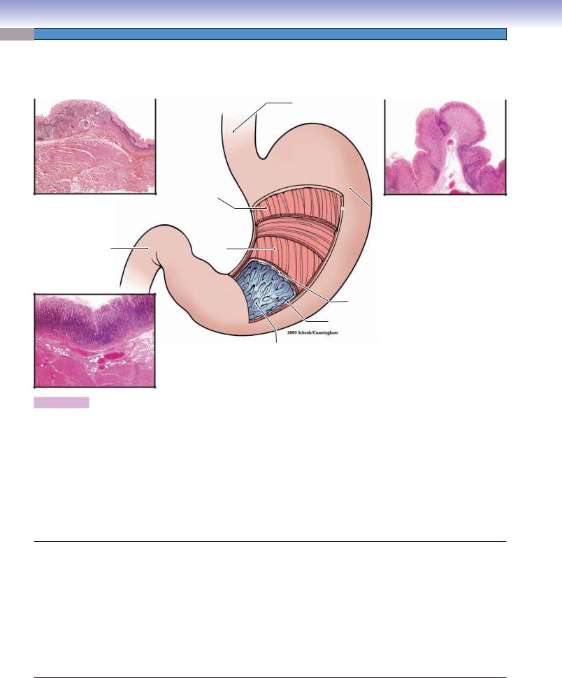
Figure 15-6. Overview of the stomach. H&E, insets (upper left) 5; (lower left) 9; (right) 7.
The stomach is a “J”-shaped hollow organ. It connects the esophagus and duodenum of the small intestine. The stomach initiates digestion of food and temporarily stores food. It can be divided into four parts: the cardia, fundus, body, and pylorus. Histologically, stomach tissue can be distinguished by its mucosal glands (glands in the mucosa). (1 and 2) The cardia connects to the esophagus, and the pylorus connects to the duodenum. These two ends of the stomach have similar histological characteristics. The mucosal glands of the cardia and pylorus are called cardiac glands and pyloric glands, respectively. Both contain many mucus-secreting cells and produce mucus, which is a thick, gel-like material that coats the surface of the stomach and protects it from acidic gastric fluid. Mucus-secreting cells contribute a small volume to the gastric juices. (3 and 4) The fundus and body of the stomach form the largest part of the stomach (about two thirds). The mucosal glands in this region are called fundic (gastric) glands. These glands are composed mainly of parietal and chief cells, which produce large volumes of gastric juices. A small number of various types of enteroendocrine cells are found at the base of the gastric glands. The gastric juices contain primarily water, HCl, mucus, pepsin, IF, rennin, and lipase. This is a highly acidic fluid and plays an important role in digesting food.
Stomach
I.Cardia
A.Cardiac glands
B.Mucus-secreting cells (produce mucus and lysozyme)
II. Fundus and Body
A.Fundic (gastric) glands
B.Mucous neck cells (secrete mucus)
C.Parietal cells (secrete HCl and gastric IF)
D.Chief cells (secrete pepsinogen, rennin, and lipase)
E.Enteroendocrine (diffuse neuroendocrine) cells (release, e.g., gastrin, histamine, and serotonin)
III. Pylorus
A.Pyloric glands
B.Mucus-secreting cells (produce mucus and lysozyme)
C.Enteroendocrine cells
D.G cells (secrete gastrin)
E.D cells (release somatostatin)
CHAPTER 15 ■ Digestive Tract
Surface mucous cells
Esophaguss u g a h p o s E
h Stomachc a m to S
Gastric
pits
A
Cardiac
glands
Mucus-secreting cells of cardiac glands
289
Figure 15-7A. Cardiac region, esophagogastric junction. H&E,
34; insets (left) 192, (right) 331
The inner surface of the stomach is lined by columnar epithelium, which forms a secretory sheath and is composed of mucus-secreting cells called surface mucous cells. These cells cover the entire inner surface of the stomach and also line the surface of the gastric pits. The gastric pits are invaginations of the epithelium and are surrounded by the lamina propria. This is an example of the cardiac region of the stomach at the esophagogastric junction. The lamina propria (loose connective tissue) contains cardiac glands. These glands are branched tubular glands (see Fig. 3-22A,B) and are composed of mucus-secreting cells. The secretions of the cardiac glands empty directly into the gastric pits. The cardiac glands consist of mucous cells, which are similar in appearance to the surface mucous cells. They have basally positioned nuclei and lightly staining cytoplasm.
Gastric
pits
Mucosa
Chief cell
Parietal cells
Parietal Epithelium cells (mucous
surface cells)
Fundic
 glands
Muscularis |
|
Muscularis |
Chief cells |
mucosae |
|
mucosae |
|
|
B |
|
Figure 15-7B. |
Fundic region of the stomach. H&E, 51; |
|
|
insets 245 |
|
|
|
The surface of the fundus region of the stomach is covered by |
|
|
|
surface mucous cells, which produce mucus to protect the epi- |
|
|
|
thelium from the acidic gastric juice. The fundic glands in the |
|
|
|
lamina propria are different from the cardiac glands but similar |
|
|
|
to the glands in the body of the stomach. Fundic glands con- |
|
|
|
tain stem cells, mucous neck cells (Fig. 15-7C), parietal cells, and |
|
|
|
chief cells. The stem cells can differentiate into other types of |
|
|
|
cells in the glands. The parietal cells are large, round cells with |
|
|
|
centrally positioned nuclei (“fried-egg” appearance). Their cyto- |
|
|
|
plasm stains paler than that of chief cells. The parietal cells are |
|
|
|
more numerous in the superior half of the fundic glands (upper |
|
|
|
inset). The chief cells are small, columnar cells and have darkly |
|
|
|
stained cytoplasm. Their nuclei are commonly located at the base |
|
|
|
of the cells. The chief cells are more numerous in the inferior half |
|
|
|
of the fundic glands (lower inset). |
|
Lumen |
C |
Gastric
pits
|
Surface |
|
mucous cells |
|
Lamina |
|
propria |
Fundic |
Mucosa |
glands |
|
Muscularis mucosae |
Submucosa Mucous neck cell |
|
Figure 15-7C. Body region of the stomach. H&E,
34; insets (left) 232; (right upper) 164; (right lower) 394
Histologically, the body and fundus of the stomach are similar to one another, and their glands are identical. They are called fundic (gastric) glands. The mucous neck cells are commonly found between the parietal cells at the neck region of the gastric glands. They secrete acidic mucus. The parietal cells secrete large quantities of HCl and gastric IF. The parietal cells are also called oxyntic (acid-forming) cells. The chief cells are often called zymogenic or peptic cells; their cytoplasm contains zymogen granules. They secrete pepsinogen and precursors of rennin and lipase. Enteroendocrine cells may also be found at the neck and base of the gastric glands. They release endocrine molecules (e.g., serotonin, gastrin, histamine). These cells are similar in appearance to the enteroendocrine cells in the small intestine (see Fig. 15-13B). Stem cells may also be found at neck regions of the gastric glands.
290 UNIT 3 ■ Organ Systems
A |
Gastric pits |
Gastric pits |
 Mucosa
Surface mucous cells
Mucus-secreting Pyloric cells glandsna
Muscularis
mucosae
Submucosa |
Pyloric |
|
glandsna |
Figure 15-8A. Pyloric region of the stomach. H&E, 68; insets 283
The pylorus is the last region of the stomach and connects to the duodenum. The mucosa of the pylorus has deep gastric pits. Pyloric glands, composed primarily of mucus-secreting cells, empty their secretory products into the base of the gastric pits. These mucussecreting cells are pale staining and have basally located nuclei, as do the cells of the cardiac glands. They produce mucus to protect the epithelium of the pylorus from acidic gastric secretions. Two types of enteroendocrine cells are found at the base of the pyloric glands. G cells release gastrin, which stimulates parietal cells to secrete HCl. Another type of enteroendocrine cell, called the D cell, releases somatostatin, which inhibits the release of gastrin by G cells. These two types of enteroendocrine cells are also found in the mucosa of the duodenum (see Fig. 15-13B). The upper inset shows a gastric pit and surface mucous cells in the superior portion of the mucosa. The lower inset shows pyloric glands and mucus-secreting cells in an inferior portion of the mucosa. Both cell types have basally positioned nuclei and clear cytoplasm containing secretory granules.
CLINICAL CORRELATIONS
Granulation
tissue
B
Fibrinopurulent
 exudate (ulcer)
Gastric mucosa (with intestinal metaplasia)
 Chronic gastritis
Figure 15-8B. Gastric Ulcer (Peptic Ulcer). H&E, 19
Peptic ulcers are chronic mucosal lesions that occur in the gastrointestinal tract. The duodenum and stomach are the most common sites for ulcers. Causes of these ulcers include Helicobacter pylori infection, long-term use of nonsteroidal anti-inflammatory drugs and corticosteroids, and cigarette smoking. Epigastric burning or pain, bleeding, and even perforation are the common signs and symptoms of the peptic ulcers. Morphologically, peptic ulcers are usually small, round to oval in shape, less than 4 cm in diameter with well-defined margins without elevation, and have a clean, smooth base. Histologically, a thin layer of necrotic fibrinoid debris with neutrophil infiltration is seen, beneath which lies granulation tissue. Treatments include using H2 receptor antagonists; antibiotics; proton pump inhibitors; and surgery for severe, refractory cases. Care must be taken to differentiate benign ulcers from malignant adenocarcinomas, which may appear ulcerated. This image shows the transition from gastric mucosa to ulcer, showing a fibrinopurulent surface with underlying granulation tissue. The gastric mucosa shows chronic gastritis with plasma cells within the lamina propria and intestinal metaplasia (note the goblet cells).
C
Normal  pancreatic parenchyma
 Gastrinoma
Figure 15-8C. Gastrinoma (Zollinger-Ellison Syndrome). H&E, 97 Gastrinomas, also called Zollinger-Ellison syndrome, are neoplasms producing the hormone gastrin, which commonly arise in the duodenum and pancreas. Hypersecretion of gastrin by the tumor leads to hypergastrinemia, resulting in excess production of gastric acid. Patients have symptoms of peptic ulcers, with clinical findings, such as epigastric tenderness, bleeding, and perforation. Pathologic findings include hyperplasia of the parietal cells that produce gastric acid within the mucosa of the stomach. Tumor cells resemble pancreatic endocrine cells, are well differentiated, and contain gastrin peptides within the secretory granules. Proton pump inhibitors and surgical removal of the tumor are the first treatment choice for this syndrome. This image shows normal pancreatic parenchyma (upper portion) and a well-circumscribed gastrinoma (lower portion). Note the relatively uniform neoplastic cells within the gastrinoma.

CHAPTER 15 ■ Digestive Tract |
291 |
Small Intestine
Jejunum
Myenteric (Auerbach) plexus
Muscularis
mucosae 
Mucosa Lamina
propria
Epithelium
Submucosa
Figure 15-9. Overview of the small intestine.
Duodenum
Stomach
Esophagus
Serosa/adventitia |
|
Outer |
|
longitudinal |
|
muscle |
Muscularis |
Inner circular |
externa |
|
|
 muscle
 Submucosal
(Meissner) plexus
Vein
Artery
Nerve
The small intestine is a very long, tubular organ, about 6 to 7 m long, with a relatively small diameter. It connects the stomach to the large intestine and can be divided into three regions based on anatomy and function. (1) The duodenum, the most proximal region of the small intestine, is a short, C-shaped segment about 20 to 25 cm long. Mucous glands called Brunner glands are present only in the duodenum. Bile and pancreatic secretions enter the duodenum through their duct systems. (2) The jejunum makes up about two fifths of the rest of the small intestine. It has a larger diameter and thicker wall than the ileum. The jejunum has long villi and has neither Brunner glands nor Peyer patches. (3) The ileum is the most distal portion of the intestine, and it makes up about three fifths of the small intestine. The ileum has a thinner wall and fewer villi than the jejunum, and it has clusters of lymphatic nodules, called Peyer patches, in the lamina propria. This illustration shows the general tunics (layers) of the small intestine. Like the other parts of the digestive tract, the small intestine consists of a mucosa (epithelium, lamina propria, and muscularis mucosae), submucosa, muscularis externa, and serosa/adventitia. Several myenteric (Auerbach) plexuses are illustrated between the two layers of the muscularis externa; submucosal (Meissner) plexuses are located in the submucosal layer.
Small Intestine
I.Duodenum
A.Mucosa
B.Submucosa (Brunner glands)
C.Muscularis externa
D.Serosa/adventitia
II.Jejunum
A.Mucosa
B.Submucosa
C.Muscularis externa
D.Serosa
III.Ileum
A.Mucosa (Peyer patches)
B.Submucosa (Peyer patches may extend into this layer)
C.Muscularis externa
D.Serosa
Cell Types in the Small Intestine
Villi: columnar absorptive cells and goblet cells
Glands (crypts) of Lieberkühn: absorptive cells, goblet cells, Paneth cells, enteroendocrine cells, and stem cells
292 UNIT 3 ■ Organ Systems
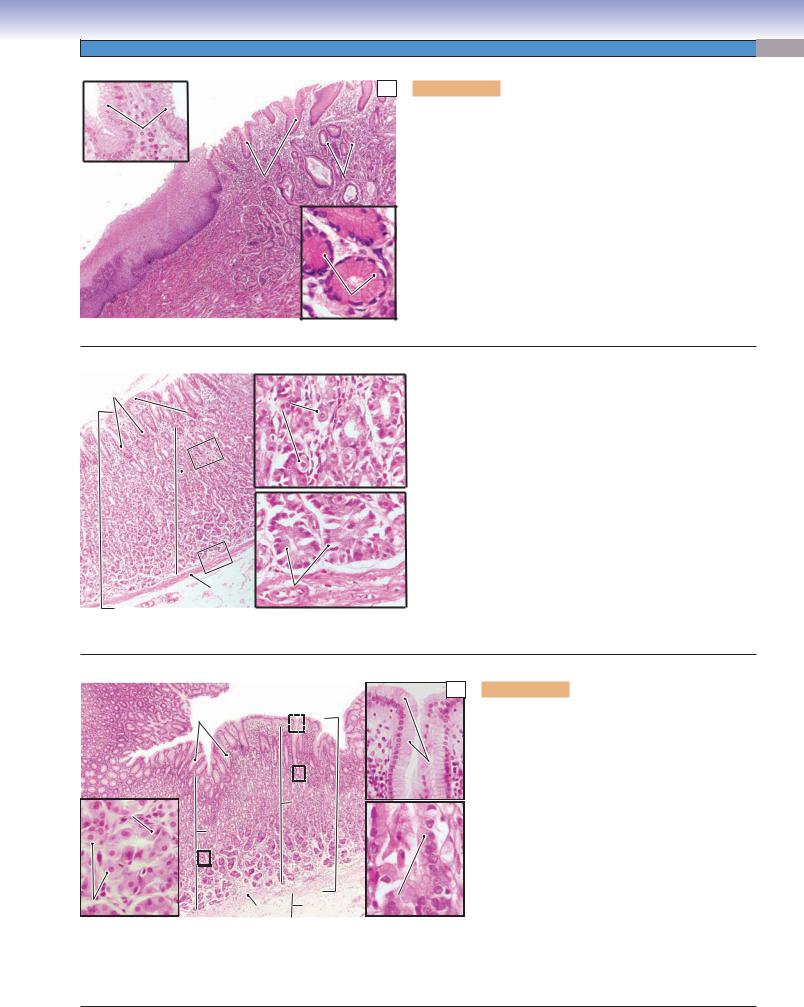
A Figure 15-10A. Duodenum, small intestine. H&E, 14
 Mucosa
 Villi
Villi
Submucosa |
Glands of |
|
Lieberkühn |
||
|
Brunner
gland
Muscularis
 externa
The duodenum connects to the stomach. The mucosa of the duodenum is composed of simple columnar epithelium, lamina propria, and muscularis mucosae. Epithelial cells lining the surface of the villi and the glands of Lieberkühn include absorptive cells, goblet cells, Paneth cells, enteroendocrine cells, and stem cells. The lamina propria is a layer of loose connective tissue, which forms the core of the villus and contains various types of connective tissue cells including fibroblasts, plasma cells, macrophages, and some leukocytes (see Fig. 4-3A). The muscularis mucosae is a thin layer of smooth muscle (Fig. 15-10B). The submucosa is a layer of dense connective tissue containing mucous glands called Brunner glands, which produce mucus to protect the duodenal wall from acidic gastric juice from the stomach. The muscularis externa consists of two layers of smooth muscle: an inner circular layer and an outer longitudinal layer. The outer layer of the duodenum is mostly covered by serosa; areas where it is attached to other organs are covered by adventitia.
Brunner |
B |
glands |
|
|
Lumen |
|
Villi |
|
|
|
|
Glands of |
|
|
|
Lieberkühn |
|
Brunner |
Submucosa |
Brunner |
|
Muscularis |
|||
glands |
glands |
||
mucosae |
|||
|
|
Figure 15-10B. Duodenum, small intestine. H&E, 45; inset
112
An example of the mucosa and submucosa of the duodenum is shown. A thin layer of muscularis mucosae lies between the lamina propria and the submucosa. Fingerlike villi project into the lumen (Fig. 15-11B). Brunner glands are distributed in the submucosa and extend into the lamina propria of the mucosa. Brunner glands produce mucus that protects the epithelium from HCl secreted in the stomach. They also secrete large numbers of bicarbonate ions, which neutralize acidic gastric juice from the stomach. Two types of enteroendocrine cells associated with the regulation of gastric secretion are also found in the glands of Lieberkühn of the duodenum: (1) G cells that release gastrin, which stimulates parietal cell secretion of HCl and (2) D cells that release somatostatin, which inhibits gastrin release. G cells and D cells are predominantly found in the pylorus of the stomach but are also found in the duodenum.
If Brunner cells are not able to produce enough mucus and bicarbonate ions over the long term, a duodenal (Brunner) ulcer may develop.
CLINICAL CORRELATION
C 

Metaplastic gastric mucosa with loss of goblet cells
Blunt, widened villous with inflammation
Figure 15-10C. Peptic Duodenitis. H&E, 48
Peptic duodenitis is an inflammatory process caused by chronic exposure of the duodenal mucosa to increased levels of gastric acid and is usually found in the first portion of the duodenum, the duodenal bulb. Symptoms of peptic duodenitis include epigastric pain and dyspepsia. Histologic features include flattening, or blunting, of the normally fingerlike villi, increased inflammatory cells within the lamina propria, Brunner gland hyperplasia, crypt hyperplasia, and gastric foveolar metaplasia of the epithelium. Metaplasia to a gastric foveolar type of epithelium is an adaptive protective response to the increased levels of acid. H. pylori may be found in the metaplastic mucosa as seen in the stomach. In time, a duodenal ulcer may result from peptic duodenitis. This photomicrograph shows duodenal mucosa with complete replacement of the normal epithelium with goblet cells by gastric foveolar epithelium. Note the widened, distorted villi and increased inflammatory cells within the lamina propria.
CHAPTER 15 ■ Digestive Tract |
293 |
A
|
Villi |
|
|
Villus |
|
|
|
|
|
|
|
Microvilli |
|
Goblet |
|
|
|
cell |
|
|
|
|
Goblet |
|
|
|
|
|
|
Lamina |
|
|
cell |
Columnar |
|
|
|
||
propria |
|
|
|
absorptive |
|
|
|
|
cell |
|
|
|
|
Capillary |
 Microvilli
(brush border)

 Lacteal
Lumen of the
gland (crypt)
of Lieberkühn
|
Gland (crypt) |
|
|
Villus |
of Lieberkühn |
Columnar |
Entero- |
|
|||
|
|
endocrine |
|
|
|
absorptive cells |
|
|
|
cell |
|
|
|
|
Stem |
cell |
Paneth
cells
Lamina propria
Plicae circulare |
Submucosa |
T. Yang |
|
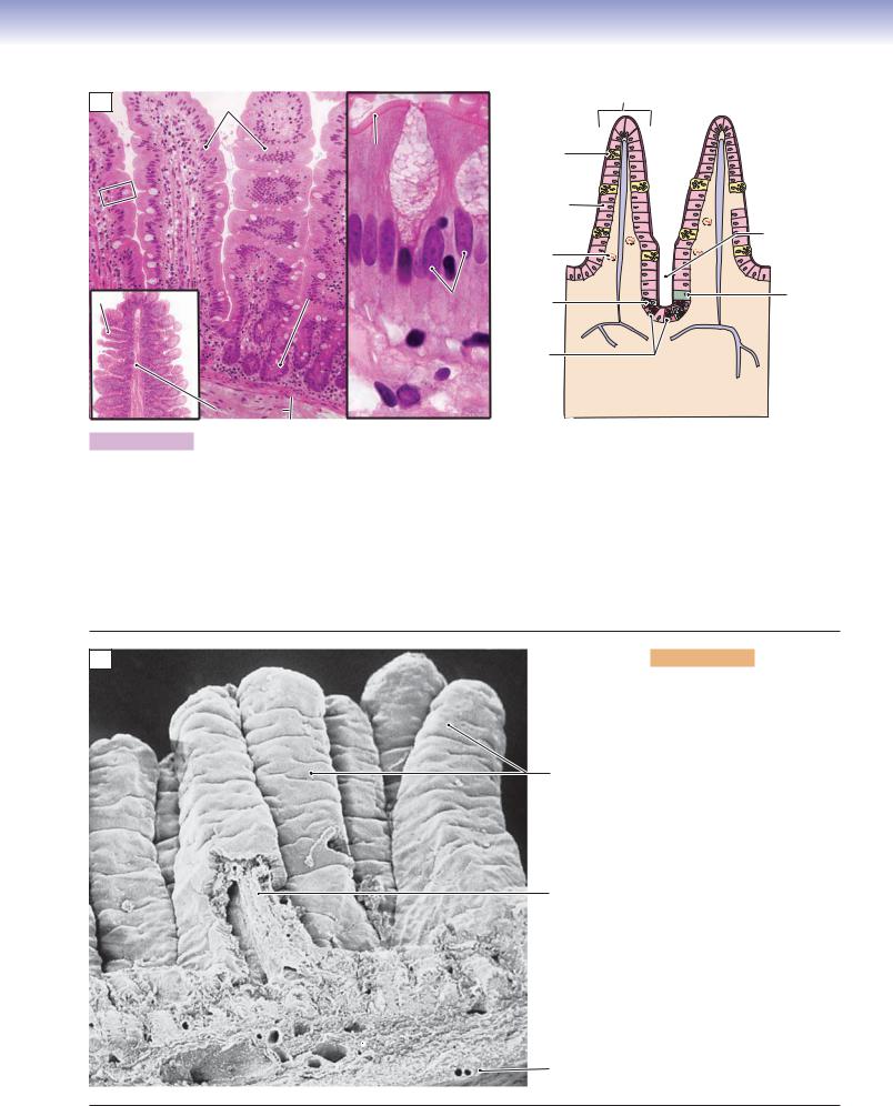
Figure 15-11A. Plicae circulares, villi, and microvilli. H&E, 124; inset (left) 15, (right) 882
The small intestine is a long tube with three levels of folds that increase the surface area for absorption. (1) Plicae circulares (valves of Kerckring) are gross folds involving the mucosa and submucosa that project into the lumen (left inset). (2) Villi are smaller folds than the plicae circulares and involve only mucosa. The central core of each villus is formed by the lamina propria; the nutrients absorbed from the lumen by absorptive cells are transported into the lamina propria. The lamina propria contains a central lacteal (a blind-ended lymphatic vessel) and many capillaries involved in the transport of absorbed nutrients. (3) Microvilli (see Fig. 15-12B) are at the apical surfaces of columnar absorptive cells, increasing the surface area at the cellular level. They appear as a pink border in light microscopy and form a brush border. The drawing on the right shows various types of cells arranged in the epithelium of the mucosa. These cells include column-shaped absorptive cells (enterocytes), goblet cells, Paneth cells, and enteroendocrine cells. The central lacteals are also illustrated.
B
Villi
Lamina propria
 Epithelium
 Submucosa
Muscularis
 externa
Serosa
Figure 15-11B. Villi of the small intestine. SEM, 170
An example of a scanning electron microscopy image showing fingerlike villi extending from the intestinal wall and projecting into the lumen is shown. Each villus is composed of mucosa (epithelium and lamina propria). The lamina propria (connective tissue) forms the central core of the villus. Epithelium with microvilli forms the outer layer of the villus. The villi are unique structures in the small intestine; they are found neither in the stomach nor in the large intestine. The villi are tallest in the duodenum and shortest in the ileum. They gradually reduce in height and size from the proximal regions to the distal regions of the small intestine. The submucosa and muscularis externa are also shown here. The outermost layer of the intestinal wall is serosa.
294 UNIT 3 ■ Organ Systems
Microvilli
Goblet
cells
|
Columnar |
|
|
absorptive |
|
|
cells |
|
A |
||
|
Figure 15-12A. Columnar absorptive and goblet cells of the small intestine. H&E, 1,422
The columnar absorptive cells of the small intestine are also called enterocytes or intestinal absorptive cells. They are tall and columnar in shape; oval-shaped nuclei lie in the basal region of the cells. The columnar absorptive cells are the predominant cells in the epithelium of the small intestine. The apical surfaces of the cells are covered by microvilli, which are coated with glycocalyx. The microvilli increase the cellular surface area for absorption. Goblet cells are interspersed among the absorptive cells. These are unicellular glands (see Fig. 3-20A,B) with a distinctive goblet shape. The goblet cells are mucus-secreting cells, and their numbers gradually increase from the proximal (duodenum) to the distal (ileum) portions of the small intestine.
Microvilli
Goblet cell
Mucus (mucinogen) granules
Microvilli
 Tight junctions
 Desmosome
B
Absorptive cells |
Mitochondria |
|
of absorptive cells |
Figure 15-12B. Goblet cells, columnar absorptive cells, and microvilli. EM, (left, large intestine) 4,831, (right, small intestine)
6,906
There are numerous mitochondria in the apical cytoplasm of the columnar absorptive cells. Junction complexes are located between neighboring cells near the lumen, and microvilli are present on the apical surfaces of the absorptive cells. A goblet cell with many mucus-secretory granules in the cytoplasm is also shown here. These granules contain mucinogen and are released onto the surface of the epithelium by exocytosis. During exocytosis, mucinogen becomes hydrated and forms mucin, which expands greatly in volume after it is released from the goblet cell.
CHAPTER 15 ■ Digestive Tract |
295 |
A |
|
Secretory |
|
Figure 15-13A. |
Paneth cells, small intestine. H&E, |
|
Gland of |
granules |
|
702; inset 1,488 |
|
|
|||||
|
|
|
|
|
|
|
Lieberkühn |
|
|
Paneth cells have basally positioned nuclei and contain |
|
|
|
|
|
||
|
|
|
|
acidophilic-secretory granules in the apical region of the |
|
|
|
|
|
cytoplasm. These granules appear bright red in H&E stains. |
|
|
|
|
|
Paneth cells are located at the base of the glands (crypts) of |
|
|
|
|
|
Lieberkühn. Their secretory granules contain lysozymes, |
|
|
|
|
|
tumor necrosis factor-a, and defensins (cryptidins). These |
|
|
|
|
|
are antibacterial enzymes that help to regulate the normal |
|
|
|
|
|
bacterial flora of the intestine. Paneth cells are protein- |
|
|
|
|
|
secretory cells and have well-developed rough endoplasmic |
|
|
|
|
|
reticulum (RER) and Golgi complexes. Like other epithelial |
|
|
|
|
|
cells, the Paneth cells are derived from stem cells located at |
|
|
|
|
|
the base of the intestinal glands of Lieberkühn. A gland of |
|
Nuclei of the |
|
|
Lieberkühn is indicated by the dashed line at left. Its lumen |
||
|
|
is filled with secretory material and is not easy to see. |
|||
paneth cells |
|
|
|||
|
|
|
|
||
B
Paneth cell
Enteroendocrine cell
Rough endoplasmic reticulum
Nucleus of enteroendocrine cell
Mitochondria
Secretory granules
Basal lamina
Endothelial cell of capillary
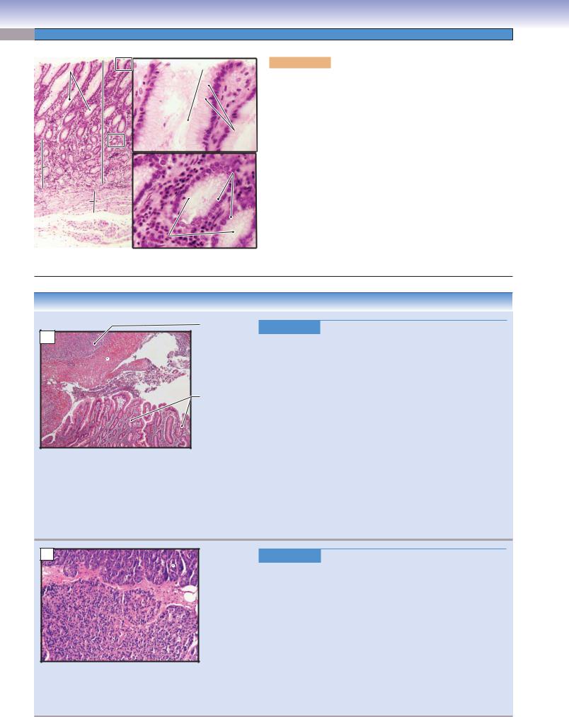
Figure 15-13B. Enteroendocrine cells, small intestine. EM, 10,611; (color) 1,028
There are many types of enteroendocrine cells, which are also called diffuse neuroendocrine cells, in the digestive tract. They are recognized as hormone-releasing cells, and the various types of enteroendocrine cells are similar in appearance. They are often found at the base of glands of Lieberkühn and have many mitochondria, abundant RER, and well-developed Golgi complexes (see Fig. 15-11A). Their secretory granules are located at the basal cytoplasm. Each type of enteroendocrine cell releases one particular hormone.
296 UNIT 3 ■ Organ Systems
A
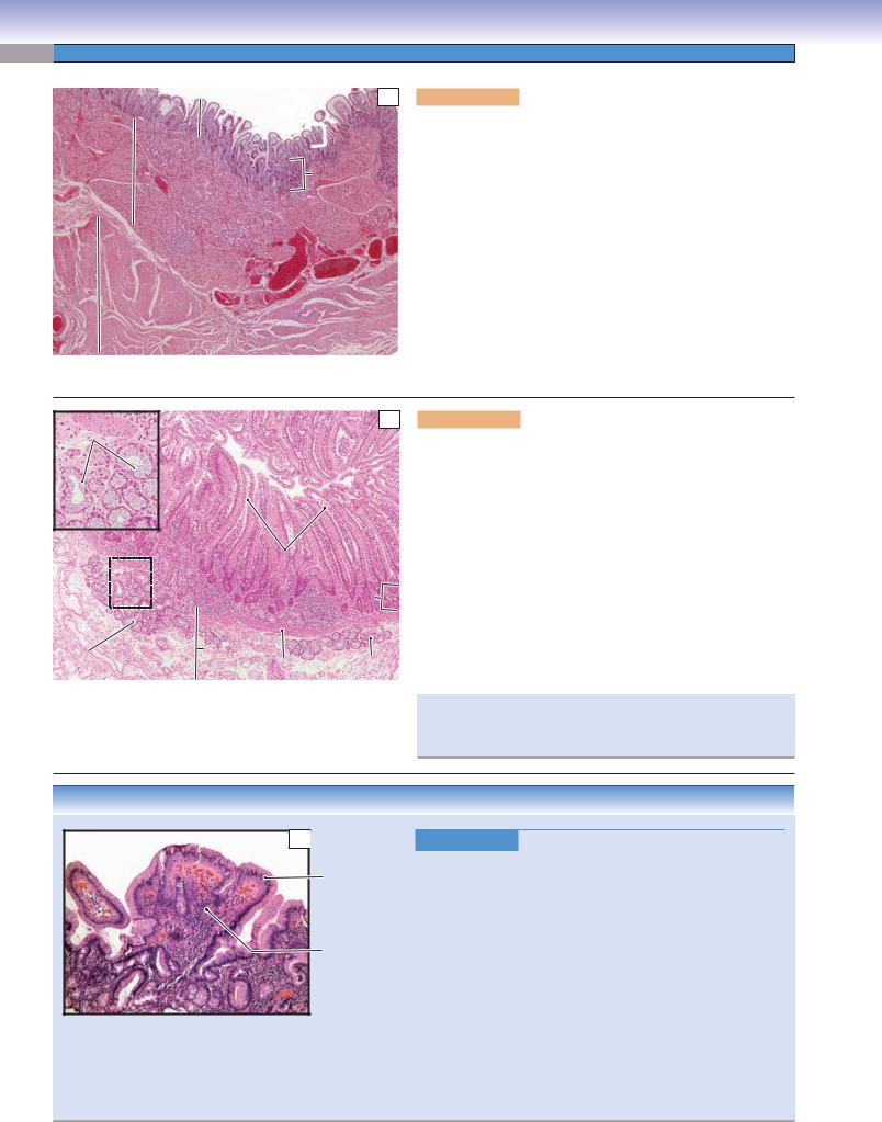
Glandular |
Figure 15-14A. |
Submucosal/Meissner plexus, small |
epithelium |
intestine. H&E, |
272; inset 544 |
Submucosa
Submucosal  plexus Muscularis mucosae
The enteric nervous system is able to operate independently, although it is usually influenced by the parasympathetic and sympathetic nervous systems. The
Neuron cell body two types of ganglia of the enteric nervous system are found in the wall of the digestive tract. (1) Submu-
cosal plexuses (Meissner plexuses) are located in the submucosal layer. (2) Myenteric plexuses (Auerbach plexuses) are located between the two layers of the muscularis externa. Here is an example of a submucosal plexus in the submucosa of the wall of the small intestine. Submucosal plexuses are scattered small groups of neuron cell bodies (sensory and motor neurons and interneurons) and unmyelinated nerve fibers. The axons of the sensory neurons receive mechanical and chemical signals from the glandular epithelium; the axons of the motor neurons innervate the muscularis mucosae and glandular epithelium.
B |
|
Auerbach plexus |
|
Figure 15-14B. |
Muscularis externa, small intestine. |
|
Submucosa |
|
|
H&E, 68; inset 354 |
|
|
|
||||
|
|
|
|
|
|
|
|
|
|
Here is an example of the muscularis externa of the |
|
|
|
|
|
small intestine, which contains two layers of smooth |
|
|
|
|
|
muscle. (1) Inner circular muscle layer: When the |
|
|
Inner circular muscle |
|
|
smooth muscle fibers in this layer contract, the diame- |
|
|
|
|
ter of lumen of the small intestine decreases. (2) Outer |
||
Muscularis |
|
|
|||
|
|
longitudinal muscle layer: This layer surrounds the |
|||
enterna |
|
|
inner circular muscle layer. When the smooth muscle |
||
|
|
|
|
||
|
|
|
|
fibers in this layer contract, the length of the intestine |
|
|
Outer longitudinal muscle |
|
|
is reduced. These two layers of muscle work together |
|
|
|
|
|
to make successive waves of involuntary movements |
|
|
|
|
|
called peristalsis, which force digestive contents to |
|
|
|
|
|
move downward into the large intestine. The inner |
|
|
Auerbach |
|
|
circular and outer longitudinal muscles are innervated |
|
|
plexus |
|
|
by axons from neurons in the myenteric/Auerbach |
|
plexuses of the enteric nervous system (see Fig. 7-14).
C |
Glial cells |
|
Inner circular muscle |
|
Neuron cell bodies |
|
Auerbach |
Outer longitudinal muscle |
plexuses |
Figure 15-14C. Myenteric/Auerbach plexus, muscularis externa of the small intestine. H&E, ×136; inset ×317
Myenteric plexuses (Auerbach plexuses) are found between the inner circular muscle and the outer longitudinal muscle layers. Myenteric (Auerbach) plexuses are much larger than submucosal plexuses. The inset shows several neuron cell bodies and enteric glial cells surrounded by connective tissues in a myenteric plexus. Neurons in both the submucosal plexuses and the myenteric plexuses are multipolar in shape. Like submucosal plexuses, they may have little or no encapsulation and contain unmyelinated nerve fibers and ganglionic neurons mainly belonging to the enteric nervous system. The enteric nervous system coordinates peristaltic reflexes, which evoke waves of contraction and relaxation of the gut wall, moving the contents toward the anus and also regulates enteroendocrine cells.
CHAPTER 15 ■ Digestive Tract |
297 |
Villus |
|
A |
|
Figure 15-15A. |
Jejunum, small intestine. H&E, 34; inset |
|
103 |
|
|||
|
Villi |
|
|||
Mucosa |
|
The jejunum is a segment of small intestine between the duode- |
|||
|
|
|
|||
|
|
|
num and the ileum. It is similar in general structure and layers to |
||
|
Glands of |
|
|||
|
|
the other regions of the small intestine. It contains mucosa, sub- |
|||
|
Lieberkühn |
|
|||
|
|
mucosa, muscularis externa, and serosa. The jejunum has neither |
|||
|
|
|
|
||
|
Submucosa |
|
Brunner glands nor Peyer patches. The cells of the epithelium of |
||
Muscularis |
|
the mucosa are similar to those of the epithelium of other regions |
|||
|
|
|
|||
mucosae |
|
|
|
of the small intestine (see Figs. 15-12A to 15-13B). Goblet cells |
|
|
|
|
|
||
|
|
|
|
steadily increase in number along the entire length of the small |
|
Inner circular muscle |
|
|
|
intestine from the duodenum to the ileum. Paneth cells are often |
|
Muscularis externa |
|
found at the base of the glands of Lieberkühn (lower inset). The |
|||
Glands of |
|
glands of Lieberkühn are intestinal glands (simple tubular glands), |
|||
|
|
|
|||
Lieberkühn |
|
|
|
which extend from the spaces between the bases of the villi deep |
|
Outer longitudinal muscle |
|
|
|
into the lamina propria. |
|
 Peyer
Peyer
patches
Mucosa
Serosa Submucosa
Muscularis externan
|
|
Figure 15-15B. |
Ileum with Peyer patches, small intestine. |
B |
|
||
|
H&E, 13 |
||
|
|
||
|
|
The ileum is the longest segment of the small intestine making up |
|
|
|
three fifths of the 6 to 7 m length of the small intestine. One of the |
|
|
|
unique features of the ileum is the presence of clusters of lymphatic |
|
|
|
nodules called Peyer patches. These are most numerous in the dis- |
|
|
|
tal portion of the ileum. Some isolated lymphatic nodules may be |
|
|
|
found in other parts of the digestive tract but not in aggregations |
|
|
|
of clusters of nodules like Peyer patches. The villi in the ileum are |
|
|
|
shorter and smaller than in other parts of the small intestine. The |
|
|
|
numbers of goblet cells are greatly increased in the ileum. The inset |
|
|
|
shows an endoscopic image of the ileum with its relatively smooth |
|
|
|
surface. |
|
|
|
|
|
|
|
Vitamin K is absorbed in both the jejunum and the ileum, but |
|
|
|
vitamin B12 is only absorbed in the ileum (especially the ter- |
|
|
|
minal ileum). The absorption of vitamin B12 requires coupling |
|
|
|
with gastric intrinsic factor, which is produced by parietal cells in |
|
|
|
the stomach; these two substances become intimately associated |
|
|
|
with the wall of the ileum. If a large portion of the stomach or |
|
|
|
ileum is surgically removed, vitamin B12 deficiency may result, |
|
|
|
leading to megaloblastic anemia and neurologic symptoms. |
|
|
|
|
|
|
|
|
|
Figure 15-15C. |
Mucosa of the ileum, small intestine. H&E, 68 |
Goblet |
|
C |
|||
|
|
|
|
||
cells |
|
|
|
Here is an example of the mucosa of the ileum showing numerous |
|
|
|
|
|||
|
|
|
|
||
|
|
|
|
goblet cells in the surface epithelium. In this section, two lym- |
|
|
|
|
|
phatic nodules are located in the lamina propria. These lymphatic |
|
|
|
|
|
nodules have germinal centers and are, therefore, secondary |
|
|
|
|
|
lymphatic nodules. These nodules may extend into the submu- |
|
|
|
|
|
cosa (Fig. 15-15B). The lymphatic nodules and Peyer patches |
|
|
|
|
|
are locations where lymphocytes can interact with antigens and, |
|
|
|
|
|
therefore, play important roles in immunological function. Naive |
|
|
|
|
|
B cells (B lymphocytes) within these lymphoid patches are primed |
|
|
|
|
|
and awaiting exposure to unique epitopes. When stimulated by a |
|
|
Germinal center |
|
|
specific antigen from the intestinal mucosa, they differentiate into |
|
|
|
|
plasma cells and memory B cells. In response, the plasma cells pro- |
||
|
of lymphatic nodules |
|
|
||
|
|
|
duce large quantities of immunoglobulins ([Ig] antibodies), espe- |
||
|
|
|
|
||
|
|
|
|
cially IgA to combat mucosal infection. The memory B cells live |
|
|
|
|
|
on in the Peyer patches to retain immunity to a specific antigen. |
|
|
|
|
|
|
|
298 UNIT 3 ■ Organ Systems
Large Intestine
|
|
Transverse colon |
Colon |
|
|
|
|
Descending |
|
Ascending |
colon |
|
Sigmoid |
|
|
colon |
|
|
|
colon |
|
Cecum |
Rectum |
|
|
|
|
|
Appendix |
|
Adipose tissue |
Rectum |
|
|
|
Serosa/adventitia |
Teniae coli |
|
|
|
|
|
|
Vein |
Muscularis |
|
Artery |
mucosae |
|
Nerve |
Mucosa |
Lamina |
Submucosa |
|
|
|||
propria |
Circular muscle |
||
|
|||
|
|
||
|
|
Muscularis |
|
|
Epithelium |
externa |
|
|
Longitudinal muscle |
||
|
|
||
|
|
band (teniae coli) |
|
|
|
Teniae coli |
Figure 15-16. Overview of the large intestine. H&E (left), 14; (right) 36
The large intestine connects the small intestine to the anal canal. The large intestine is about 1.5 m long, much shorter than the small intestine. It consists of the cecum, appendix, colon, rectum, and anal canal. (1) The cecum is a small blind pouch of the large intestine, at the junction of the ileum and the ascending colon. The ileum and cecum are separated by the ileocecal valve, which prevents feces from backing up into the small intestine. (2) The appendix is a very short, small-diameter blind end tube that attaches to the posterior-medial wall of the cecum. It contains aggregates of lymphatic nodules in the lamina propria. (3) The colon is the longest part of the large intestine and includes ascending, transverse, descending, and sigmoid colons. (4) The rectum connects the sigmoid colon to the anal canal. (5) The anal canal is externally surrounded by a layer of skeletal muscle called the exterior sphincter. The junction between the rectum and the anal canal is called the anorectal junction, also called the dentate line, which marks the transitional epithelium change from simple columnar epithelium to stratified squamous epithelium. The large intestine has the same general structure of mucosa, submucosa, muscularis externa, and serosa/adventitia as the small intestine. However, the large intestine has a large lumen (excepting the appendix) and a large number of goblet cells lining the surface of the mucosa. It has crypts (intestinal glands) but no villi, and the outer longitudinal muscle layer of the muscularis externa has become three narrow bands called teniae coli. Functions of the large intestine include the absorption of water and salts and the formation, storage, and elimination of feces.
Large Intestine
I. |
Cecum |
C. Muscularis externa (inner circular muscle; teniae) |
|
A. Mucosa (crypts/glands; no villi) |
D. Serosa/adventitia |
|
B. Submucosa |
IV. Rectum |
|
C. Muscularis externa (inner circular muscle; teniae coli) |
A. Mucosa |
|
D. Serosa |
B. Submucosa |
II. |
Appendix |
C. Muscularis externa (inner circular and outer longitudinal |
|
A. Mucosa (aggregated lymphatic nodules) |
muscles) |
|
B. Submucosa |
D. Adventitia |
|
C. Muscularis externa (inner circular and outer longitudinal |
V. Anal Canal |
|
muscles) |
A. Mucosa (stratified squamous) |
|
D. Serosa |
B. Submucosa |
III. Colon: ascending, transverse, descending, and sigmoid portions |
C. Muscularis externa (internal and external sphincters) |
|
|
A. Mucosa (crypts/glands; no villi) |
D. Adventitia |
|
B. Submucosa |
|
|
|
|

CHAPTER 15 ■ Digestive Tract
A
Mucosa
Submucosa
 Mucosa
Submucosa
Circular muscle of
 muscularis externa
B |
Glands of |
|
Lieberkühnu |
Submucosa |
Glands of |
|
Lieberkühn |
||
|
Blood vessels
299
Figure 15-17A. Colon, large intestine. H&E, 15
The colon is the longest part of the large intestine. It contains an ascending colon, transverse colon, descending colon, and sigmoid colon. The colon receives digestive contents from the small intestine and absorbs a large volume of water and electrolytes from the contents. Bacteria in the colon make large quantities of vitamins K and B12, but absorption is limited. The colon also forms and stores feces, the waste matter leftover after the digestion process has been completed. The movement of the contents in the large intestine is slower than in the small intestine, taking 8 to 15 hours to move the chyme (thick, semifluid mass) from the cecum to the rectum, where feces are stored. The mucosa of the colon has a smooth surface (no villi) and contains glands of Lieberkühn. The submucosa contains blood vessels, lymphatic vessels, and nerve fibers as well as submucosal plexuses, but no glands are present. The muscularis externa includes the circular muscle (shown here). The longitudinal muscle is aggregated into three bands called teniae coli (Fig. 15-16). The myenteric (Auerbach) plexuses are located between these two muscle layers.
Figure 15-17B. Colon, large intestine. H&E,
×68 (left); mucous stain, ×44 (upper right)
The glands of Lieberkühn are intestinal glands that are straight tubular glands and are located in the lamina propria of the mucosa. The glands of Lieberkühn in the large intestine are similar to
Normal colon those of the small intestine, but they contain no Paneth cells. They are composed of great numbers
of goblet cells, columnar absorptive cells, and some enteroendocrine cells. A thin layer of the muscularis mucosae is found beneath the lamina propria; it is part of the mucosa. The upper right image shows the goblet cells, which appear red because of the mucous stain.
C |
Columnar |
|
absorptiveabsorptivecellsce l |
|
Goblet
 cell
cell
Goblet
cells
Lumen of the gland of Lieberkühn
Figure 15-17C. Mucosa of the colon, large intestine.
H&E, 272
Upper: The superior part and surface of the mucosa of the colon consists of columnar absorptive cells and goblet cells. These absorptive cells play an important role in the absorption of water and electrolytes. Water enters the absorptive cells entirely by diffusion. Most water absorption occurs in the colon, especially in the proximal colon. Goblet cells produce mucus, which protects the wall of the large intestine, glues fecal material together, and lubricates the passage. The surface of the large intestine is much smoother than the small intestine because there are no villi.
Lower: The inferior part of the mucosa in the colon contains straight tubular glands, glands of Lieberkühn, which are cut in cross section here. Most cells in these glands are goblet cells with basally positioned nuclei. The secretory (mucinogen) granules located at the apical ends of the cells appear white here. Enteroendocrine cells and stem cells also can be found. The stem cells are located at the base of the glands (crypts) of Lieberkühn and can be differentiated from other cell types of epithelia. Paneth cells are not present in the large intestine.
300 UNIT 3 ■ Organ Systems
CLINICAL CORRELATIONS
Pseudostratified columnar epithelium
A
Colon Stalk polyp
Figure 15-18A. Colon Polyps. H&E, 97; inset (upper) 1.3
Colon polyps are tissue masses that grow on the inner mucosal surface and protrude into the lumen of the colon. Polyps may contain a stalk or may be sessile, without a stalk. There are several types of polyps, the most common of which are adenomatous and hyperplastic. Adenomatous polyps are benign neoplasms, subdivided based on morphologic features into tubular adenomas, tubulovillous adenomas, and villous adenomas. Although adenomatous polyps are themselves benign, they should be considered as having malignant potential, because adenocarcinoma may arise in these polyps. Hyperplastic polyps, which are common in the descending colon and rectum, are considered benign with minimal risk for progression to cancer. Pseudopolyps may be seen in inflammatory bowel disease, particularly ulcerative colitis. Polyps may be removed during colonoscopy by endoscopic mucosal resection and sent for pathologic evaluation. These images show a large, pedunculated tubular adenoma (inset) and a higher power view of the adenomatous epithelium featuring pseudostratification of the crypt cells.
B
Adenocarcinoma  (malignant glands)
Figure 15-18B. Colorectal Cancer. H&E, 97
Colorectal cancer is a malignant neoplasm of the colon or the rectum. Risk factors include genetics, inflammatory bowel disease—especially ulcerative colitis—adenomatous polyps, high-fat and low-fiber diets, and excessive red meat consumption. Adenocarcinoma is the most common type of colon cancer (98% of cases), arising from the mucosal glandular epithelium, often in adenomatous polyps. Colorectal carcinomas invade through the layers of the intestinal wall and metastasize predominantly through the lymphatic system. Depending on the location, colorectal cancers may be asymptomatic for years. Presenting symptoms may be a change in bowel habits due to bowel obstruction, blood in the stool, or iron deficiency anemia. Surgical resection is the first choice for early-stage cancer, although chemotherapy may be considered. This photomicrograph shows a moderately differentiated adenocarcinoma of the colon infiltrating the muscularis propria.
C |
Ileal |
|
mucosa |
|
Ectopic |
|
gastric |
|
mucosa |
|
Meckel |
|
diverticulum |
Figure 15-18C. Meckel Diverticulum. H&E, ×19
Meckel diverticulum is a congenital abnormality characterized by an outpouching in the small bowel due to failure of the vitelline duct to close or involute. As a true diverticulum, it contains all three layers (mucosa, submucosa, and muscularis propria) of the normal bowel wall and is found on the antimesenteric aspect of the bowel. Meckel diverticula occur in 2% of the population, are usually located within 2 ft of the ileocecal valve, and are about 2 in length. Some Meckel diverticula contain heterotopic rests of pancreatic or gastric mucosa. Most people with a Meckel diverticulum are asymptomatic. Bleeding, inflammation, and peptic ulceration and perforation can occur, producing signs and symptoms similar to appendicitis. Surgery is the appropriate treatment for symptomatic patients. This image shows a section of a Meckel diverticulum showing normal ileal mucosa with goblet cells on the left side and ectopic gastric mucosa on the right side. This gastric mucosa increases the risk of perforation because of the elaboration of acid.
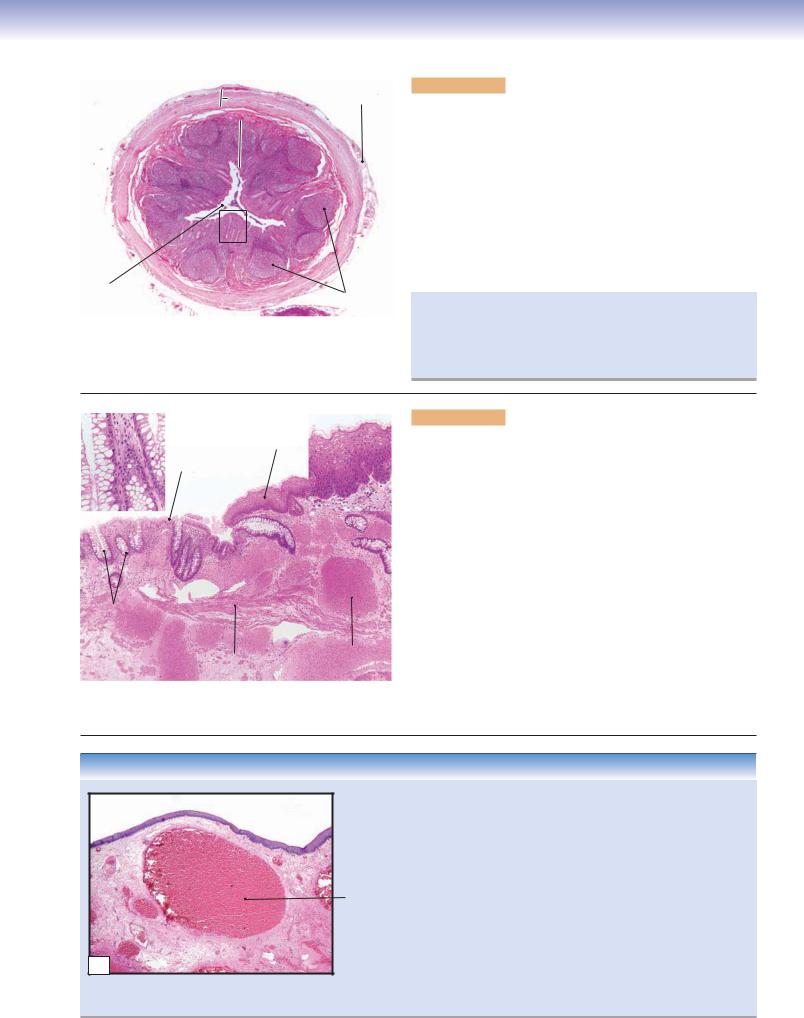
CHAPTER 15 ■ Digestive Tract |
301 |
Muscularis externa |
Serosa |
 Submucosa
 Mucosa
Lumen |
Lymphatic |
|
|
|
|
A |
|
nodules |
Figure 15-19A. Appendix and cecum. H&E, 17
The appendix is a fingerlike blind-ended tubular structure, about 10 cm in length, extending from the cecum distal to the ileocecal valve. The cecum is the first segment of the large intestine, where it connects with the ileum. The small and large intestines are separated by the ileocecal valve. The appendix has a small, irregular lumen and many lymphatic nodules in the mucosa. Although its role in digestion is not clear, it may participate in immunological function and help maintain normal bacteria flora in the large intestine. The epithelium of the appendix is simple columnar epithelium, and lymphatic nodules are located in the lamina propria of the mucosa, which may extend into the submucosa. The muscularis externa contains inner circular and outer longitudinal smooth muscles.
Appendicitis usually results from a bacterial infection and may develop slowly or acutely. A cardinal sign is pain in the lower right abdominal quadrant or when pressure is applied to the McBurney point. The most common treatment is surgical excision of the appendix.
Rectum |
|
Stratified |
Anal canal |
|
squamous |
|
|
|
|
|
|
|
Simple |
epithelium |
|
|
columnar |
|
|
|
epithelium |
|
|
|
|
|
|
Glands (crypts) of |
|
|
|
|
Lieberkühn |
|
|
|
|
Muscularis |
Large |
|
|
vein |
|
B |
|
mucosae |
|
|
|
|
|
|
|
|
|
Figure 15-19B. Anorectal junction. H&E, 34; insets (left)
131, (right) 87
The junction between the rectum and the anal canal is called the anorectal junction. The rectum connects the sigmoid colon to the anal canal. The rectum is the distalmost portion of the large intestine and functions mainly to store feces until elimination. The mucosa of the rectum is similar to that of the colon, but it has fewer intestinal glands of Lieberkühn. The anal canal is a continuation of the large intestine and has longitudinal folds (anal columns) in the gross view. The mucosa of the anal canal is covered by stratified squamous epithelium. The lamina propria contains many large veins (venous plexus). The muscularis mucosae is visible here. The muscularis externa contains inner circular and outer longitudinal smooth muscle layers (not shown here). The inner circular smooth muscle becomes thicker and forms the internal anal sphincter. The external anal sphincter surrounds the anal canal. It is a thick layer of skeletal muscle that voluntarily controls the elimination of feces. On the left in the photomicrograph is the distal end of the rectum; on the right is the proximal portion of the anal canal. Sebaceous glands and apocrine glands may be found in the distal end of the anal canal.
CLINICAL CORRELATION
C
|
Figure 15-19C. |
Hemorrhoids. H&E, 19 |
|
|
Hemorrhoids are swollen, inflamed veins in the anal region deep |
||
|
in the anal mucosa and are classified as internal hemorrhoids and |
||
|
external hemorrhoids, based on whether the hemorrhoids are |
||
|
above (internal) or below (external) the anorectal (line) junction |
||
Dilated |
(dentate/pectinate line). Causes include repeated pregnancy, con- |
||
stipation, and portal hypertension. Common symptoms include |
|||
anorectal |
|||
vein |
bleeding, itching, and pain. Pathologically, hemorrhoids are thin- |
||
|
walled varicose veins that extend into the submucosal layer. In time, |
||
|
thrombosis (clotting) may occur with resultant infarct and fissure |
||
|
formation. Treatments include surgical removal of hemorrhoids, |
||
|
coagulation therapy, and a procedure in which a rubber band is |
||
|
used to tie off the hemorrhoids. This photomicrograph shows anal |
||
|
squamous mucosa with underlying dilated anorectal veins. |
||

302 UNIT 3 ■ Organ Systems
CLINICAL CORRELATIONS
A
Acute and  chronic
inflammation
Crypt abscess
Figure 15-20A. Ulcerative Colitis. H&E, 97
Ulcerative colitis is an inflammatory bowel disease that causes ulcers in the lining of the colon and rectum characterized by intermittent exacerbations alternating with complete symptomatic remission. Major symptoms include abdominal pain, diarrhea, anemia, weight loss, bleeding from the rectum, and passage of mucus. Pathologically, the inflammation is predominantly confined to the mucosa and submucosa in contrast to Crohn disease, which is transmural. Grossly, ulcerative colitis produces shallow ulcers and pseudopolyps. Histologic features include acute and chronic colitis, crypt abscesses, atrophy of glands, and loss of mucin in goblet cells. Treatment includes anti-inflammatory drugs and immunosuppressants to control the symptoms and achieve remission. The risk for the development of adenocarcinoma is high in patients with ulcerative colitis, so surveillance colonoscopy with biopsy is necessary to detect early dysplasia of the glandular epithelium. Surgical removal of the colon is necessary in severe refractory cases or in cases where severe dysplasia or adenocarcinoma is detected.
B
Noncaseating granulomas
 Cryptitis
Figure 15-20B. Crohn Disease. H&E, 97
Crohn disease is a chronic autoimmune inflammatory disease of the gastrointestinal tract that may affect any location, from the oral cavity to the anus, but mostly involves the distal small intestine and colon. Crohn disease is characterized by asymmetric and segmental inflammation extending through the intestinal wall (transmural) from the mucosa to the serosa. Crohn disease characteristically involves areas of the bowel separated by intervening uninvolved areas or “skip” lesions. Symptoms include abdominal pain, diarrhea, vomiting, and weight loss. Pathologic changes include mucosal neutrophil and mononuclear cell infiltration, ulceration, mucosal fissures, fistulae, serosal adhesions, abscesses, pseudopolyps, and noncaseating granulomas. Treatment focuses on relieving symptoms through immunosuppressive agents to prevent relapse and complications. This image shows colonic mucosa with depletion of goblet cells, noncaseating granulomas within the lamina propria, chronic inflammation, and neutrophils invading the crypt cells.
SYNOPSIS 15 - 1 Pathological and Clinical Terms for the Digestive Tract
■Intestinal metaplasia: the reversible change of one mature type of epithelium to an intestinal type epithelium; may be seen in chronic gastritis when goblet cells are present within the gastric mucosa; considered a risk factor for the development of gastric adenocarcinoma (Fig. 15-8B).
■Dyspepsia: general term for abdominal pain or indigestion associated with the intake of food (Fig. 15-10C).
■Gastric metaplasia: the reversible change of one mature type of epithelium to a gastric type epithelium; seen in peptic duodenitis when the normal epithelial lining with goblet cells is replaced with a gastric foveolar–type mucosa in response to exposure to increased levels of stomach acid (Fig. 15-10C).
■Pseudopolyp: found in cases of chronic inflammatory bowel disease; consists of polypoid mounds of mucosa created by regenerating glandular epithelium; not considered true polyps, hence the name (Figs. 15-18A and 15-20A,B).
■Crypt abscess: an aggregate of neutrophils present within a colon crypt, usually associated with inflammatory bowel disease, particularly ulcerative colitis (Fig. 15-20A).
■Cryptitis: an indicator of acute colitis; appreciated histologically by the infiltration of neutrophils with the crypt cells of the colon (Fig. 15-20B).

CHAPTER 15 ■ Digestive Tract |
303 |
TABLE 15 - 1 Digestive Tract
Regions |
Mucosa (Epithelium, |
Submucosa |
Muscularis Externa |
Serosa/ |
Main Functions |
|
Lamina Propria, |
(Submucosal |
(Myenteric Plexuses |
Adventitia |
|
|
Muscularis Mucosae) |
Plexuses Present |
Present between |
|
|
|
|
in this Layer) |
Smooth Muscle Layers) |
|
|
|
|
|
|
|
|
Esophagus |
|
|
|
|
|
|
|
|
|
|
|
Upper |
Nonkeratinized stratified |
Esophageal glands |
Inner circular and outer |
Adventitia |
Transport food bolus |
esophagus |
squamous |
|
longitudinal skeletal |
|
|
|
|
|
muscles |
|
|
|
|
|
|
|
|
Middle |
Nonkeratinized stratified |
Esophageal glands |
Inner circular and outer |
Adventitia |
Transport food bolus |
esophagus |
squamous |
|
longitudinal mixed skeletal |
|
|
|
|
|
and smooth muscles |
|
|
|
|
|
|
|
|
Lower |
Nonkeratinized stratified |
Esophageal glands |
Inner circular and outer |
Serosa |
Transport food bolus |
esophagus |
squamous; esophageal |
|
longitudinal skeletal |
|
|
|
cardiac glands in lamina |
|
muscles |
|
|
|
propria |
|
|
|
|
|
|
|
|
|
|
Stomach (Rugae and Gastric Pits) |
|
|
|
|
|
|
|
|
|
|
|
Cardia |
Surface mucous cells |
No glands |
Inner oblique, middle |
Serosa |
Add mucus and |
|
and gastric pits; mucus- |
|
circular, and outer |
|
lysozyme; churn food |
|
secreting cells of cardiac |
|
longitudinal smooth |
|
by peristalsis |
|
glands in lamina propria |
|
muscles |
|
|
|
|
|
|
|
|
Fundus and |
Surface lining cells and |
No glands |
Inner oblique, middle |
Serosa |
Same as above; add |
body |
gastric pits; parietal |
|
circular, and outer |
|
acidic gastric juice (e.g., |
|
and chief cells of gastric |
|
longitudinal smooth |
|
HCl, mucus, enzymes); |
|
glands in lamina propria |
|
muscles |
|
process food into chyme |
|
|
|
|
|
|
Pylorus |
Surface lining cells and |
No glands |
Inner oblique, middle |
Serosa |
Add mucus; churn |
|
gastric pits; mucus- |
|
circular, and outer |
|
food and pass chyme to |
|
secreting cells of pyloric |
|
longitudinal smooth |
|
duodenum |
|
glands in lamina propria |
|
muscles |
|
|
|
|
|
|
|
|
Small Intestine (Villi and Crypts) |
|
|
|
|
|
|
|
|
|
|
|
Duodenum |
Columnar absorptive |
Brunner glands |
Inner circular and outer |
Serosa/ |
Allow bile and pancreatic |
|
and goblet cells; Paneth, |
|
longitudinal smooth |
adventitia |
juice to enter small |
|
enteroendocrine, goblet, |
|
muscles |
|
intestine; release mucus; |
|
and absorptive cells in |
|
|
|
regulate rate of emptying |
|
glands (crypts) of Lieberkühn |
|
|
|
of stomach; absorption |
|
|
|
|
|
|
Jejunum |
Similar to above |
No glands |
Inner circular and outer |
Serosa |
Absorption of |
|
|
|
longitudinal smooth |
|
carbohydrates, proteins, |
|
|
|
muscles |
|
lipids, and vitamin K |
|
|
|
|
|
|
Ileum |
Similar to above with |
Peyer patches may |
Inner circular and outer |
Serosa |
Similar to above; |
|
increased goblet cells; Peyer |
extend into this |
longitudinal smooth |
|
absorption of vitamins |
|
patches in lamina propria |
layer |
muscles |
|
K and B12 and bile salts |
|
|
|
|
|
|
Large Intestine (Crypts, no Villi)
Cecum/colon |
Goblet cells and |
No glands |
Inner circular smooth |
Serosa/ |
Absorption of water |
|
columnar absorptive |
|
muscle; outer longitudinal |
adventitia |
and electrolytes; |
|
cells; goblet, columnar, |
|
smooth muscle layer forms |
|
formation of feces |
|
and enteroendocrine cells |
|
three teniae coli |
|
|
|
in glands of Lieberkühn |
|
|
|
|
|
|
|
|
|
|
Rectum |
Similar to above; fewer |
No glands |
Inner circular and outer |
Adventitia |
Store feces; sensory |
|
glands of Lieberkühn |
|
longitudinal smooth |
|
receptors signal brain of |
|
|
|
muscles |
|
the need to evacuate |
Anal canal |
Nonkeratinized to |
Sebaceous glands |
Inner circular (thickened to |
Adventitia |
Internal sphincter and |
|
keratinized stratified |
(distal portion); |
become internal sphincter) |
|
external sphincter |
|
squamous |
venous |
and thin outer longitudinal |
|
(skeletal muscle); relax |
|
|
|
smooth muscles |
|
to release feces |
|
|
|
|
|
|
Note: Simple columnar epithelium lines most of the digestive tract, including the stomach and the small and large intestines. It does not line the esophagus and the anal canal, however.
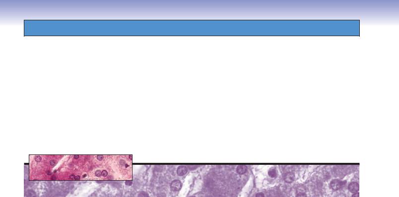
16 Digestive Glands
and Associated
Organs
Introduction and Key Concepts for the Digestive Glands and Associated Organs
Figure 16-1 |
Overview of the Digestive Glands and Associated Organs |
Figure 16-2 |
Orientation of Detailed Digestive Glands and Associated Organs Illustrations |
Salivary Glands |
|
Figure 16-3A |
General Structure of the Major Salivary Glands |
Figure 16-3B |
Parotid Gland |
Figure 16-4A |
Parotid Glands |
Figure 16-4B |
Clinical Correlation: Pleomorphic Adenoma |
Figure 16-4C |
Clinical Correlation: Parotid Cyst |
Figure 16-5A |
Submandibular Glands |
Figure 16-5B |
Serous Acinus, Submandibular Gland |
Figure 16-6A |
Mucous Acinus, Submandibular Gland |
Figure 16-6B |
Clinical Correlation: Squamous Cell Carcinoma of the Tongue |
Figure 16-7A |
Striated Duct, Submandibular Gland |
Figure 16-7B |
Intralobular Duct, Submandibular Gland |
Figure 16-8A,B |
Sublingual Glands |
Figure 16-8C |
Clinical Correlation: Sialadenitis |
Pancreas |
|
Figure 16-9A |
A Representation of the Exocrine Pancreatic Duct System |
Figure 16-9B |
Exocrine and Endocrine Pancreas |
Figure 16-9C |
Clinical Correlation: Acute Pancreatitis |
Figure 16-10A |
Pancreatic Acinar Cells |
Figure 16-10B |
Centroacinar Cells, Pancreas |
Liver |
|
Figure 16-11 |
General Structure of the Liver |
Synopsis 16-1 |
Functions of the Liver |
304
CHAPTER 16 ■ Digestive Glands and Associated Organs |
305 |
|
Figure 16-12A |
Liver Acinus |
|
Figure 16-12B |
Portal Triad, Liver |
|
Figure 16-12C |
Clinical Correlation: Alcoholic Fatty Liver (Steatosis) |
|
Figure 16-13A |
Hepatocytes and Hepatic Sinusoids, Liver |
|
Figure 16-13B |
Space of Disse, Hepatocyte |
|
Figure 16-14A |
A Representation of Bile Canaliculi and Hepatocytes |
|
Figure 16-14B |
Bile Canaliculus, Hepatocytes |
|
Gallbladder |
|
|
Figure 16-15A |
Gallbladder |
|
Figure 16-15B |
Epithelial Cells Lining the Gallbladder |
|
Figure 16-16A |
Clinical Correlation: Hepatitis C |
|
Figure 16-16B |
Clinical Correlation: Gallstones |
|
Synopsis 16-2 |
Pathological and Clinical Terms for the Digestive Glands and Associated Organs |
|
Introduction and Key Concepts
for the Digestive Glands and Associated Organs
Digestive glands and associated organs include the major salivary glands, pancreas, liver, and gallbladder. These organs are located outside the wall of the digestive tract. Their secretory products are delivered into the digestive tract via a duct system.
Major Salivary Glands
The major salivary glands produce saliva and empty into the oral cavity. Saliva is 99% water and contains protein, enzymes, glucose, cholesterol, urea, uric acid, ions (e.g., Na+, K+, Ca++, HCO3-), and antibacterial agents (lactoferrin, lysozyme, and IgA). Saliva is produced by both serous and mucous cells in the salivary glands. It plays important roles in aiding digestion, lubrication, protection, buffering, wound healing, maintaining the integrity of the esophagogastric epithelium, perception of taste, and in hardening of the enamel of the teeth. There are three major salivary glands: the parotid, submandibular, and sublingual glands. These are paired glands and have similar structures of secretory units (acini) and duct systems, including intralobular ducts (intercalated and striated ducts), interlobular ducts, lobar ducts, and a main duct (Figs. 16-1 and 16-8B).
1.The parotid gland is the largest of the three major salivary glands. This gland is surrounded by a connective tissue capsule. Its secretory unit is composed of only serous cells, which produce watery proteinaceous fluid (Figs. 16-3B and 16-4A).
2.The submandibular gland is the second largest salivary gland. This gland is surrounded by a connective tissue capsule. It is a mixed gland, although the majority of cells are serous cells. The secretory unit is composed of both serous and mucous cells. Serous demilunes (serous cell caps on the mucous cells) are present in the submandibular gland (Figs. 16-5A to 16-7B).
3.The sublingual gland is the smallest salivary gland. This gland is not surrounded by a connective tissue capsule. This
is also a mixed gland, but is predominately mucous. Acini that are completely serous are few, but serous demilunes are commonly present. Striated ducts are not as obvious as in the other two types of salivary glands (Fig. 16-8A,B).
Pancreas
The pancreas has endocrine and exocrine portions. The endocrine portion (islets of Langerhans) secretes blood glucose– regulating hormones (insulin, glucagon, somatostatin, and pancreatic polypeptide), which are released into the bloodstream (see Chapter 17, “Endocrine System”). The exocrine portion produces pancreatic secretions (juice), which are carried by pancreatic ducts. Most of these secretions go to the main pancreatic duct, which joins the hepatopancreatic ampulla (ampulla of Vater) and then enters the duodenum through the major duodenal papilla (papilla of Vater). The ampulla is surrounded by smooth muscle called the sphincter of the ampulla (sphincter of Oddi [Fig. 16-9A]). A small portion of the pancreatic secretion is carried by the accessory pancreatic duct and enters the duodenum through the minor duodenal papilla. The pancreatic duct system includes intralobular ducts, interlobular ducts, and a main duct. The pancreas does not have striated ducts, and the smallest intralobular ducts are intercalated ducts. The initial portions of the intercalated ducts are lined by centroacinar cells. The initial pancreatic secretions (enzymes) are produced by pancreatic acinar cells, and a large volume of fluid (water, sodium, and bicarbonate) is added by intercalated duct cells. Pancreatic secretions contain enzymes for digesting proteins, carbohydrates, and fats. The major components include water, sodium, bicarbonate ions, trypsinogen, chymotrypsinogen, procarboxypolypeptidase, amylase, lipase, cholesterol esterase, phospholipase, and nucleases. The main enzymes for digesting protein are trypsinogen, chymotrypsinogen, and procarboxypolypeptidase; the main enzyme for digesting carbohydrates is amylase; and the main enzymes for digesting fat are lipase, cholesterol esterase, and phospholipase. The nucleases degrade nucleic acids. When the pancreatic digestive enzymes are first synthesized by the pancreatic cells, they are in an inactive stage. They become activated after they enter the duodenum and come into

306 UNIT 3 ■ Organ Systems
contact with enterokinase in the glycocalyx. The glycocalyx is |
up nutrients and transport wastes, such as bilirubin, into the |
an extracellular polymeric material (glycoprotein), which coats |
bile. The central veins collect the exchanged blood from sinu- |
the surface of microvilli in the small intestines (Figs. 16-9A to |
soids and drain into the sublobular veins and then into the large |
16-10B). |
hepatic veins. The basic structure of the liver includes the classic |
Liver |
lobule, the portal lobule, and the liver acinus (Figs. 16-11 to |
16-12A). |
|
The liver also plays both endocrine and exocrine roles. Its |
Gallbladder |
endocrine role is in synthesizing and releasing plasma proteins, |
|
such as fibrinogen, prothrombin, lipoproteins, and albumins, |
The gallbladder is a pear-shaped, saclike organ closely associ- |
into the bloodstream. Its exocrine role is the production of bile. |
ated with the liver. It stores, concentrates, and releases bile. |
Bile is important in emulsifying and degrading fat into smaller |
When the smooth muscle (muscularis) of the gallbladder |
molecules and in carrying wastes out of the body. Bile contains |
contracts, bile is released from the gallbladder through the |
water, bile salts, bilirubin, cholesterol, fatty acids, lecithin, and |
cystic duct into the bile duct. This joins the hepatopancreatic |
electrolytes. Bile is produced by hepatocytes and is collected |
ampulla and drains into the duodenum at the major duode- |
by bile canaliculi; it drains into the hepatic duct, then into the |
nal papilla. The wall of the gallbladder is composed of three |
cystic duct, and finally enters the gallbladder (Figs. 16-11 and |
layers: (1) mucosa, which includes epithelium (simple colum- |
16-15A). Other functions of the liver include detoxification; |
nar epithelium) and lamina propria; (2) muscularis, which |
involvement in lipid, carbohydrate, and protein metabolism; |
contains interlacing smooth muscle bundles and contracts in |
and storage of iron, blood, glycogen, triglycerides (lipid drop- |
response to cholecystokinin, which is released by enteroen- |
lets), and vitamins A, D, and B12. The liver is supplied by the |
docrine cells in the small intestine (the smooth muscle of the |
portal veins and the hepatic arteries at the portal triads. The |
gallbladder extends from the body into the neck where it is |
hepatic arteries carry oxygen-rich blood, and the portal veins |
known as the spiral valve of Heister and is vital in controlling |
carry nutrient-rich blood to the hepatic sinusoids through their |
gallbladder flow); and (3) serosa/adventitia, a connective layer |
branches (hepatic arterioles and portal venules). The hepatocytes |
with a lining of mesothelium (serosa) or without mesothelium |
are not in direct contact with the blood stream. A small space |
(adventitia). Most of the outer surface of the gallbladder is |
between the hepatocyte and the endothelium of the sinusoids |
covered with serosa, which is continuous with the peritoneum |
is called the perisinusoidal space or space of Disse. The discon- |
that covers the liver. A small region of the gallbladder is cov- |
tinuous endothelium of the sinusoids allows proteins, nutrients, |
ered by adventitia, which attaches the gallbladder to the liver |
wastes, and plasma components (but not blood cells) from the |
(Fig. 16-15A,B). |
hepatic sinusoids to enter the space of Disse. Hepatocytes take |
|
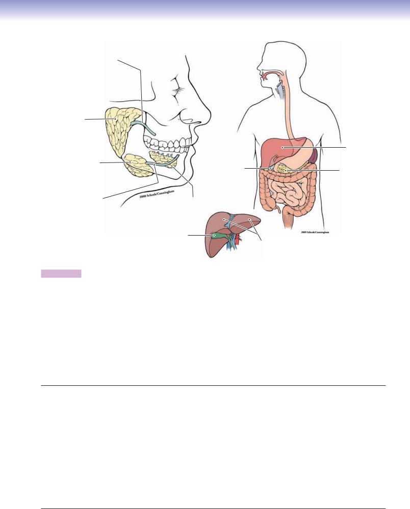
CHAPTER 16 ■ Digestive Glands and Associated Organs |
307 |
Main duct of parotid gland
Parotid |
|
|
gland |
|
|
|
|
Liver |
Submandibular |
|
|
gland |
Gallbladder |
Pancreas |
|
||
|
|
|
Main duct of |
|
|
submandibular |
Sublingual |
|
gland |
gland |
|
|
Gallbladder |
|
|
|
Liver |
Figure 16-1. Overview of the digestive glands and associated organs.
The major digestive glands include the salivary glands, pancreas, and liver. Although the gallbladder is not a gland, it is closely associated with the liver and digestive system. The digestive glands and gallbladder are located outside of the digestive tract. Their ducts carry their products (saliva) into the oral cavity or into the lumen of the duodenum of the small intestine (pancreatic secretions and bile). The salivary glands include the major salivary glands and the minor salivary glands. The parotid, submandibular, and sublingual glands are the three major salivary glands. These glands are either serous glands or mixed glands (both serous and mucous). Most of the minor salivary glands are mucous or mixed glands, with only von Ebner glands being entirely serous glands (see Fig. 14-6A). For details of classification of the glands see Chapter 3, “Epithelium and Glands.” The pancreas includes an endocrine portion (islets of Langerhans) and an exocrine portion. The endocrine portion secretes hormones into the bloodstream (see Chapter 17, “Endocrine System”), whereas the exocrine portion has ducts that carry enzyme products into the lumen of the duodenum (see Figs. 16-9A to 16-10B). The liver produces bile, which is carried by the bile duct system into the gallbladder (see Figs. 16-11 and 16-15A). The liver also aids in the metabolism of lipids, carbohydrates, and proteins; stores iron, glycogen, triglycerides, and vitamins A, D, and B12; and detoxifies certain toxic substances in the blood. The gallbladder stores, concentrates, and releases bile into the duodenum (see Fig. 16-15A,B).
Digestive Glands and Associated Organs
I.Major Salivary Glands
A.Parotid glands (serous)
B.Submandibular glands (mixed, predominately serous)
C.Sublingual glands (mixed, predominately mucous)
II.Pancreas
A.Exocrine portion
B.Endocrine (islets of Langerhans) portion
III.Liver
A.Central artery
B.Portal triad
C.Liver lobules
1.Classic lobule
2.Portal lobule
3.Hepatic acinus
IV. Gallbladder
A.Mucosa
B.Muscularis
C.Serosa
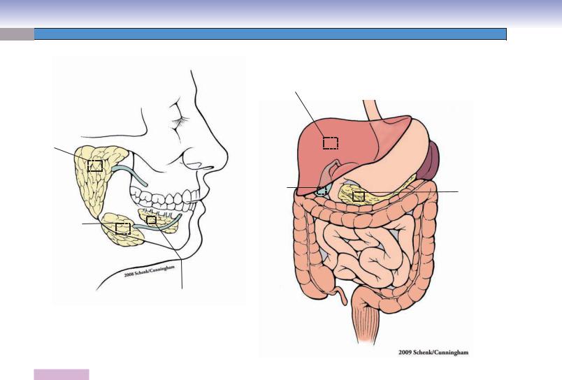
308 UNIT 3 ■ Organ Systems
Fig. 16-12A,B
Fig. 16-13A,B
Fig. 16-14A,B
Fig. 16-3B
Fig. 16-4A
Fig. 16-15A,B
Fig. 16-9B
Fig. 16-10A,B
Fig. 165A,B
Fig. 166A,B
Fig. 167A,B
Fig. 16-8A,B
Figure 16-2. Orientation of detailed digestive glands and associated organs illustrations.
Digestive Glands and Associated Organs with Figure Numbers
Parotid Glands |
Pancreas |
|
Figure 16-3B |
Figure 16-9A |
|
Figure 16-4A |
Figure 16-9B |
|
Figure 16-4B |
Figure 16-9C |
|
Figure 16-4C |
Figure 16-10A |
|
Submandibular Glands |
Figure 16-10B |
|
|
||
Figure 16-5A |
Liver |
|
Figure 16-5B |
Figure 16-11 |
|
Figure 16-6A |
Figure 16-12A |
|
Figure 16-6B |
Figure 16-12B |
|
Figure 16-7A |
Figure 16-12C |
|
Figure 16-7B |
Figure 16-13A |
|
Sublingual Glands |
Figure 16-13B |
|
Figure 16-14A |
||
Figure 16-8A |
||
Figure 16-14B |
||
Figure 16-8B |
||
Figure 16-16A |
||
Figure 16-8C |
||
|
||
|
Gallbladder |
|
|
Figure 16-15A |
|
|
Figure 16-15B |
|
|
Figure 16-16B |
|
|
|

CHAPTER 16 ■ Digestive Glands and Associated Organs |
309 |
Salivary Glands
Secretory portion
|
Serous |
Intralobular duct |
||
|
demilune |
|||
Serous |
(multiple branches) |
|||
|
||||
|
|
|
||
cells |
|
Intercalated |
Striated |
|
|
|
|||
|
|
ducts |
ducts |
|
Serous
acinus
Lumen of the
Intercalated ducts
D. Cui |
Mucous |
|
cells |
||
A |
||
|
Duct system
Interlobular duct |
Lobar duct |
(multiple branches) |
(a few branches) |
Figure 16-3A. General structure of the major salivary glands.
The parotid, submandibular, and sublingual glands are structurally very similar to one another, although they produce various secretions. Each unit of the salivary glands can be divided into a secretory portion and a duct system. The secretory portion contains serous cells, mucous cells, or a mixture of both. These secretory cells are arranged into acini resembling grapes on a stem (intercalated duct). Several serous cells form a cap, called a serous demilune, on the outer aspect of mucous cells; this arrangement can be found in the mixed glands. A capsule (dense connective tissue layer) surrounds an entire gland. Connective tissue septa penetrate the gland and subdivide it into lobes and lobules. The duct system includes intralobular ducts (located within the lobules), interlobular ducts (outside or between the lobules), lobar ducts, and a main duct. The intralobular ducts include intercalated ducts and striated ducts. The main duct empties the secretory products (saliva) into the oral cavity.
B
Serous acini
Striated
Septa ducts
Adipose
cells
Striated
duct
Figure 16-3B. Parotid gland. H&E, 130 skin, palm.
The parotid glands are paired glands and are the largest of the major salivary glands. They are located anterior to and below the lower half of the ear, and superior, posterior, and deep to the ramus of the mandible. The main duct of each parotid gland passes through the cheek and opens into the oral cavity near the second upper molar tooth (see Fig. 16-1). The parotid glands are composed of serous acini and ducts. They are classified as compound (branched) acinar glands based on their duct shape and secretory units (see Fig. 3-27A). This photomicrograph shows striated ducts (intralobular ducts) located in the lobules. The striated ducts are lined by taller cuboidal (or columnar) cells with centrally located nuclei (see Fig. 16-7A). Connective tissue septa divide the gland into small lobules, and there are some adipose cells distributed in among the serous acini. Each acinus is formed by several serous cells with basally positioned dark nuclei (Fig. 16-4A).
Salivary Gland Duct System
Small intralobular duct
(Serous, mucous, or mixed cells) (Intercalated duct)  (striated duct) (Simple low cuboidal epithelium)
|
|
|
|
Interlobular duct |
Main duct |
|
(Stratified columnar epithelium) |
|
|
|
|
|
|
|
|
|
|
|
|
D. Cui
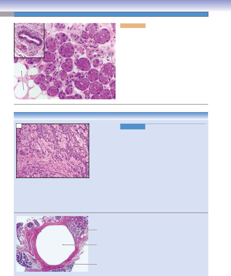
310 UNIT 3 ■ Organ Systems
A |
|
Intercalated |
|
|
duct |
|
|
Serous
cells
Interlobular
duct
Intercalated
duct
Adipose 
cells
Figure 16-4A. Parotid glands. H&E, 295; inset 97
Parotid glands contain only serous secretory cells. There are some adipose cells scattered throughout the gland. All three types of salivary glands have similar duct systems. The intercalated ducts are shown here; they have a small lumen and are lined by lower cuboidal cells with basally located nuclei. The inset shows a large interlobular duct that is lined by stratified columnar cells and is surrounded by a large amount of connective tissue. The diameter of the ducts gradually increases from the intercalated ducts to the large interlobular ducts, and their lining cells increase in height and in number of layers (see Fig. 16-3A). The main duct (duct of Stenson) of the parotid gland traverses the buccinator muscles and opens opposite the secondary upper molars (see Fig. 16-1). All three types of salivary glands receive parasympathetic innervation. The parotid gland is innervated by the glossopharyngeal nerve (cranial nerve [CN] IX).
CLINICAL CORRELATIONS
B
 Myoepithelial
cells
Myxoid  background
Figure 16-4B. Pleomorphic Adenoma. H&E, 55 Pleomorphic adenoma, also called benign mixed tumor, is the most common benign salivary gland tumor. The majority, approximately 80%, occur in the parotid gland, most of which are in the superficial lobe, and are benign pleomorphic adenomas. The tumor may also involve submandibular, sublingual, and minor salivary glands. It is characterized by a slow-growing, mobile, and painless parotid mass. Most patients are not aware of the tumor for years. The mass itself is typically well demarcated, but may be nodular in appearance. Histologically, the neoplasm is composed of epithelial and myoepithelial cells in a chondromyxoid background. Fine needle biopsy is useful in the diagnosis of pleomorphic adenoma. After surgery, pleomorphic adenomas may recur. Rapid growth of a mass in the same area after surgery may signify malignant transformation of the residual adenoma called carcinoma expleomorphic adenoma. This photomicrograph shows nests of myoepithelial cells in a myxoid background.
|
C |
|
|
Figure 16-4C. |
Parotid Cyst. H&E, 11 |
|
|
|
|
|
A parotid cyst is a fluid-filled closed cavity occurring |
||
|
|
|
Parotid |
within the parenchyma of the parotid gland and is usu- |
||
|
|
|
gland |
ally caused by trauma, infections, salivary gland stones, |
||
|
|
|
|
|||
|
|
|
|
or tumors. Physical exam usually reveals a painless |
||
|
|
|
|
lump or swelling. As the cyst enlarges, it may interfere |
||
|
|
|
Cyst |
with chewing, swallowing, and speaking. Additionally, |
||
|
|
|
|
parotid cysts may become infected. Histologically, the |
||
|
|
|
|
cyst is lined by an epithelium, and the cavity is filled |
||
|
|
|
|
with fluid or mucus. The surrounding stroma shows |
||
|
|
|
Stroma |
dense fibrosis and may be infiltrated by aggregates of |
||
|
|
|
lymphocytes, as shown in this slide. If necessary, surgical |
|||
|
|
|
|
|||
|
|
|
|
removal of the cyst is recommended. |
||
|
|
|
|
|
|
|
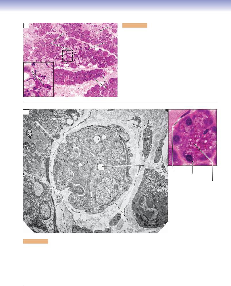
CHAPTER 16 ■ Digestive Glands and Associated Organs |
311 |
A |
|
Striated |
|
|
duct |
|
Mucous |
Serous |
|
demilune |
|
|
cells |
|
|
|
|
Serous |
|
|
demilunes |
|
Septum |
|
|
Septum |
Serous |
|
acini |
Figure 16-5A. Submandibular glands. H&E, 136; inset 408
Submandibular glands are also paired glands. They are smaller than the parotid glands, but larger than the sublingual glands. Each submandibular gland is divided into superficial (large) and deep (small) lobes and is located under the floor of the oral cavity adjacent to the mandible (see Fig. 16-1). The main duct of the submandibular gland drains saliva into the oral cavity at the sublingual caruncles on both sides of the frenulum linguae. The submandibular glands are mixed glands that contain predominantly serous cells but also some mucous cells. They are classified as compound (branched) tubuloacinar glands (see Fig. 16-3A; see also Chapter 3, “Epithelium and Glands,” Fig. 3-28A). Serous cells are arranged into many acini, and mucous cells are arranged as acini or tubular structures, which may have caps of serous cells (serous demilunes). Submandibular glands are innervated by parasympathetic nerve fibers from branches of the facial nerve (CN VII).
B
RER
Lumen
Myoepithelial
cell Serous acinar cell
Secretory granules
Nucleus of the
acinar cell
Figure 16-5B. Serous acinus, submandibular gland. EM, 3,937; inset (color), H&E, 1,079
Several serous acinar cells arranged in an acinus and sharing a common lumen are shown. The serous lumens are smaller than those of a mucous acinus (see Fig. 16-6A). Each secretory cell has a relatively large nucleus and numerous rough endoplasmic reticulum (RER) cisterns in the basal portion of the cytoplasm. These features indicate their active protein synthesis. Secretory granules are usually located on the apical region of the cells (color inset). Secretory granules are not seen in this electron micrograph because they have already been discharged from these particular cells. The inset shows an acinus with secretory cells having round nuclei located basally and many secretory granules in the apical portions of the secretory cells. A myoepithelial cell with a flat nucleus is present here, located beneath the serous cell. It is a contractile cell and contains smooth muscle myosin. The myoepithelial cells contract and move the secretory products toward the intercalated duct.
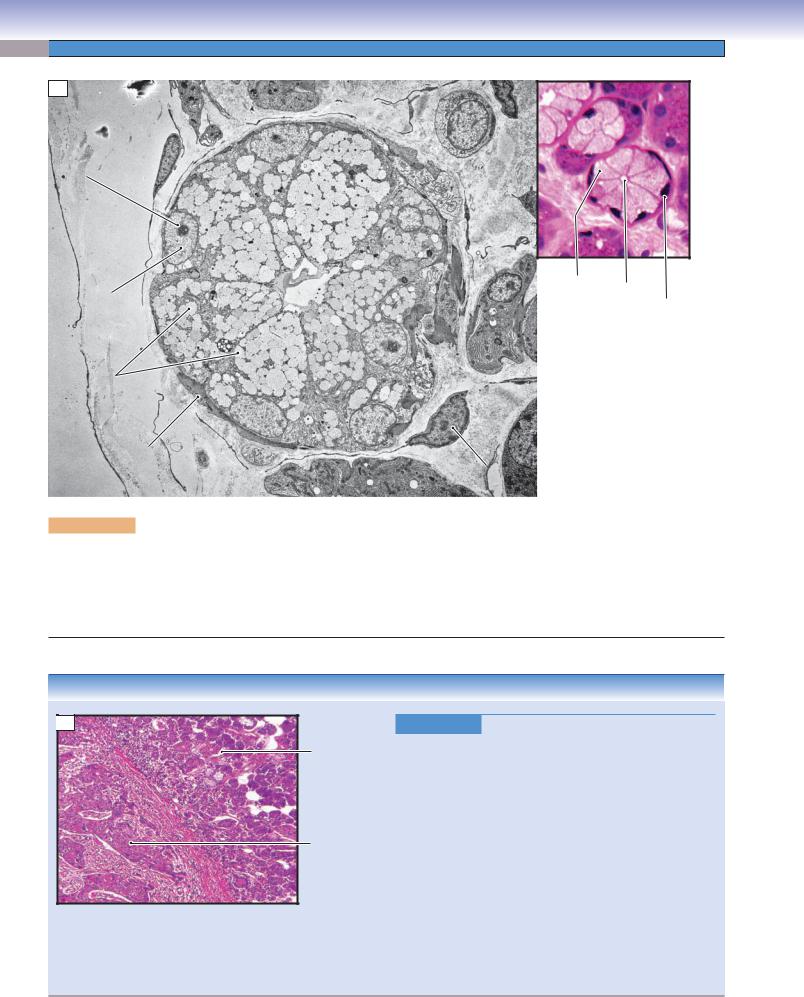
312 UNIT 3 ■ Organ Systems
A
Nucleolus
|
|
Mucous |
Lumen |
|
Nucleus of mucous |
Lumen |
acinar cell |
||
Nucleus of |
||||
|
|
|||
acinar cell |
|
|
||
|
|
mucous |
||
|
|
|
||
|
|
|
acinar cell |
|
Secretory |
|
|
|
|
granules |
|
|
|
Process of
myoepithelial cell
Nucleus of the myoepithelial cell
Figure 16-6A. Mucous acinus, submandibular gland. EM, 6,818; inset (color) H&E, 598
This is an example of a mucous acinus, showing numerous large secretory granules in the apical region of the cells. These granules contain mucins, which are synthesized within the cells. The nuclei of the mucous cells are flattened and lie against the basement membrane. Mucous cells have less RER than serous cells. Myoepithelial cells share the basement membrane with mucous cells. Myoepithelial cells have long cellular processes that can contract, promoting mucous cells to discharge their secretory products into the lumen of the intercalated ducts.
CLINICAL CORRELATION
 B
B
Submandibular gland
Squamous cell carcinoma
Figure 16-6B. Squamous Cell Carcinoma of the Tongue.
H&E, 83
Squamous cell carcinoma of the tongue is a malignant oral neoplasm representing the most common intraoral cancer. Squamous cell carcinoma is usually asymptomatic in the early stages, but local pain, difficulty swallowing, and dysphagia are common in the late stages. Clinically, the neoplasm may appear as a red or white nonhealing ulcer or exophytic mass, most commonly found on the lateral aspect of the tongue. This image shows a squamous cell carcinoma on the floor of the mouth invading the submandibular glands. The major risk factors for oral and tongue squamous cell carcinoma include smoking and alcohol consumption, chronic irritation, and chewing tobacco. Pathologically, the cancer cells show large, irregular and darkly stained nuclei. This cancer tends to metastasize though the lymphatic system. Surgical removal of the primary growth and related lymphatic nodes, chemotherapy, and radiation therapy are treatment options.

CHAPTER 16 ■ Digestive Glands and Associated Organs |
313 |
A
Na+ |
|
Cl_ |
Lumen |
|
|
Na+ K+ |
HCO3_ Cl_ |
Striated |
|
|
duct |
|
|
D. Cui |
Interstitial |
|
|
|
tissue |
|
Serous |
Na+ |
Cl_ |
|
|
|
|
Interstitial |
acinus |
|
|
Striations |
|
|
|
tissue |
|
|
Figure 16-7A. Striated duct, submandibular gland. H&E, 845
Striated ducts are lined by simple cuboidal or columnar cells with centrally positioned nuclei. Their cytoplasm shows striations in the basolateral region, hence the name. The striations are due to deep indentations in the plasma membrane (basolateral folds), which increase the surface area occupied by pumps involved in ion and fluid transport. The folds contain many mitochondria that provide adenosine triphosphate for active transport. The striated ducts collect primary saliva (produced by acinar cells) from the intercalated ducts. The main function of the striated ducts is modulation of the pH and ion composition of the saliva. The striated ducts remove Na+ and Cl− ions in the primary saliva from the lumen and transport these ions to the interstitial tissues. Striated ducts also pump K+ and HCO3− into the saliva fluid. This results in a hypotonic alkaline saliva (secondary saliva), which moves from the striated ducts to the interlobular ducts, then to the main duct, and eventually enters the oral cavity. Saliva plays important roles in moistening and cleansing the oral cavity, helping to repair oral tissues, influencing the pH (buffering) in the oral cavity, stimulating the taste buds, helping to digest food, helping in the mineralization and hardening of the enamel of posterupted teeth, and destroying bacteria by antimicrobial action.
B
Basement membrane
Nucleus
|
Basal |
||
|
membrane |
||
Lumen |
enfolding |
||
|
|
Mitochondrion |
|
|
|
|
|
|
|
|
|
|
|
|
|
|
|
|
|
|
|
|
|
Figure 16-7B. Intralobular duct, submandibular gland. EM, 3,382; inset
6,779
The duct shown here appears to be at the transition between a striated intralobular duct and an interlobular duct. Some of the lining cells, such as the one shown in the inset, have features of a striated duct in that there are extensive basolateral folds containing numerous mitochondria. These are the same features that account for the eosinophilic striations seen in these cells in H&E-stained sections for light microscopy.

314 UNIT 3 ■ Organ Systems
A |
|
Septum |
|
Figure 16-8A. |
Sublingual glands. H&E, 123 |
|
Sublingual glands are the smallest of the three major salivary |
||||
|
|
|
|
||
Interlobular |
|
|
glands. They are paired glands and are located deep in the floor |
||
|
|
of the oral cavity, anterior to the submandibular glands. They |
|||
|
ducts |
|
|
are covered by the oral mucosa, but have no capsule (dense con- |
|
|
|
|
|
||
|
|
|
|
nective tissue) enclosing them as the other two major salivary |
|
|
|
|
|
glands do. Sublingual glands have about 8 to 20 small ducts, |
|
|
|
|
|
which open into the crest of the sublingual fold on the floor |
|
|
|
|
|
of the oral cavity. They are mixed glands, and contain serous |
|
|
|
|
|
and mucous secretory cells, but predominantly mucous cells. As |
|
|
|
|
|
in submandibular glands, serous demilunes are present; sublin- |
|
|
|
|
|
gual glands are also classified as compound (branched) tubu- |
|
|
|
|
|
loacinar glands (see Figs. 16-3A and 3-28A). Two intralobular |
|
|
|
|
Mucous |
ducts located inside connective tissue septa are labeled here. |
|
|
|
|
acini |
Sublingual glands are innervated by parasympathetic nerve |
|
|
|
|
|
fibers from branches of the facial nerve (CN VII). |
|
|
|
|
|
|
|
|
|
|
|
|
|
|
|
|
|
Figure 16-8B. |
Sublingual gland. H&E, 179; inset 408 |
B |
|
Mucous acini |
Capillary |
||
|
Mucous cells make up the majority of the cells in the sublingual |
||||
|
|
|
|||
|
|
|
|
||
|
|
|
|
glands and are distributed throughout the gland. Some mucous |
|
|
|
|
|
cells may be capped with serous cells (serous demilunes); very |
|
|
|
|
|
occasionally complete serous acini may be present. Mucous cells |
|
Intralobular |
|
|
stain lighter than serous cells and they are often arranged in an |
||
|
|
elongated tubular structure (mucous acinus) with a flattened or |
|||
duct |
|
|
round lumen. These mucous cells have dark nuclei located at the |
||
|
|
|
|
||
Serous demilune |
|
|
basal end of the cells. Nuclei of the mucous cells are smaller and |
||
|
|
|
|
flatter than in serous cells. The large intralobular ducts in the |
|
|
|
|
|
sublingual glands are short, and the striations of the ducts are |
|
|
|
|
|
not particularly obvious. The small intralobular ducts (interca- |
|
|
|
|
|
lated ducts) are similar to those of the other two major salivary |
|
|
|
|
|
glands; they receive secretions directly from secretory cells. |
|
|
Mucous cell |
Serous demilune |
|
|
|
|
|
|
|
|
|
|
|
|
|
|
|
CLINICAL CORRELATION
 C
C
 Lymphocytes
Sublingual
 gland tissue
Figure 16-8C. Sialadenitis. H&E, 109
Sialadenitis is the inflammation of salivary tissues or salivary glands caused by injuries, viral or bacterial infection, autoimmune disease, or stones within the salivary gland ducts. Ductal obstruction due to stones (sialolithiasis) may lead to painful gland enlargement and abscess, most often because of bacteria such as Staphylococcus aureus. Histologic findings in acute sialadenitis would show infiltration of glandular parenchyma by abundant neutrophils. The most common cause of viral sialadenitis is mumps, often affecting the parotid glands. Sjögren syndrome is an autoimmune disease characterized by periductal and periacinar lymphocytic infiltrates, formation of lymphatic nodules, periductal fibrosis, and destruction of the glands. This image shows chronic sialadenitis with a lymphocytic infiltrate within the glandular parenchyma.
CHAPTER 16 ■ Digestive Glands and Associated Organs |
315 |
Pancreas
Pancreatic |
Large |
Interlobular |
Main |
|
acini |
duct |
duct |
||
intralobular |
||||
|
|
|
||
|
duct |
|
|
D.Cui |
Small intralobular |
(intercalated) |
|
|
ducts |
A |
Centroacinar |
cells |
Figure 16-9A. A representation of the exocrine pancreatic duct system.
Minor duodenal papilla
Gallbladder
Bile duct
T. Yang
Major duodenal papilla
Interlobular
duct
Main
duct
 Tail
Tail
 Body
Body
 Head
Head
Main
Hepatopancreatic duct ampulla
The pancreas is an elongated gland, which lies mostly posterior to the stomach (see Fig. 16-1). It can be divided into a head, body, and tail. The pancreatic duct begins in the tail of the pancreas, passes through the body, and enters the head of the pancreas. Most exocrine pancreatic secretions are carried by the main duct, which joins the bile duct of the gallbladder at the hepatopancreatic ampulla and empties the secretions into the duodenum through the major duodenal papilla. A small portion of the pancreatic secretion is released into the duodenum through the minor duodenal papilla. The exocrine portion of the gland has a duct system that is similar to that of the major salivary glands, except the pancreas does not have striated ducts and lobar ducts. Secretory products are released into the smallest portions of the intercalated ducts, formed by centroacinar cells, and then drained into the intralobular ducts, the interlobular ducts, and finally into the main duct.
B |
Islet of |
Intralobular |
|
Langerhans |
ducts |
|
|
Centroacinar
cell
Pancreatic
acini
Figure 16-9B. Exocrine and endocrine pancreas. H&E, 272
The pancreas consists of exocrine and endocrine portions. The exocrine pancreas has many serous secretory cells, which stain darkly, as in the major salivary glands. These secretory cells are often called pancreatic acinar cells and are arranged as acini. Each pancreatic acinar cell has a round nucleus and its cytoplasm contains many zymogen granules (see Fig. 16-10A). These cells secrete enzymes that help in the digestion of proteins, lipids, and carbohydrates. The pancreas is innervated by parasympathetic nerve fibers from the right branch of the vagus nerve (CN X).The endocrine pancreas, known as the islets of Langerhans, is found within the exocrine pancreas. The islets produce the hormones insulin and glucagon, which are released into the bloodstream to regulate blood glucose level. The endocrine portion of the gland does not have a duct system (see Chapter 17, “Endocrine System”).
CLINICAL CORRELATION
C |
Acute |
inflammation |
(neutrophils) |
Fat |
necrosis |
Pancreatic |
parenchyma |
tissue |
Figure 16-9C. Acute Pancreatitis. H&E, 50
Acute pancreatitis is an acute inflammatory disease of the pancreas characterized by severe upper abdominal pain, nausea and vomiting, and elevated serum pancreatic enzymes, amylase and lipase. Acute pancreatitis may be caused by hypertriglyceridemia, alcohol ingestion, infections, trauma, drugs, and gallstones. Gallstones may block the pancreatic duct, resulting in autodigestion of the pancreatic parenchyma by enzymes released because of disrupted cell membranes. Pathologic changes include interstitial and peripancreatic edema, fat necrosis with saponification, inflammatory infiltration of neutrophils, and necrosis of the pancreatic parenchyma. Based on the severity of the disease, treatment includes pain control, intravenous fluids, nasogastric suction, and reduction of food intake. In very severe cases, surgical removal of the damaged tissue may be necessary. This image shows acute pancreatitis with fat necrosis, an acute inflammatory infiltrate consisting of neutrophils, and inflamed pancreatic parenchyma.

316 UNIT 3 ■ Organ Systems
A
Islet of
Langerhans
Lumen
RER
 Junctional
complexes
Pancreatic acinar cells
Zymogen granules
Golgi complex
Nucleus of pancreatic acinar cell
Nucleolus |
Rough endoplasmic |
|
|
|
reticulum (RER) |
Figure 16-10A. Pancreatic acinar cells. EM, 9,509; inset (color), H&E, 157
Pancreatic acinar cells are protein-synthesizing cells and contain many secretory (zymogen) granules in the apical region of the cytoplasm and plentiful RER in the basal cytoplasm of the cells. A Golgi complex is seen close to the nucleus of the cell. A lumen is located at the apical region of the acinar cells, where zymogen granules are released. Several junctional complexes near the lumen indicate the fused borders between the neighboring acinar cells. The inset color microphotograph shows pancreatic cells arranged in acini as a secretory unit. These cells have basally positioned nuclei.
B |
Nucleolus of acinar cell |
Nucleus
Zymogen 

granules Centroacinar cells
Lumen
Zymogen
 granules
Centroacinar cells
Figure 16-10B. Centroacinar cells, pancreas. EM, 5,891; inset (color), H&E, 628
Centroacinar cells are not proteinsynthesizing cells and they do not have the features of active protein synthesis and secretion. They have many mitochondria but do not have much RER. These cells form the initial portions of the intercalated ducts and are located at the center of each acinus, hence their name. The centroacinar cells, along with intercalated ducts, play roles in transporting primary pancreatic secretions. They also secrete a large volume of fluid (rich in sodium and bicarbonate) into the pancreatic secretions to help neutralize acidic food contents entering the duodenum from the stomach.

CHAPTER 16 ■ Digestive Glands and Associated Organs |
317 |
Right |
Falciform |
Left lobe |
ligament |
||
lobe |
|
|
Central
vein
Aorta
Bile Gallbladder duct
Portal Inferior vein
vena cava
Portal lobule (3 central veins +1 portal triad)
Sublobular |
Hepatic acinus ( 2 central veins +2 portal triads) |
|
vein |
||
|
|
|
Central vein |
1 |
2 |
3 |
|
Portal vein |
|
|
|
Bile ductule |
Portal triad |
|
|
Hepatic artery |
|
Classic lobule
( 6 portal triads + 1 central vein)
|
Central |
|
vein |
|
Hepatocytes |
Bile |
(hepatic plate) |
|
|
canaliculus |
Venous |
|
(hepatic) |
|
sinusoids |
|
Bile ductule |
|
Portal triad |
Portal vein |
|
|
Hepatic artery |
Hepatic |
|
arteriole |
Figure 16-11. General structure of the liver.
The liver is the largest gland and the largest visceral organ in the body. It has left and right lobes and is covered by a thin capsule called the Glisson capsule. The liver is located in the superior right and superior left quadrants of the abdominal cavity (see Fig. 16-1). The liver contains numerous hepatocytes arranged in plates; venous sinusoids (hepatic sinusoids) run between these hepatic plates. The liver has a rich vascular supply; it receives blood from both the portal veins and the hepatic artery. Secretory products and waste drain out of the liver by three routes: (1) the hepatic venous system, in which blood drains from the hepatic sinusoids (white arrows in detail view above) to the central vein, then to the sublobular vein, and finally to the large hepatic veins; (2) the duct system for bile, in which bile drains from bile canaliculi (green arrow in the detail view above) to the bile ductules, from the ductules into the hepatic ducts, which then join the cystic duct from the gallbladder; and (3) the lymphatic vessel system, in which lymph from the liver drains into the hepatic lymph vessels and passes through the lymph nodes near the liver to then drain into the thoracic duct. The liver has many lobules. These can be classified into three types based on their structure and function. (1) Classic lobule (hexagon shape) is the traditional way to describe liver lobules based on the direction of the blood flow. Each lobule contains six portal triads and one central vein; blood flows from portal triads into a central vein. (2) Portal lobule (triangular shape) emphasizes the exocrine functions of the liver (production of the bile). This classification is based on the direction of the bile flow. Each lobule contains three central veins and one portal triad in the center. The bile is produced by hepatocytes and enters the bile canaliculi to then drain into the bile ductule in the portal triad. (3) Hepatic acinus (diamond shape), which includes two central veins and two portal triads in each lobule. This classification is based on the blood oxygen level, nutrient supply, and metabolic activity. It is an important concept for liver pathology. The hepatic acinus can be subdivided into three zones: zone 1, zone 2, and zone 3 (Fig. 16-12A).
SYNOPSIS 16 - 1 Functions of the Liver
■Exocrine function: secretion of bile into the duodenum to help digest fat and eliminate waste products, such as bilirubin and excess cholesterol. The main components of bile include water, bile salts, bilirubin, cholesterol, fatty acids, lecithin, and electrolytes.
■Endocrine function: synthesis of majority of plasma proteins such as fibrinogen, prothrombin, lipoproteins, and albumins, and their release into the bloodstream.
■Metabolism and detoxification: breakdown of proteins, toxic substances, and many drugs; oxidation and conjugation of toxins, estrogens, and other hormones; and elimination via bile or urine.
■Storage: storage of iron; blood; glycogen; triglycerides (lipid droplets); and vitamins A, D, and B12.

318 UNIT 3 ■ Organ Systems
Liver
A |
|
Central |
Portal triad |
|
|
vein |
|
|
Portal |
Central |
|
|
|
vein |
|
|
triad |
|
|
|
|
Zone 1 |
|
|
|
Zone 1 |
|
|
|
Zone 2 |
Portal triad |
Central |
Zone 3 |
|
|
|
vein |
Portal triad |
Central |
|
|
||
|
|
Portal triad |
vein |
Figure 16-12A. Liver acinus. Silver stain, 89 (left); H&E, 42 (right)
Left: An example of the architecture and general pattern of the liver is shown. The hepatocytes are supported by a network of reticular fibers (black). The red dotted line indicates a portion of the classic lobule. The diamond-shaped hepatic acinus (blue dotted line) is located between two central veins and two portal triads. The hepatic acinus has three zones. Zone 1 is closer to the portal triads and receives the most blood flow from the portal veins and hepatic arteries in the portal triads. Hepatocytes in this region are more likely to survive than cells in zone 3 in case of insufficient oxygen and nutrient supply in the liver, such as in heart failure. However, hepatocytes in zone 1 are also exposed to blood-borne toxins first and are more likely to be damaged than the cells in zone 3. Zone 3 is far from the portal triads and close to the central veins; it has a poor oxygen and nutrient supply, but is also less exposed to blood-borne toxins. In case of heart failure, hepatocytes in zone 3 lack oxygen and appear necrotic first. Zone 2 has an intermediate response to oxygen and toxins.
Right: This image shows the general pattern of organization of the liver at a lower magnification. The classic lobule is indicated by the white dotted line.
Lymphatic |
B |
|
Figure 16-12B. |
Portal triad, liver. H&E, 277 |
vessel |
|
|
|
|
Bile ductule |
|
|
Hepatic artery |
Portal |
|
vein |
||
|
Six portal triads make up one hexagon-shaped, classic lobule. Each portal triad is composed of a portal vein, a hepatic artery, and a bile ductule. These structures are surrounded by connective tissues, which usually contains a lymphatic vessel. (1) The portal vein has a large lumen and thin vessel wall. The branches (portal venules) of the portal vein feed the hepatic sinusoids. (2) The hepatic artery has a small lumen and a wall of smooth muscle that is 2 to 3 cell layers thick. The hepatic artery also has branches (hepatic arterioles), which feed into the hepatic sinusoids. The hepatic sinusoids, therefore, receive glucose-rich blood from the portal vein and oxygen-rich blood from the hepatic artery (see Fig. 16-11). (3) The bile ductule is lined by cuboidal cells with dark, round nuclei. The bile ductule collects bile from the bile canaliculi and drains it into the hepatic duct.
CLINICAL CORRELATION
C 
Portal area with neutrophils
Mallory
body
Steatosis
Figure 16-12C. Alcoholic Fatty Liver (Steatosis). H&E, 186
Alcoholic fatty liver, or steatosis, is usually an asymptomatic and reversible liver condition associated with mild to moderate alcohol use in which lipid accumulates within hepatocytes. Severe steatosis may lead to hepatomegaly and mildly elevated serum bilirubin and alkaline phosphatase levels. Grossly, a fatty liver appears yellow and greasy compared to the normal red-brown appearance of liver tissues. Chronic alcohol abuse, particularly heavy binge drinking, may lead to alcoholic hepatitis, an inflammatory condition of the liver characterized by abdominal discomfort, malaise, hepatomegaly, and abnormal liver tests. Severe cases may result in fulminant hepatic failure. Histologic features of alcoholic hepatitis include a neutrophilic infiltrate, hepatocyte edema and necrosis, and the presence of Mallory bodies, which are cellular accumulations of cytokeratin intermediate filaments. Some patients will progress to alcoholic cirrhosis characterized by regenerating nodules of hepatocytes with intervening fibrous septae. This image shows portal fibrosis, infiltration by neutrophils, fatty change, and Mallory bodies.
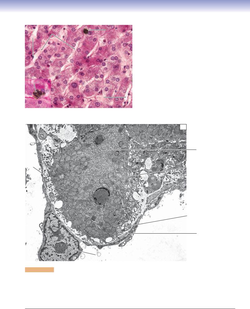
CHAPTER 16 ■ Digestive Glands and Associated Organs |
319 |
Hepatic |
Kupffer |
A |
|
Figure 16-13A. |
Hepatocytes and hepatic sinusoids, liver. |
|
Endothelial sinusoids |
cell |
|
|
H&E, 523; inset |
593 |
|
|
|
|||||
cell |
|
|
|
|
|
|
|
|
|
|
Hepatocytes are large polygonal cells with round nuclei posi- |
||
|
|
|
|
tioned at the center of the cells. These cells have numerous |
||
|
|
|
|
functions that include modification and storage of ingested |
||
|
|
|
|
nutrients, production of many blood proteins, drug metabo- |
||
|
|
|
|
lism, and exocrine secretion of bile salts. This higher magnifi- |
||
|
|
|
|
cation view shows hepatocytes arranged in plates that are one |
||
|
|
|
|
or two cells thick. The hepatic sinusoids are located between |
||
|
|
|
|
the plates of hepatocytes. The endothelial cells that line the |
||
|
|
|
|
sinusoids are discontinuous, which allows proteins and other |
||
Kupffer |
|
|
|
materials to pass through the walls of the sinusoids. Kupffer |
||
cell |
|
|
|
|||
|
|
|
cells are specialized macrophages in the liver. They are irregu- |
|||
|
|
|
|
|||
|
|
Hepatic |
|
lar in shape with ovoid nuclei and often have ingested mate- |
||
|
|
|
rials in their cytoplasm, making them distinguishable from |
|||
|
|
sinusoid |
|
|||
|
|
|
hepatocytes and other cells. They move along the luminal sur- |
|||
|
|
|
|
|||
|
|
|
|
face of the hepatic sinusoids and clear (phagocytose) debris |
||
|
|
|
|
and damaged erythrocytes from the bloodstream. |
||
|
|
|
|
|
|
|
B
Smooth endoplasmic
reticulum of hepatocyte
Space
of Disse Mitochondria  of hepatocyte
Lumen of hepatic sinusoid
Microvilli in Space of  space of Disse
Disse
Endothelial cell
Lumen of
hepatic sinusoid
Nucleus of lymphocyte
Figure 16-13B. Space of Disse, hepatocyte. EM, 7,991
The perisinusoidal space known as the space of Disse is shown here. This space is formed by the surfaces of the hepatocytes and endothelial cells of the hepatic sinusoid. A nucleus of a lymphocyte is seen in the lumen of the hepatic sinusoid. The space of Disse contains many microvilli, which are extensions from the hepatocyte surface. Hepatocytes contain many mitochondria and much smooth endoplasmic reticulum (SER). Hepatocytes are not directly in contact with the bloodstream; the space of Disse provides an environment for the exchange of proteins, plasma, and other material between the hepatocytes and blood in the hepatic sinusoid (Fig. 16-14A). The perisinusoidal space of Disse is also the main extracellular compartment from which liver lymph is derived.
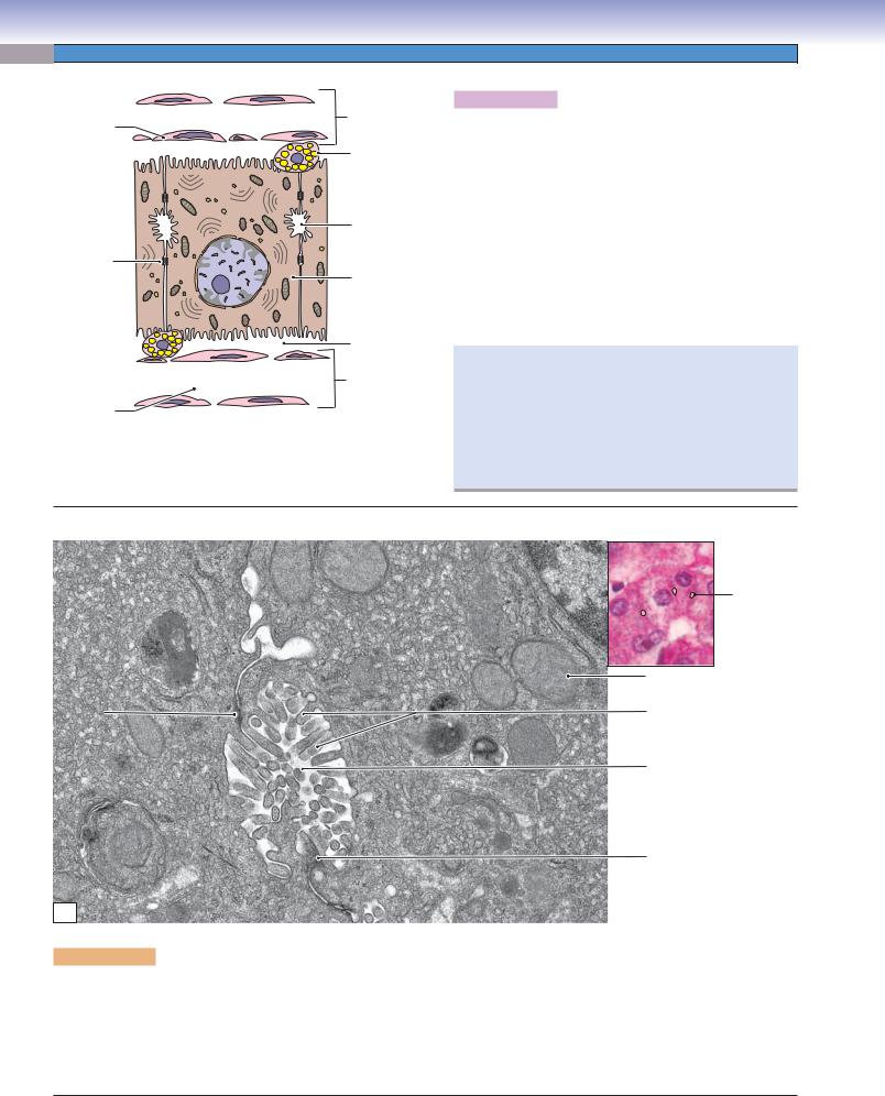
320 UNIT 3 ■ Organ Systems
|
|
|
Figure 16-14A. A representation of bile canaliculi and |
|
Endothelial |
Lumen of sinusoid |
Hepatic sinusoid |
hepatocytes. |
|
cell |
|
|
|
|
|
Space of Disse |
Ito cell |
The bile canaliculi are enlarged intercellular spaces, located |
|
|
|
|
between two adjacent hepatocytes. They receive bile after |
|
|
|
|
it is produced by hepatocytes and transport bile into the bile |
|
|
|
|
ductules (see Fig. 16-11). The hepatic sinusoid wall is lined |
|
|
|
Bile |
by a thin, discontinuous endothelium. The hepatic sinusoids |
|
|
|
canaliculus |
carry glucose-rich and oxygen-rich blood to supply the hepato- |
|
Tight |
|
|
cytes through the space of Disse. This is the space between the |
|
junction |
|
Hepatocyte |
endothelial cells of the hepatic sinusoid and the hepatocytes. |
|
|
|
Short microvilli of the hepatocytes extend into the space of |
||
|
|
|
Disse; fat-storing cells called Ito cells or hepatic stellate cells |
|
|
|
|
are also located in the space. These cells contain many lipid |
|
T. Yang |
|
Space of Disse |
droplets or vacuoles in their cytoplasm, which store vitamin A. |
|
|
Jaundice is a condition in which the skin and sclera become |
|||
|
|
|
||
A |
Lumen of sinusoid |
Hepatic sinusoid |
markedly yellow. It results from a high level of bilirubin in |
|
the bloodstream. Bilirubin is normally removed from blood |
||||
|
|
|||
Endothelial |
|
|
by hepatocytes and then modified and excreted into the |
|
|
|
bile. When excess bilirubin is released into the bloodstream |
||
cell |
|
|
||
|
|
|
(because of destruction of a large number of erythrocytes), |
|
|
|
|
or the elimination of bilirubin is interrupted (as in liver dis- |
|
|
|
|
ease or gallstones), jaundice can develop. |
Hepatocyte #1 |
Hepatocyte #2 |
Nucleus |
|
||
|
|
|
|
|
Bile |
|
|
canaliculus |
SER |
SER |
|
|
|
|
Mitochondria |
Tight |
Microvili |
junction |
|
|
Lumen of bile |
|
canaliculus |
Tight
junction
B
Figure 16-14B. Bile canaliculus, hepatocytes. EM, 6,380; inset (color), H&E, 618
This image shows the lumen of a bile canaliculus between two neighboring hepatocytes. A small lumen of the canaliculus is filled with the microvilli of the two hepatocytes. A small portion of the nucleus, belonging to one of the hepatocytes, is visible in the upper right corner. The SER and mitochondria are shown in the cytoplasm of both hepatocytes. Tight junctions (zonulae occludens) seal each side of the bile canaliculus, preventing initial bile from leaking out of the canaliculus. The inset color photomicrograph shows the pink borders between neighboring hepatocytes. The positions of the bile canaliculi are indicated by small circles on the pink borders. The bile canaliculi are the smallest channels that collect bile in the hepatocyte plates.
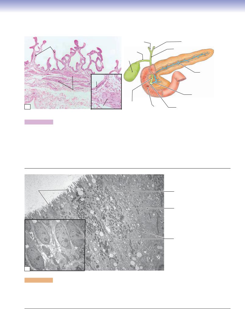
CHAPTER 16 ■ Digestive Glands and Associated Organs |
321 |
Gallbladder
A
|
Right hepatic |
Left hepatic |
Mucosa |
duct |
duct |
|
Common |
|
|
Cystic |
|
|
hepatic duct |
|
|
duct |
|
|
Gallbladder |
|
|
|
|
Main |
|
Columnar |
|
|
pancreatic |
|
|
|
duct |
||
epithelium |
|
|
|
|
Muscularis |
|
T. Yang |
Main pancreatic |
|
|
|
duct |
||
|
|
|
||
Adventitia |
Bile |
|
Hepatopancreatic |
|
Smooth muscle |
duct |
Major |
||
ampulla (of Vater) |
||||
|
|
duodenal papilla
Figure 16-15A. Gallbladder. H&E, 34; inset 99
The gallbladder is a pear-shaped, saclike organ that stores, concentrates, and releases bile. It connects directly to the cystic duct, which is an extension of the gallbladder. Bile from the right and left hepatic ducts drains into the common hepatic duct, which connects to the cystic duct and enters the gallbladder. The gallbladder releases bile in response to cholecystokinin. The gallbladder has a thin wall, which is composed of three layers. (1) The mucosa is the innermost layer, lined by simple columnar epithelium, with many microvilli on the apical surfaces and a lamina propria (loose connective tissue) beneath the epithelium. The mucosa has many branching folds. (2) The muscularis consists of interlacing longitudinal and obliquely oriented bundles of smooth muscle fibers. The contraction of these muscle fibers helps empty bile through the cystic duct into the bile duct. The spiral valve of Heister (smooth muscle at neck of the gallbladder) controls the opening or closing of the gallbladder. The bile duct joins the pancreatic duct at the hepatopancreatic ampulla, and bile enters the duodenum through the major duodenal papilla. (3) The serosa is a connective tissue that covers most of the gallbladder. It contains mesothelium and is continuous with the covering of the liver. The adventitia attaches the gallbladder to the liver and lacks a mesothelium.
Lumen
Microvilli |
Mitochondria |
Interdigitating lateral membranes
Nuclei of |
Columnar cells |
columnar cells |
|
B
Basal  lamina
Figure 16-15B. Epithelial cells lining the gallbladder. EM, 5,291; inset 2,278
There are many short microvilli on the apical surfaces of the columnar cells that line the gallbladder. Numerous microvilli indicate the function of these cells, which is to absorb water from bile in the lumen and transport it into the interstitial tissue. Concentrating bile is one of the main functions of the gallbladder. The interdigitating lateral membranes at the lateral borders of the columnar cells are typical of water-transporting cells. Many mitochondria are in the cytoplasm of these cells, and are more numerous in the superior region. The inset shows the basal region of the epithelium. Oval-shaped nuclei are located close to the basal lamina.
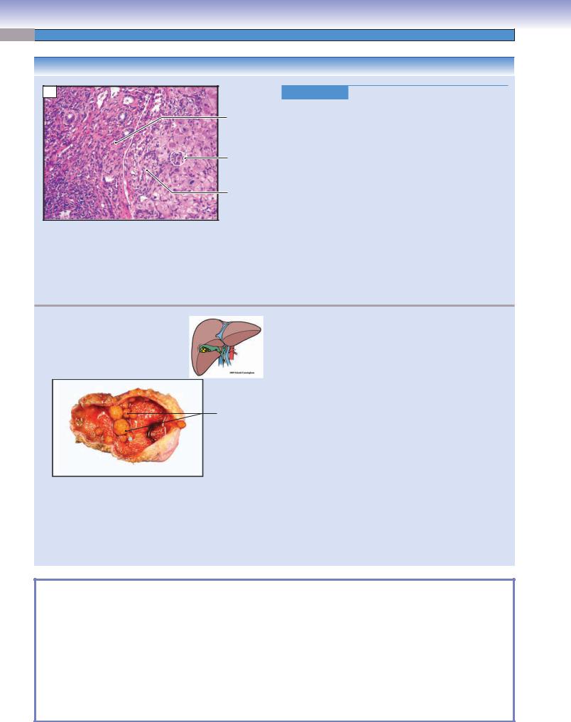
322 UNIT 3 ■ Organ Systems
CLINICAL CORRELATIONS
A
Portal area with chronic inflammation and fibrosis
Necrotic hepatocyte
Periportal hepatitis
Figure 16-16A. Hepatitis C. H&E, 53
Hepatitis C is an infectious liver disease caused by the hepatitis C virus, which may result in chronic hepatitis and cirrhosis. The hepatitis C virus is spread primarily by blood-to-blood contact. Common signs and symptoms may include fatigue, nausea, poor appetite, muscle and joint pains, jaundice, low fever, and tenderness in the liver. In some patients, the disease is self-limiting, but in others it becomes chronic with serious consequences. In the later stages of the disease, cirrhosis may ensue with resultant liver failure and ascites. Pathologic changes to the liver include portal tract expansion with lymphocytes, portal fibrosis, periportal hepatitis, steatosis (fatty change), and lobular parenchymal inflammation. In time, fibrosis becomes marked, surrounding nodules of regenerating hepatocytes to produce hepatic cirrhosis. Treatment options include injections of pegylated interferon-a, the antiviral drug ribavirin, and liver transplantation. This image shows a portal area and adjacent hepatocytes with portal fibrosis and a lymphocytic inflammatory infiltrate along with periportal hepatitis (or “piecemeal” necrosis) and scattered necrotic hepatocytes.
|
|
|
|
|
Figure 16-16B. |
Gallstones. |
|
|
|
|
|
|
Gallstones are a condition in which stones are formed in |
||
|
|
|
|
|
the gallbladder or in the bile ducts. There are two major |
||
|
|
|
|
|
types of gallstones: cholesterol stones and pigment stones. |
||
|
|
|
|
|
Cholesterol stones are far more common than pigment stones |
||
|
|
|
|
|
in Western countries and the United States. Risk factors for |
||
|
B |
|
|
|
gallstones include female gender, obesity, oral contraceptives, |
||
|
|
|
|
|
and being of Northern European, Mexican American, or |
||
|
|
|
Cholesterol |
|
Native American descent. Most patients are asymptomatic |
||
|
|
|
|
until stones obstruct the cystic or common bile ducts, causing |
|||
|
|
|
stones |
|
|||
|
|
|
|
|
severe pain, called biliary colic, because of smooth muscle |
||
|
|
|
|
|
contraction of the duct against the stone. The pain is colicky |
||
|
|
|
|
|
(wavelike) because of the intermittent nature of the contrac- |
||
|
|
|
|
|
tion. Gallstones can cause acute or chronic cholecystitis, and |
||
|
|
|
|
|
blockage of the hepatopancreatic ampulla (ampulla of Vater) |
||
|
|
|
|
|
may lead to acute pancreatitis. The size of gallstones can |
||
|
|
|
|
|
range from that of a grain of sand to a golf ball. Cholesterol |
||
|
|
|
|
|
stones are pale yellow, radiolucent, and large (1–3 cm), |
||
|
|
|
|
|
whereas pigment stones are black, radiopaque, and smaller |
||
|
|
|
|
|
(<1 cm). Open surgical or laparoscopic removal of the gall- |
||
|
|
|
|
|
bladder is the most common treatment for gallstones. This |
||
|
|
|
|
|
gross photograph shows a fresh cholecystectomy specimen |
||
|
|
|
|
|
containing multiple cholesterol stones. |
||
|
|
|
|
|
|
|
|
SYNOPSIS 16-2 Pathological and Clinical Terms for the Digestive Glands and Associated Organs
■Sialolithiasis: the presence of sialoliths, or stones within the ducts of salivary glands; may cause obstruction resulting in inspissated secretions and swelling as well as infection (see Fig. 16-8C).
■Autodigestion: in reference to acute pancreatitis, the destruction of pancreatic tissues and surrounding adipose tissues by the release of pancreatic enzymes as a result of damage to exocrine pancreas cells (see Fig. 16-9C).
■Saponification: in reference to acute pancreatitis, the necrosis of adipose tissues by lipolytic pancreatic enzymes released as a result of damage to exocrine pancreas cells; free fatty acids react with calcium to form insoluble salts (see Fig. 16-9C).
■Mallory body: an intracellular accumulation of intermediate keratin filaments seen in hepatocytes in cases of alcoholic hepatitis (see Fig. 16-12C).
■Periportal hepatitis: also called “piecemeal” necrosis in cases of hepatitis; inflammatory process in a portal area transgresses the limiting plate and involves the surrounding hepatocytes causing cell injury and death (see Fig. 16-16A).
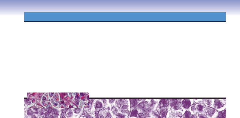
17 Endocrine System
Introduction and Key Concepts for the Endocrine System
Figure 17-1 |
Overview and Orientation of Detailed Endocrine Organ Illustrations |
Pituitary Gland |
|
Figure 17-2 |
Overview of Hormone Regulation by the Pituitary Gland |
Figure 17-3A–C |
Overview of the Pituitary Gland |
Figure 17-4A |
Pars Distalis, Adenohypophysis (Anterior Pituitary Gland) |
Figure 17-4B |
Cells in the Pars Distalis, Anterior Pituitary Gland |
Figure 17-5A |
Pars Intermedia, Anterior Pituitary Gland |
Figure 17-5B |
Blood Supply of the Pituitary Gland |
Figure 17-6A |
Pars Nervosa, Neurohypophysis (Posterior Pituitary Gland) |
Figure 17-6B |
Pars Nervosa, Posterior Pituitary Gland |
Figure 17-7A,B |
Clinical Correlation: Pituitary Adenoma |
Figure 17-7C |
Clinical Correlation: Pituitary Adenoma in Magnetic Resonance Imaging |
Thyroid Gland |
|
Figure 17-8A |
Thyroid Follicles, Thyroid Gland |
Figure 17-8B |
Parafollicular Cells, Thyroid Gland |
Figure 17-8C |
Clinical Correlation: Hashimoto Thyroiditis |
Parathyroid Glands |
|
Figure 17-9A |
Overview of the Parathyroid Glands |
Figure 17-9B |
Chief Cells and Oxyphil Cells of the Parathyroid Glands |
Figure 17-9C |
Clinical Correlation: Parathyroid Adenoma |
Adrenal Glands (Suprarenal Glands) |
|
Figure 17-10A |
Overview of the Adrenal Glands |
Figure 17-10B |
Cortex of the Adrenal Gland |
Figure 17-11A |
Adrenal Cortical Cells, Adrenal Cortex |
323