

Биоинженерия / Биомеханика_микрофлюидные_устройства / статьи / b804795b
.pdfPublished on 23 July 2008. Downloaded on 11/18/2019 11:51:43 AM.
|
View Article Online / Journal Homepage / Table of Contents for this issue |
|
PAPER |
www.rsc.org/loc | Lab on a Chip |
|
|
|
|
Microfluidic devices for studies of shear-dependent platelet adhesion
Edgar Gutierrez,a Brian G. Petrich,b Sanford J. Shattil,b Mark H. Ginsberg,b Alex Groisman*a and Ana Kasirer-Friedeb
Received 26th March 2008, Accepted 19th June 2008
First published as an Advance Article on the web 23rd July 2008
DOI: 10.1039/b804795b
Adhesion of platelets to blood vessel walls is a shear stress dependent process that promotes arrest of bleeding and is mediated by the interaction of receptors expressed on platelets with various extracellular matrix (ECM) proteins that may become exposed upon vascular injury. Studies of dynamic platelet adhesion to ECM-coated substrates in conventional flow chambers require substantial fluid volumes and are difficult to perform with blood samples from a single laboratory mouse. Here we report dynamic platelet adhesion assays in two new microfluidic devices made of PDMS. Small cross-sections of the flow chambers in the devices reduce the blood volume requirements to <100 ll per assay, making the assays compatible with samples of whole blood obtained from a single mouse. One device has an array of 8 flow chambers with shear stress varying by a factor of 1.93 between adjacent chambers, covering a 100-fold range from low venous to arterial. The other device allows simultaneous high-resolution fluorescence imaging of dynamic adhesion of platelets from two different blood samples. Adhesion of platelets in the devices to three common ECM substrate coatings was verified to conform with published results. The devices were subsequently used to study the roles of extracellular and intracellular domains of integrin aIIbb3, a platelet receptor that is a central mediator of platelet aggregation and thrombus formation. The study involved wild-type mice and two genetically modified mouse strains and showed that the absence of the integrin impaired adhesion at all shear stresses, whereas a mutation in its intracellular domain reduced the adhesion only at moderate and high stresses. Because of small sample volumes required, the devices could be employed in research with genetically-modified model organisms and for adhesion tests in clinical settings with blood from neonates.
Introduction
Regulated reactivity of platelets to extracellular matrices (ECM) is fundamental to thrombosis and stoppage of bleeding. Vascular injury and atherosclerotic plaque rupture expose ECM on which platelets may be captured from bulk blood transport and adhere through multiple receptors. These receptors, including GPIb-IX-V for von Willebrand factor (VWF) and GPVI and a2b1 for collagen, cooperate to stimulate complex “inside-out” signaling networks that activate integrin aIIbb3 (reviewed in ref. 1), resulting in stable platelet capture to ECM and thrombus growth.2,3 Atherosclerotic lesions have been reported to contain adsorbed fibrinogen,4 which can be recognized by non-activated platelet aIIbb3.5 Furthermore, platelet-vessel wall interactions may promote leukocyte recruitment, which has an impact on atherogenesis, inflammation and pathological thrombosis.6,7
Because of the importance of inside-out aIIbb3 signaling for platelet function, it has been investigated in some detail. A current model holds that a final step in effecting the requisite conformational change and activation of the integrin involves the binding of talin to the b3 cytoplasmic tail of the integrin.8,9
aDepartment of Physics, UCSD, 9500 Gilman Drive, MC 0374, La Jolla, CA, 92093, USA. E-mail: agroisman@ucsd.edu; Fax: (858)534-7697; Tel: (858)822-1838
bMedicine, University of California San Diego, La Jolla, CA, USA
Support for this model is provided by recent studies of a strain of knock-in mice (b3Y747A) harboring a point mutation in the b3 tail (Y747A) that abrogates interaction with talin and other integrin binding proteins.10 Platelets from these mice exhibit deficient agonist-induced platelet activation in vitro and impaired thrombus formation in vivo. However, no detailed study has been made on how this mutation affects the adhesion of platelets under physiologically relevant shear conditions.
In the circulation, platelets experience a wide range of shear stresses from approximately 0.8–8 dyn cm−2 in the venous circulation to 10–60 dyn cm−2 in the arterial circulation.11 Various flow devices, particularly parallel plate flow chambers, have been used in ex vivo platelet studies to mimic flow conditions occurring in vivo.12,5 Conventional flow chambers often have a depth of 0.1–0.3 mm and a width of 2–10 mm, requiring a relatively large volume of blood (milliliters) for an assay. For tests with mouse blood, this volume is often obtained by mixing blood samples drawn from several laboratory animals. In addition, conventional flow chambers typically test adhesion at a single shear stress or in a small range of shear stresses per experiment. Therefore, studies of platelet adhesion over the entire physiological range of shear stresses are costly in terms of time and laboratory animals. The sample volume requirements are dramatically reduced in microfluidic flow chambers. Microfluidic perfusion chambers have been applied to capturing different sub-populations of lymphocytes to substrates with
1486 | Lab Chip, 2008, 8, 1486–1495 |
This journal is © The Royal Society of Chemistry 2008 |

Published on 23 July 2008. Downloaded on 11/18/2019 11:51:43 AM.
View Article Online
various coatings from continuous flow13,14 and testing the |
s. Furthermore, b3−/− and b3Y747A platelets that adhered to |
|||||
strength of adhesion of different cells to a substratum.15,16 Re- |
collagen did not form aggregates and remained as a monolayer, |
|||||
cently, microfluidic devices were used to study initiation of blood |
thus enabling the use of the microfluidic devices for exploring |
|||||
clotting,17 particle adhesion in complex channel networks,18 |
the rolling and arrest of granulocytes on platelet monolayers. |
|||||
the dynamics of adhesion of infected erythrocytes to different |
Experimental |
|
|
|||
substrata19 and of neutrophils to endothelial monolayers.20 |
|
|
||||
However, the microfluidic technology has not yet been applied |
Construction and operation of microfluidic devices |
|
||||
to studies of shear-dependent trends in platelet adhesion. |
|
|||||
|
|
|
|
|
||
Here we report platelet adhesion assays in two novel microflu- |
Microfluidic devices (Fig. 1) consisted of PDMS chips sealed |
|||||
idic devices (Fig. 1). Both devices have flow chambers with a |
with #1.5 microscope coverglasses. The chips were cast out of |
|||||
small cross-section, resulting in a blood consumption of 2–3 |
PDMS (Sylgard 184 by Dow Corning) using master molds fab- |
|||||
orders of magnitude less than in conventional flow chambers and |
ricated with a two-step protocol described in detail elsewhere.21 |
|||||
making it possible to run tests with blood samples obtained from |
The mold fabrication involved two consecutive coatings of a |
|||||
a single laboratory mouse. Device 1 (Fig. 1A) has an array of 8 |
silicon wafer with different formulations of a UV-curable epoxy, |
|||||
identical rectilinear flow chambers with shear stresses, s, varying |
SU8 (Microchem, Newton, MA), and their exposure to UV- |
|||||
by a factor of 1.93 between adjacent chambers, thus covering a |
light through two different photomasks. All test chambers (flow |
|||||
100-fold range in s, representative of low venous to arterial blood |
chambers) in both microfluidic devices (Fig. 1) had a depth h = |
|||||
flow. Device 2 (Fig. 1B) is similar to device 1, but consists of two |
24 lm and a width w = 200 lm. The substratum shear rate, |
|||||
separate mirror-symmetric microchannel networks, permitting |
c˙ , in an internal region of a test chamber (away from the side |
|||||
observation of dynamic adhesion of platelets from two different |
walls) is found as c˙ ≈ 6m¯ /h, where m¯ is the mean flow velocity in |
|||||
blood samples in a single field of view of a high-resolution |
the chamber. The substratum shear stress is given by s ≈ 6mg¯ /h, |
|||||
fluorescence imaging setup. |
where g ≈ 0.038 P is the viscosity of blood. (A shear rate c˙ = |
|||||
|
1 s−1 corresponds to a shear stress s = 0.038 dyn cm−2.) The |
|||||
|
volumetric flow rate, |
, through a test chamber is found as |
Q = |
|||
|
2 |
c˙ /6 |
2Q |
|
||
|
wh¯m ≈ wh |
= wh s/(6g). The rate of consumption of blood |
||||
|
at a given shear stress is proportional to the chamber width and |
|||||
|
to the chamber depth squared and is 340 times lower for the |
|||||
|
microfluidic devices than for a 0.125 × 2.5 mm commercial flow |
|||||
|
chamber (GlycoTech Inc., MD). |
|
||||
|
The microfluidic device 1 (Fig. 1A) had one inlet, one outlet |
|||||
|
and an array of 8 parallel test chambers. All test chambers, except |
|||||
|
for test chamber 1, were connected at their exits to resistance |
|||||
Fig. 1 Drawings of microchannel networks of the two microfluidic |
channels (24 lm deep and 40 lm wide). A test chamber and |
|||||
devices used in the study: (A) device 1 and (B) device 2. 24 lm and |
its resistance channel constituted a channel line. Each channel |
|||||
250 lm deep channels are shown in dark and light grey, respectively. |
line was connected to the feeder channel on the upstream side |
|||||
The 250 lm deep feeder channels minimize the shear stress on the way |
||||||
and to the collector channel on the downstream side (Fig. 1A). |
||||||
from the inlet to the test chamber and, just as the collector channels, |
||||||
The feeder and collector channels were both 250 lm deep |
||||||
have low flow resistance and nearly uniform pressure in them. Flow rate |
||||||
through the 24 lm test chambers is set by the flow resistance of the |
and 500 lm wide, making their flow resistances negligible |
|||||
resistance channels connected in series with the test chambers. The flow |
compared with those of the 24 lm deep channel lines, and |
|||||
providing equal pressures at the entrances and equal pressures |
||||||
rate is highest for the test chamber 1 and lowest for the test chamber |
||||||
8. The bypass channels help synchronize the injection of blood into |
at the exits of all 8 channel lines. Thus the differential pressures, |
|||||
different test chambers. |
DP, across the 8 channel lines were all equal to each other |
|||||
|
and equal to the difference in pressure between the inlet and |
|||||
To test the devices and validate their application to platelet |
outlet of the device. The volumetric flow rate, Q, through a |
|||||
adhesion assays, we first verified that the dynamic adhesion |
test chamber is found as Q = DP/R where R is the cumulative |
|||||
of platelets to substrates coated with three commonly used |
hydrodynamic resistance of the channel line. The values of R |
|||||
ECM molecules (fibrinogen, collagen, and VWF) occurred in |
were designed to vary by a factor of 1.93 between adjacent |
|||||
agreement with previous reports. To demonstrate the utility |
channel lines, thus providing a 1.93-fold change in Q and shear |
|||||
of the proposed microfluidic devices for biological studies, |
stress, s, between adjacent test chambers, with a total 100-fold |
|||||
we used the devices to explore the role of integrin aIIbb3 in |
variation between the test chambers 1 and 8. We note that the |
|||||
dynamic adhesion of platelets to fibrinogen and collagen in the |
microchannel architecture of device 1 was essentially different |
|||||
physiological range of shear stresses, s. The study was performed |
from the architectures of some perfusion devices described |
|||||
with blood from normal mice (b3+/+) and mice whose platelets |
earlier, where shear stress variations were achieved by varying |
|||||
either lack aIIbb3 (b3−/− ), or have normal extracellular domains, |
the width of the flow chambers, limiting the variations to about |
|||||
but are activation-defective by virtue of a point mutation in |
one order of magnitude.12,13,15,16 |
|
||||
the b3 cytoplasmic domain (b3Y747A). The results showed |
Because of the large cross-section of the feeder channel as |
|||||
that the dynamic adhesion of platelets that lack aIIbb3 was |
compared with the test chambers, the shear stress in the feeder |
|||||
strongly impaired at all s, whereas the adhesion of the activation |
channel was 2 times lower than the lowest shear stress in the |
|||||
defective mutant was only reduced at intermediate and high |
test chambers (found in the chamber 8), resulting in a minimal |
|||||
|
|
|
|
|
|
|
This journal is © The Royal Society of Chemistry 2008 |
|
|
|
Lab Chip, 2008, 8, 1486–1495 | |
1487 |
|

Published on 23 July 2008. Downloaded on 11/18/2019 11:51:43 AM.
View Article Online
activation of platelets by shear stress prior to their arrival at the |
mate controls. The platelet counts of mice of all three genotypes |
||||||||||||||||
test chambers. Bypass channels at the edges of the test chamber |
were similar. |
|
|
|
|||||||||||||
array (Fig. 1) had relatively low flow resistance and were added |
|
|
|
|
|
|
|
||||||||||
to suppress the formation of air bubbles in the corners and to |
Experimental protocol |
|
|
||||||||||||||
better synchronize the arrival of blood at different test chambers. |
|
|
|||||||||||||||
|
|
|
|
|
|
|
|||||||||||
The microfluidic device 2 (Fig. 1B) had a set of two identical |
Platelets were visualized by adding 2 lM mepacrine5 to whole |
||||||||||||||||
(mirror-symmetric) disconnected microchannel networks that |
blood to label dense granules. At high concentrations, mepacrine |
||||||||||||||||
were placed in close proximity of each other. Each network |
(which also labels leukocytes) may inhibit phospholipase A2. |
||||||||||||||||
had an inlet, an outlet, and a vent port, marked by numbers |
Nevertheless at 2 lM, platelet and leukocyte functions are |
||||||||||||||||
1 and 2 for networks 1 and 2, respectively. Each network had |
maintained.5,30 For studies of platelet–granulocyte interactions, |
||||||||||||||||
two separate channel lines identical to two of the channel lines of |
granulocytes in whole blood were additionally labeled with 16 lg |
||||||||||||||||
device 1 (cf . Fig. 1A). For the device in Fig. 1B, these two channel |
mL−1 of a phycoerythrin (PE)-conjugated anti-Gr-1 antibody. |
||||||||||||||||
lines were 1 and 3, and test chambers 1 of the two microchannel |
No bleed-through occurred between the green fluorescence of |
||||||||||||||||
networks were adjacent to each other with a 40 lm partition |
mepacrine and red fluorescence of phycoerythrin. All inhibitors |
||||||||||||||||
between them. (We also used two other versions of device 2, |
were added 25 min prior to onset of blood flow through the |
||||||||||||||||
in which the adjacent test chambers were from channel lines 3 |
device, except ethanol control and prostaglandin E1 (PGE1), |
||||||||||||||||
and 5.) |
|
|
|
|
|
|
|
that were added 5 min prior to flow onset. |
|
|
|||||||
|
|
|
|
|
|
|
|
|
|
To coat the glass substrata of the microfluidic devices with |
|||||||
Flow control |
|
|
|
|
|
physiological matrices, the microchannels were filled with 20 lg |
|||||||||||
Flow in the |
microfluidic devices was driven by differential |
mL−1 |
fibrinogen, 300 lg mL−1 acid-soluble Type I collagen or |
||||||||||||||
10 lg mL−1 |
VWF, and incubated for 1 h at room temperature. |
||||||||||||||||
hydrostatic pressure, DP, applied between the inlet and outlet, |
|||||||||||||||||
Subsequently, the devices were rinsed with an excessive amount |
|||||||||||||||||
generated by gravity, and controlled within 10 Pa.21 Care was |
|||||||||||||||||
of Hepes buffer (150 mM NaCl, 20 mM Hepes, pH 7.4) or |
|||||||||||||||||
taken to prevent platelet activation by avoiding exposure of |
rinsed with buffer and then blocked with 1% BSA for another |
||||||||||||||||
blood to glass or metal components: blood and buffer solutions |
|||||||||||||||||
30 minutes, with similar results. The flow through the device was |
|||||||||||||||||
used in the experiments were kept in plastic syringes (1 mL and |
|||||||||||||||||
stopped by clamping the Tygon tubing, and the outlet syringe |
|||||||||||||||||
10 mL) connected to the device through flexible Tygon tubing |
|||||||||||||||||
was placed at a level of 10 cm above the microscope stage. |
|||||||||||||||||
(0.5 mm inner diameter) and short polyimide capillaries that |
|||||||||||||||||
were inserted into the device ports. |
|
200 ll of human or mouse blood with or without indicated |
|||||||||||||||
|
inhibitors was gently drawn into a 1 cc plastic syringe through |
||||||||||||||||
|
|
|
|
|
|
|
|
|
|
||||||||
Reagents and blood preparation |
|
a Tygon tubing line by pushing the plunger to 1/5 of the |
|||||||||||||||
|
|
|
|
|
|
|
|
|
|
syringe length, immersing the tubing into blood, pulling the |
|||||||
Function-blocking monoclonal antibody 1B5 against murine |
plunger to the end of the syringe (thus creating a gauge pressure |
||||||||||||||||
a |
|
b |
|
from Dr Barry Coller (Rockefeller University, New |
of −1/5 atm), waiting till the amount of blood in the syringe |
||||||||||||
IIb |
3 was 22 |
Monoclonal antibody AP-1 against human GP |
|||||||||||||||
York, NY). |
reached 100 lL (plus 100 lL in the tubing), and then quickly |
||||||||||||||||
Iba was from Dr Thomas Kunicki (Scripps Research Institute, |
removing the plunger. The tubing was then connected to the |
||||||||||||||||
La |
Jolla, CA).23 Monoclonal antibody 5H1 against mouse P- |
device inlet. The inlet was pressurized at |
D |
P = 2.5 kPa with |
|||||||||||||
|
|
24 |
was from Dr Rodger McEver (Oklahoma Medical |
|
|||||||||||||
selectin |
|
respect to the outlet by raising the syringe with the blood so |
|||||||||||||||
Research Foundation, Oklahoma City, OK), and a polyclonal |
that the level of blood was 25 cm above the level of the buffer |
||||||||||||||||
anti-mouse PSGL-1 blocking antibody25was from Dr Bruce |
in the outlet syringe. Alternatively, 100 lL of mouse blood |
||||||||||||||||
Furie (Harvard University, Boston, MA). |
Integrilin, a selective |
were loaded into a 0.5 mL Eppendorf tube that was sealed by a |
|||||||||||||||
antagonist to human or murine aIIbb326 was from Dr David |
PDMS plug with two openings, for a luer stub connecting the |
||||||||||||||||
Phillips (Portola Pharmaceuticals, Inc., South San Francisco, |
tube to a source of compressed air with a pressure P = 2.5 kPa, |
||||||||||||||||
CA). Non-function-blocking antibodies to aIIb and granulocyte |
and for PE 10 polyethylene tubing. One end of the tubing line |
||||||||||||||||
Ly-6 (Gr-1) were from Invitrogen/BD Biosciences (Carlsbad, |
(that was 10 cm long) was touching the bottom of the tube, and |
||||||||||||||||
CA), as were control IgG antibodies. Human plasma VWF was |
its other end was directly inserted into the device port. Because |
||||||||||||||||
a gift from Dr Zaverio Ruggeri (Scripps Research Institute, La |
of the small volume of blood in the polyethylene tubing line |
||||||||||||||||
Jolla CA).27 Fibrinogen was from Enzyme Research Co. (South |
( 6 lL), almost the entire blood sample loaded into the |
||||||||||||||||
Bend, IN). All other reagents were from Sigma Chemical Co. |
Eppendorf tube could be used for perfusion experiments. |
||||||||||||||||
(St. Louis, MO). 1.9 lm fluorescent beads, used to measure |
For the device 1 (Fig. 1A), flow of blood through the |
||||||||||||||||
the flow rates by particle tracking, were purchased from Bangs |
device was initiated by removing the clamp from the outlet |
||||||||||||||||
Laboratories (Fishers, IN). |
|
tubing. Because of relatively low volume of buffer between the |
|||||||||||||||
Human blood obtained from normal, drug-free donors was |
test channels and the tubing with the blood ( 0.5 lL) and |
||||||||||||||||
anticoagulated with 20 |
U mL−1 heparin, which maintains |
relatively high total volumetric flow rate through the device |
|||||||||||||||
normal calcium |
concentrations and does not interfere with the |
( 3.7 |
l |
L min |
−1), the transient time of injection of blood into the |
||||||||||||
|
28 |
Mouse blood was drawn by cardiac |
|
|
|
|
|||||||||||
platelet adhesion assays. |
|
test channels was only 8 s, which was substantially shorter than |
|||||||||||||||
puncture into heparin-containing syringes. Mice deficient in |
the duration of adhesion assays. In the device 2 (Fig. 1B), there |
||||||||||||||||
integrin b3 (b3−/− )29 were obtained from Jackson Laboratories |
were four syringes with buffer connected to the outlets and vents |
||||||||||||||||
(Bar Harbor, Maine), and b3 knock-in mice (b3Y747A) were |
of each of the two microchannel networks (marked by numbers |
||||||||||||||||
generated at UCSD.10 Wild-type mice (b3+/+) represented litter- |
1 and 2 in Fig. 1B) through four separate Tygon tubing lines. The |
||||||||||||||||
|
|
|
|
|
|
||||||||||||
1488 | |
Lab Chip, 2008, 8, 1486–1495 |
|
|
|
This journal is © The Royal Society of Chemistry 2008 |
||||||||||||
Published on 23 July 2008. Downloaded on 11/18/2019 11:51:43 AM.
View Article Online
lines connected to both outlets were initially clamped, and when |
Image acquisition and analysis |
the syringes with blood were connected to the inlets, the flow |
In the experiments with device 1 (Fig. 1A), the microfluidic |
of blood was from the inlets to the vents. Once the buffer was |
|
purged from the channels connecting the inlets with the vents, |
device was mounted on a mechanical stage of a Nikon Diaphot |
and the channels were filled with whole blood, the vent tubing |
inverted fluorescence microscope that was equipped with a |
lines were clamped, completely stopping the flow through the |
Newport 850G linear actuator. The actuator was driving the |
device. The adhesion assay was started by simultaneous removal |
stage in the direction perpendicular to the direction of flow |
of the clamps from both outlet lines, thus starting flow of blood |
in the test chambers and enabled moving the field of view of |
from inlets 1 and 2 to outlets 1 and 2, respectively (Fig. 1B). |
the microscope between different test chambers with a 5 lm |
At that moment, blood in both networks was separated from |
positioning accuracy. Fluorescence microscopy was performed |
the test chambers by small and equal volumes of buffer in |
with a Nikon 100 W mercury light source and a GFP filter set |
the feeder channels. Therefore, the arrival of blood at the test |
(Ex470/Em525). The images of the platelets were acquired with |
chambers was synchronized within less than a second and |
a 63×, NA = 1.4 oil immersion objective lens, a 0.42× video |
occurred within less than a second from the moment of clamp |
adapter, and a Sony SX900 IEEE1394 camera with a 1/2”, |
removal. The duration of a perfusion experiment was <10 min, |
1280 × 960 pixel CCD array. An ND8 neutral density filter |
and the total consumption of blood during an experiment was |
was used to reduce the intensity of fluorescence illumination |
<40 lL (<100 lL with occasional sample loss during tubing |
and minimize platelet photoactivation.31 Motion of the stage |
reconnection). Cells were counted as stably attached if they |
and image acquisition were controlled through RS232 and |
moved by less than one cell diameter in 10 s. To fix cells after an |
IEEE1394 interfaces, respectively, using a code in LabView7.1 |
adhesion assay, the device was perfused with 3.7% formaldehyde |
(National Instruments, Austin, TX). The stage was programmed |
and incubated for 10 min. The device was then disassembled and |
to move in periodic scanning loops between test chambers 1–8, |
coverglasses were rinsed with Hepes buffer. |
with eight stops to take a fluorescence image of each chamber. |
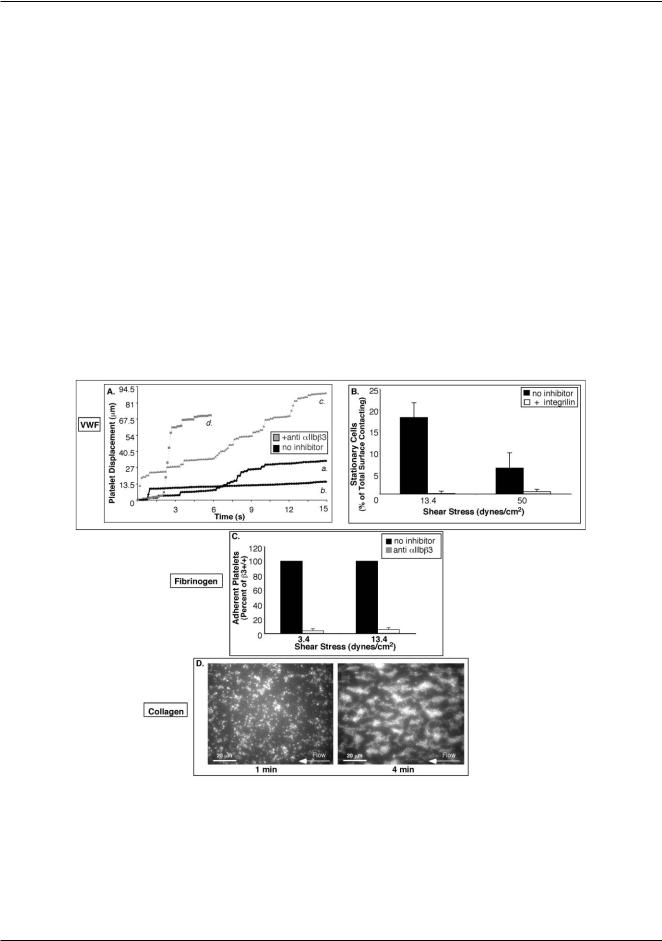
Fig. 2 Validation of platelet adhesion to physiologic matrices in microfluidic devices. (A) Representative trajectories of individual platelets on VWF without inhibitor (black symbols; a and b) and with 20 lM integrilin, an anti-aIIbb3 antagonist (grey symbols; c and d). Long plateaus correspond to prolonged periods of rest (a) that may lead to stable attachment (b) and are only observed for untreated cells. All platelet surface interactions are abolished in the presence of 10 lg mL−1 AP-1, an anti-GPIba antibody (not shown). (B) Histogram of the fraction of untreated cells and cells treated with integrilin that become stably attached to VWF at 3.4 and 50 dyn cm−2 after 1 min of flow. (C) Adhesion to fibrinogen is aIIbb3 dependent. Whole blood was incubated with or without 15 lg mL−1 Ib5, an anti-murine aIIbb3 monoclonal antibody, and the platelet adhesion to fibrinogen was determined after 1 min of flow at s = 3.4 and 13.4 dyn cm−2 . Average values of 3 separate experiments ± SEM are shown. (D) Fluorescence micrographs of a test chamber with collagen matrix in the device 1 at 13.4 dyn cm−2 showing normal thrombus growth from whole blood. After 1 minute of flow (left panel), there is a monolayer of adherent platelets serving as nucleation centers that develop into thrombi after 4 min of flow (right panel).
This journal is © The Royal Society of Chemistry 2008 |
Lab Chip, 2008, 8, 1486–1495 | 1489 |
View Article Online
One scanning loop took 16 s that set the interval between |
microscopy, we used a Nikon 100 W mercury light source, and |
|
consecutive images of individual test chambers. The images were |
either a FITC filter set (Ex470/Em535) for stained platelets or |
|
acquired at 400 lm from the beginning of the test chambers. |
a TRITC-HQ filter (Ex545/Em620) for stained granulocytes. |
|
In the experiments with device 2, when the adhesion of |
Quantification of adherent platelets, as well as velocity |
|
platelets from two different blood samples was simultaneously |
measurements by platelet tracking were performed using Image |
|
monitored, the device was mounted on a Nikon TE2000 inverted |
ProPlus (Media Cybernetics, Silver Spring, MD) at the UCSD |
|
fluorescence microscope. The imaging was performed with |
Neurosciences Core Microscopy Center (NINDS grant no. |
|
a 40×, NA= 1.3 PlanFluor oil immersion objective lens, a |
NS047101). |
|
0.42× Diagnostic Instruments video adapter, and a Hamamatsu |
Results |
|
C4742–95 IEEE1394 camera with a 2/3”, 1280 × 1024 pixels |
||
CCD array. The field of view of this video microscopy setup |
Characterization of flow |
|
was 500 × 375 lm that allowed imaging the entire width of two |
||
|
||
adjacent test chambers (440 lm including a 40 lm partition) |
Flow velocity in the test chambers was measured using a 50% |
|
with different blood samples in them (Fig. 4B). For fluorescence |
aqueous solution of ethylene glycol, with viscosity matching the |
Published on 23 July 2008. Downloaded on 11/18/2019 11:51:43 AM.
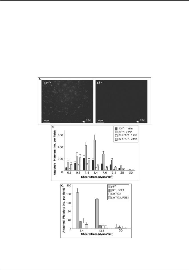
Fig. 3 Dynamic adhesion of platelets to fibrinogen depends on the presence of aIIbb3 and its cytoplasmic tail associations. (A) Fluorescence micrographs showing adhesion of b3+/+ (left panel) and b3−/− platelets (right panel) to fibrinogen at 3.4 dyn cm−2 , after 1 min of flow. (B,C) Histograms of numbers of b3+/+ and b3Y747A platelets attached to fibrinogen within the field of view at different shear stresses in the device 1.
(B) Attachment to fibrinogen after 1 and 2 min of flow (average of 4 separate experiments). (C) Whole blood from b3+/+ or b3Y747A mice was pre-incubated with PGE1 or ethanol vehicle control and then perfused over fibrinogen. Average values of 3 separate experiments ± SEM after 1 min of flow are shown.
1490 | Lab Chip, 2008, 8, 1486–1495 |
This journal is © The Royal Society of Chemistry 2008 |

Published on 23 July 2008. Downloaded on 11/18/2019 11:51:43 AM.
View Article Online
Fig. 4 Dynamic adhesion of platelets to collagen depends on the presence of aIIbb3 and its cytoplasmic tail associations. (A) Fluorescence micrographs of test chambers with b3+/+ (left panel) and b3−/− (right panel) blood after 2 min of flow at 3.4 dyn cm−2 . b3+/+ but not b3−/− platelets exhibit characteristic thrombus growth. (B). Adhesion of b3+/+ and b3−/− platelets to collagen in the two adjacent test chambers of the device 2 after 2 min of flow at 50 dyn cm−2 as evaluated from a fluorescence micrograph. Surface coverage by mepacrine-labeled platelets is represented by color-coded fluorescence intensity (red corresponds to highest platelet density). Data shown is representative of at least 3 separate experiments. (C) Histogram of the levels of attachment of b3+/+ and b3Y747A platelets to collagen after 1 minute of flow. Data are depicted as the total integrated fluorescence intensity for the field in arbitrary units (a.u.), and are an average of 3 separate experiments ± SEM.
standard viscosity of mouse blood, g = 0.038 P, by seeding the solution with fluorescent beads and analyzing their streaklines. The measurements were done at the driving pressure, DP = 2.5 kPa, used in all platelet adhesion assays. The ratios between the values of maximal flow velocity, mmax, in adjacent test chambers in the device 1 were close to the target ratio of 1.93 (Table 1). Shear stresses in the test chambers, calculated as s = 4mmaxg/h, assuming a fully developed laminar shear flow, covered a range of 0.5–50 dyn cm−2. The Reynolds number in the test chambers can be calculated as Re = qm¯ h/g, where q = 1.05 g cm−3 is the density of blood. The values of Re in the test chambers were always low, 0.1 in the chamber 1 and <0.1 in the other chambers, suggesting that the flow was always laminar with negligible non-linear effects. The total volumetric flow rate through the device 1 was 3.7 lL min−1.
In a steady laminar flow in a microchannel driven by differential pressure, shear stress is a function of the pressure,
DP, and channel geometry only and does not depend on the viscosity of the fluid. Therefore, the actual viscosity of the blood samples (that could be different from the standard value of 0.038 P) was not measured. For example, in a wide and shallow rectilinear channel, such as the test chamber 1 (Fig. 1A), in a region away from the side walls, the surface shear stress, s, is found from the equation (DP/L)h = 2s, where L is the channel length. The equation predicts a shear stress s = DPh/(2L) = 50 dyn cm−2 for the 6 mm long test chamber 1, the same as the value of s measured experimentally. Because the surface shear stress is proportional to the channel depth, adhesion of platelets to the substrate is expected to reduce the shear stress. An adherent platelet has a height of 1 lm, which is 4% of the channel depth. All quantitative results reported in this paper were obtained when platelets covered only a part of the substrate surface and before they started aggregating (platelet monolayer), thus limiting the expected
This journal is © The Royal Society of Chemistry 2008 |
Lab Chip, 2008, 8, 1486–1495 | 1491 |

Published on 23 July 2008. Downloaded on 11/18/2019 11:51:43 AM.
View Article Online
Table 1 Results of characterization of device 1 with flow of 50% aqueous solution of ethylene glycol (g = 0.038 P) driven by a differential pressure DP = 2.5 kPa between the inlet and outlet. Experimentally measured maximal flow velocities, mmax , in the test chambers 1–8 are shown along with the values of substrate shear rate, c˙ , and shear stress, s, calculated from mmax as c˙ = 4mmax /h and s = 4mmax g/h, respectively. The experimental values of s are contrasted with shear stress values derived theoretically using the geometrical parameters of the channel lines and equations for laminar flow in rectangular channels. The uncertainty in the measurements of mmax was 3% and it propagated into uncertainties of the shear rate and shear stress
Test chamber |
1 |
2 |
3 |
4 |
5 |
6 |
7 |
8 |
|
|
|
|
|
|
|
|
|
mmax /mm s−1 |
7.8 |
4.3 |
2.11 |
1.12 |
0.54 |
0.279 |
0.135 |
0.082 |
Shear rate/s−1 |
1310 |
720 |
351 |
184 |
89 |
47 |
22.5 |
13.6 |
Shear stress/dyn cm−2 |
50 |
27.5 |
13.4 |
7.0 |
3.4 |
1.77 |
0.86 |
0.50 |
Theoretical shear stress/dyn cm−2 |
50 |
25.8 |
13.4 |
6.9 |
3.6 |
1.86 |
0.97 |
0.50 |
reduction of the surface shear stress due to the platelet adhesion to <4%.
Validation of platelet adhesion to common physiological matrices
Almost no platelets adhered to the glass substratum coated with BSA (≤5 platelets in a 180 × 240 lm field of view after 3 min perfusion at any of the shear stresses tested), indicating minimal non-specific adhesion (cf . Fig. 2D and Fig. 3A, left). No platelets attached to PDMS walls of the test chambers. Platelet adhesion to physiological substrata may involve different braking, stabilization or thrombus growth events depending on the matrix presented. Therefore in order to assess the suitability of microfluidic devices for platelet adhesion studies, we tested whether the adhesion to three common physiologic matrices, VWF, fibrinogen, and collagen, occurred in a manner consistent with previous reports.
Adhesion to VWF. Platelets interacting with VWF-coated substrata first translocate on VWF via GPIb-IX-V tethering5 and subsequently induce signaling pathways that activate aIIbb332 for stable attachment. We used substrata coated with human VWF and assayed platelet adhesion to the substrata using human whole blood, because mouse platelets do not recognize human VWF and mouse VWF was not available. For an untreated blood sample, trajectories of individual platelets on VWF displayed intermittent intervals of translocation and rest (Fig. 2A). After 1 min of flow at 13.4 and 50 dyn cm−2, respectively, 18.2 ± 8 and 6.6 ± 3% of platelets that interacted with the substratum within the field of view were stably attached to the substratum (Fig. 2B). Pretreatment of blood with an aIIbb3 antagonist, integrilin, abolished stable attachment (Fig. 2B), and platelets showed increased translocation and shortened rest periods (Fig. 2A). No platelet-VWF surface interactions occurred in the presence of an antibody against GPIba (not shown), confirming that initial platelet interactions with VWF were dependent on GPIb-IX-V.
Adhesion to fibrinogen and collagen. As we subsequently intended to use mouse models to investigate aIIbb3-dependent adhesion to fibrinogen and collagen, further validations were performed using mouse blood. We assayed the adhesion dynamics of mouse platelets to fibrinogen-coated substrata using human fibrinogen, which mouse platelets are known to recognize via aIIbb3;33 purified murine fibrinogen is not readily available. At both shear stresses tested, 3.4 and 13.4 dyn cm−2, wild-type platelets were captured from the bulk of flowing blood and were immediately arrested on immobilized fibrinogen. As expected, this platelet adhesion to fibrinogen was aIIbb3 dependent, since
it was almost completely inhibited (% I = 89.0 ± 2.4%) by a function-blocking antibody against aIIbb3 (Fig. 2C). Thus, the adhesion assay in the microfluidic device specifically reports on aIIbb3-dependent platelet arrest on a fibrinogen matrix.
Collagen stimulates platelets through the two primary collagen receptors, GP VI and integrin a2b1, and induces aIIbb3 activation that is required for aggregation of platelets and their incorporation into thrombi.34,35 In agreement with the existing literature36 on platelet adhesion to collagen-coated substrata, wild-type platelets initially formed monolayers that induced platelet aggregation and thrombus formation (Fig. 2D).
The results of the assays described above agree with previous reports on platelet interactions with VWF, fibrinogen, and collagen-coated substrata, and thus indicate that the proposed microfluidic devices are appropriate tools to study sheardependent platelet adhesion.
The role of aIIbb3 extracellular domains and cytoplasmic tail associations in dynamic platelet adhesion to fibrinogen and collagen
Similar to human platelets,5,37 wild-type mouse platelets (b3+/+) attached to fibrinogen in a shear stress dependent manner, with maximal attachment found at s between 3.4 and 7 dyn cm−2 (Fig. 3A,B). Platelets from knockout littermate mice lacking aIIbb3 (b3−/− )29 minimally attached to fibrinogen at any of the shear stresses tested (Fig. 3A), confirming that the adhesion of b3+/+ platelets to fibrinogen was critically dependent on aIIbb3.
To study how the reduced aIIbb3 activation and loss of cytoplasmic associations of aIIbb3 due to the b3 Tyr 747 mutation modulate platelet adhesion to fibrinogen, we repeated the same flow experiments using blood from knock-in b3Y747A mice. In sharp contrast to b3−/− platelets, b3Y747A platelets were able to attach to fibrinogen at low shear stresses, attaining 50–80% of the attachment observed for b3+/+ littermate controls at s ≤ 1.8 dyn cm−2 (Fig. 3B). This result was consistent with the close to 100% adhesion of b3Y747A platelets under static conditions.10 Nevertheless, the loss of b3 intracellular linkages became a more crucial factor at higher shear stresses: after 1 min of flow, the attachment of b3Y747A platelets was only 25.4 ± 6.8% of b3+/+ controls (n = 5, p < 0.01) at s = 3.4 dyn cm−2 and was minimal at s > 7 dyn cm−2. Furthermore, b3+/+ platelets treated with PGE1 to inhibit aIIbb3 activation had similar levels of adhesion to fibrinogen and shear stress dependence as b3Y747A platelets (e.g. 19.5 ±3.9% of untreated b3+/+ platelets at 3.4 dyn cm−2; p < 0.01, n = 3) (Fig. 3C). Therefore, linkage of the b3 cytoplasmic tail to intracellular
1492 | Lab Chip, 2008, 8, 1486–1495 |
This journal is © The Royal Society of Chemistry 2008 |
Published on 23 July 2008. Downloaded on 11/18/2019 11:51:43 AM.
View Article Online
proteins that promote its activation and stabilize it serves to |
characteristic of wild-type platelets. The presence of a normal |
|
enhance the platelet attachment to fibrinogen at s = 3.4– |
extracellular aIIbb3 domain on b3Y747A platelets provided |
|
13.4 dyn cm−2. |
wild-type levels of attachment to collagen at low shear stresses |
|
In contrast to the major differences observed in adhesion |
(s ≤ 1.8 dyn cm−2). Nevertheless, the attachment of b3Y747A |
|
to fibrinogen, b3+/+, b3−/− , and b3Y747A platelets were all |
platelets was still greatly reduced relative to b3+/+ platelets at |
|
able to establish initial monolayers on collagen (Fig. 4A, right |
s ≥ 3.4 dyn cm−2 (Fig. 4C). This finding suggests that the |
|
panel). Nevertheless, b3−/− and b3Y747A adherent platelets |
contribution of integrin aIIbb3 to primary platelet attachment |
|
remained solitary, whereas most of the adherent b3+/+ platelets |
to collagen at intermediate and high shear stresses strongly |
|
seeded recruitment of platelets from flow into small aggregate |
depends on the presence of normal b3 cytoplasmic tail linkages |
|
islands (thrombi) that expanded into a sheet-like structure |
to intracellular proteins that permit aIIbb3 activation and |
|
(Fig. 4A, left panel). The aggregate formation was abolished |
cytoskeletal associations. |
|
when b3+/+ platelet activation was inhibited with PGE1 (not |
|
|
shown). Together with the lack of aggregate formation by b3−/− |
Quantification of platelet–granulocyte interactions in |
|
and b3Y747A platelets, this result suggested that aIIbb3 must |
||
microfluidic devices |
||
be present and retain intact cytoplasmic linkages that promote |
||
|
||
its activation in order to bind soluble ligands and form platelet |
To test whether the microfluidic devices could be used to study |
|
aggregates on collagen. |
heterocellular interactions, we examined dynamic attachment |
|
In addition to its critical role in thrombus formation, the |
of PE-anti-Gr1-labeled granulocytes to platelets adherent to |
|
presence of aIIbb3 strongly influenced the rate of primary |
collagen. Importantly, in our assay, platelet–granulocyte inter- |
|
attachment of platelets to collagen (Fig. 4B). The reduced |
actions evolved naturally over time from cellular levels present |
|
rate of primary attachment of b3−/− platelets to collagen was |
in blood and no exogenous agonists were added to activate cells. |
|
observed at all shear stresses, and the disparity between b3−/− |
Granulocytes were often entrapped in growing platelet thrombi |
|
and b3+/+ platelets increased with shear stress. For instance, |
in b3+/+ blood (Fig. 5A), making it difficult to quantify granulo- |
|
after 1 min of flow, the surface coverage by b3−/− at s = |
cyte interactions with platelets. No thrombi formed from b3−/− |
|
0.8, 13.4, and 50 dyn cm−2 was reduced compared with b3+/+ |
blood, however. Therefore, rolling velocities and the subsequent |
|
platelets by 56, 78, and 94%, respectively, (n = 3; p < 0.05). |
attachment of granulocytes on b3−/− collagen-adherent platelet |
|
The extent of platelet adhesion was also severely compromised |
monolayers could both be determined. Transient b3−/− platelet– |
|
even after several minutes of flow at higher shear stresses. |
granulocyte interactions were observed even at s > 30 dyn cm−2 |
|
Thus, in the absence of aIIbb3, interactions through GPVI and |
and were often accompanied by extension and retraction of |
|
a2b1 alone cannot provide the strong attachment to collagen |
granulocyte tethers (Fig. 5A). |
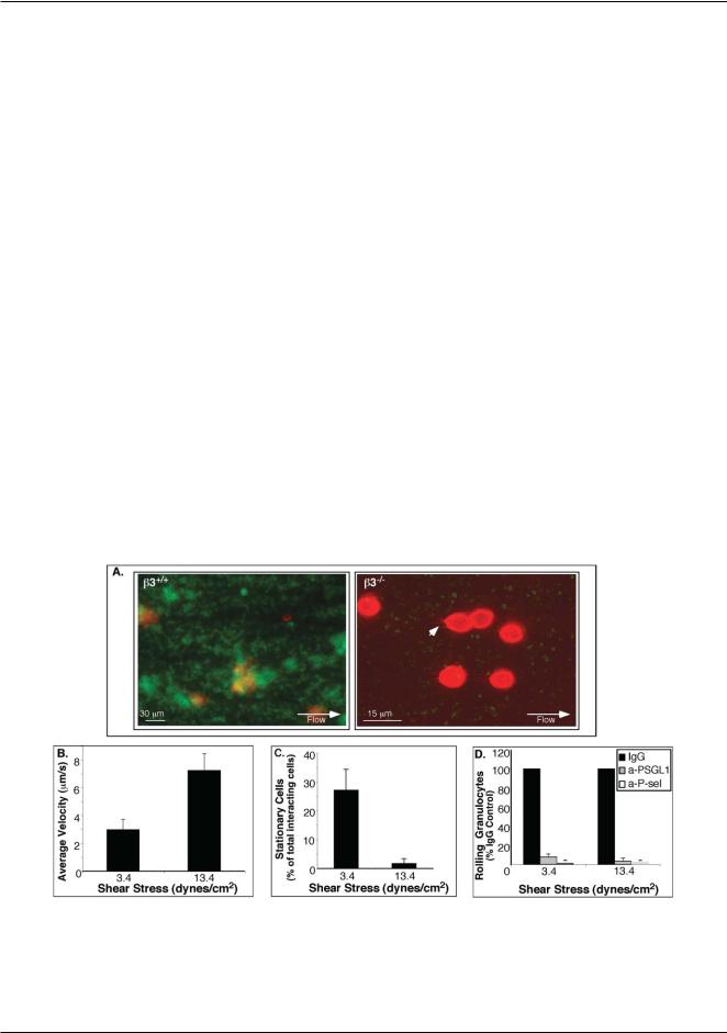
Fig. 5 Platelet–granulocyte interactions. (A) Fluorescence micrographs of a test chamber with whole blood from wild-type (left panel) and b3−/− mice (right panel) labeled with PE-anti-Gr-1 antibody (granulocytes: red) and mepacrine (platelets: green) after 10 minutes of flow over collagen matrix at 3.4 dyn cm−2 . Note extended b3−/− granulocyte tethers (arrowhead). (B)–(D). Quantification of interactions between granulocytes and collagen-adherent b3−/− platelets. (B) Average translocation velocities of granulocytes on platelets. (C) Percentage of stably attached granulocytes.
(D) Inhibition of granulocyte–platelet interactions. Whole blood from wild type and b3−/− mice was incubated with antibodies against P-selectin (a-P-sel), PSGL-1 (a-PSGL1) or with control IgG for 30 min, prior to flow. Histogram shows the percentage (± SEM) of granulocytes rolling on platelets from blood treated with each inhibitor versus IgG control antibody.
This journal is © The Royal Society of Chemistry 2008 |
Lab Chip, 2008, 8, 1486–1495 | 1493 |

Published on 23 July 2008. Downloaded on 11/18/2019 11:51:43 AM.
View Article Online
Mean rolling velocities of granulocytes on collagen-adherent |
made comparisons between the two blood samples particularly |
|
b3−/− platelets increased with shear stress, from 3.0 ± 0.7 lm s−1 |
simple and straightforward. |
|
at 3.4 dyn cm−2 to 7.2 ± 1.2 lm s−1 at 13.4 dyn cm−2 (n = |
The experiments helped elucidate the roles of extracellular |
|
5, p <0.01) (Fig. 5B). At 3.4 dyn cm−2, initial interactions led |
and intracellular domains of integrin aIIbb3 in platelet adhesion |
|
to stable attachment of a substantial portion of granulocytes |
to fibrinogen and collagen at different shear stresses, resulting |
|
(27.1 ± 7.2%), but at 13.4 dyn cm−2 the attachment was minimal |
in the following main findings: (a) similar to human platelets, |
|
(Fig. 5C). Interactions of granulocytes with b3−/− platelets |
wild-type mouse platelets adhere to fibrinogen most efficiently at |
|
were abolished by antibodies against P-selectin and PSGL-1, |
venous shear stresses; (b) as shear stresses increase, structurally |
|
but not by control antibodies (% rolling granulocytes relative |
intact extracellular aIIbb3 domains are no longer sufficient to |
|
to IgG control at 3.4 and 13.4 dyn cm−2, respectively: 1.3 ± |
mediate stable adhesion to fibrinogen in the absence of normal |
|
0.8%, and 0% for anti P-selectin; 7.9 ± 3.4% and 3.4 ± 3.0% |
b3-cytoplasmic tail linkages that enable integrin activation and |
|
for anti-PSGL-1; n = 3, p< 0.01) (Fig. 5D). Therefore, the |
stabilization; (c) the presence of intact aIIbb3 extracellular |
|
granulocyte–platelet interactions were dependent on P-selectin |
domains and intracellular linkages provides a significant incre- |
|
and PSGL-1, in agreement with previous reports.38,39 On the |
mental contribution to primary platelet adhesion to collagen at |
|
other hand, treatment of b3+/+ platelets with anti-P-selectin and |
arterial and venous shear stresses and is important for formation |
|
anti-PSGL-1 antibodies did not entirely prevent the presence of |
of thrombi at all shear stresses; (d) collagen-adherent b3−/− |
|
granulocytes in thrombi derived from b3+/+ blood (not shown), |
platelet monolayers deposited from whole blood promote P- |
|
providing further evidence for non-specific b3+/+ granulocyte |
selectin and PSGL-1 dependent granulocyte rolling and stable |
|
entrapment in the thrombi. No rolling of granulocytes was |
adhesion. Above all, these results highlight the importance of |
|
observed on platelets attached to fibrinogen for either b3+/+ or |
b3 tyrosine747-mediated linkage to intracellular proteins that |
|
b3−/− blood, presumably due to lower surface expression of P- |
promote aIIbb3 activation and stabilization for optimal platelet |
|
selectin compared to platelets adherent to collagen (not shown). |
adhesion at physiologic shear stresses. |
|
|
An interesting consequence of the absence of thrombus |
|
Discussion |
formation on collagen by b3−/− platelets was the facilitated |
|
access and recruitment of granulocytes to activated platelet |
||
|
||
Motivated by the importance of platelets for arrest of bleeding |
monolayers. Our experiments on the granulocyte recruitment |
|
(hemostasis) and thrombosis, we constructed two new microflu- |
in the microchannels confirmed that rolling of granulocytes on |
|
idic devices to study dynamic platelet adhesion at shear stresses |
collagen-adherent b3−/− platelets is mediated by P-selectin and |
|
typically found in the circulation. Due to small amounts of blood |
PSGL-1. In vivo, rolling of granulocytes on b3−/− platelets and |
|
required for assays in the device (3.7 lL min−1 and <100 lL per |
their subsequent stable attachment may increase the numbers |
|
assay for the device 1), it was possible for the first time to perform |
of extravasating granulocytes and thus contribute to the inflam- |
|
assays on dynamic platelet adhesion with whole blood samples |
mation and atherosclerosis described in the b3−/− mice.40 These |
|
obtained from a single laboratory mouse. The results of adhesion |
observations warrant further investigation of the mechanisms |
|
assays in the devices with VWF, fibrinogen, and collagen-coated |
by which aIIbb3 modulates inflammation. |
|
substrata (Fig. 2) agreed with previous reports, thus validating |
The proposed microfluidic devices are made of single casts |
|
the use of the proposed microfluidic devices for studying shear- |
of PDMS sealed with a coverglass and, as is common for this |
|
dependent platelet adhesion. To demonstrate an application of |
type of device, they can be produced at low cost in amounts |
|
the proposed adhesion assays, we performed an extensive series |
sufficient for an extended series of laboratory experiments. The |
|
of experiments with blood from wild-type, b3-deficient29 and |
devices are also easily recycled by detaching the PDMS chip |
|
activation-defective b3Y747A10 mice at shear stresses ranging |
from the coverglass, cleaning the chip in a mild detergent, and |
|
from low venous to arterial (0.8–50 dyn cm−2) with fibrinogen |
sealing it with a new coverglass. The flow in the devices is |
|
and collagen coated substrates (Fig. 3 and 4). |
driven by hydrostatic pressure, making them simple to operate, |
|
The experiments highlighted the key advantages of the |
in particular the device 1, which has a single inlet and outlet. |
|
proposed microfluidic devices as compared to traditional flow |
When a polyethylene (PE) tubing of an appropriate diameter |
|
chambers for studies of platelet adhesion. Small blood con- |
is inserted into the device inlet, the tubing is held in place and |
|
sumption and high throughput dramatically reduced the costs |
makes an instantaneous sealed connection. The insertion of the |
|
of the experiments in terms of both time and laboratory animals. |
other end of the tubing line into an artery of an anesthesized |
|
A blood sample drawn from a single mouse was sufficient |
mouse would convert the device into an ex vivo autoperfusion |
|
for several adhesion tests in different devices and could be |
flow chamber,41,42 reducing to a minimum the variation of blood |
|
used for tests with different ECM coatings or repeated tests |
between the circulation and the microfluidic test chambers. In |
|
with identical coatings. Moreover, simultaneous monitoring of |
addition to the dynamic adhesion of platelets, the proposed |
|
platelet adhesion over the entire physiological range of shear |
devices could also be used to study rolling and substrate |
|
stresses in device 1 eliminated the sample and matrix variability |
adhesion of other cell types, e.g., neutrophils,43 without the need |
|
concerns that would inevitably arise, if tests at different shear |
of sacrificing donor mice. Another possible application of the |
|
stresses were performed with different blood samples or in |
devices is studies of formation of platelet aggregates (thrombi) |
|
different flow chambers. Side-by-side observation of dynamic |
on various substrates at controlled flow conditions.44 Reduction |
|
adhesion of platelets from two different blood samples at |
of the test chamber cross-sections would further reduce the |
|
identical flow conditions and with synchronized initiation times |
sample volumes and potentially enable adhesion assays in shear |
|
under a high-resolution fluorescence microscope in device 2 |
flow with blood from genetically amenable organisms, such |
|
|
|
|
1494 | Lab Chip, 2008, 8, 1486–1495 |
This journal is © The Royal Society of Chemistry 2008 |

Published on 23 July 2008. Downloaded on 11/18/2019 11:51:43 AM.
View Article Online
as zebrafish.45 A possible clinical application of the proposed |
18 |
B. Prabhakarpandian, K. Pant, R. C. Scott, C. B. Patillo, D. Irima, |
||
devices is for testing blood of neonates and young patients, |
|
M. F. Kiani and S. Sundaram, Biomed. Microdevices, 2008, March8, |
||
where blood availability is limited. To conclude, the proposed |
|
2008 e-pub. |
||
19 |
M. Antia, T. Herricks and P. K. Rathod, PLoS Pathog., 2007, 3, e99. |
|||
microfluidic devices and dynamic adhesion assays could find |
||||
20 |
U. Y. Schaff, M. M. Xing, K. K. Lin, N. Pan, N. L. Jeon and S. I. |
|||
multiple applications in research and in medical laboratories. |
|
Simon, Lab Chip, 2007, 7, 448–456. |
||
|
|
21 |
C. Simonnet and A. Groisman, Anal. Chem., 2006, 15, 5653–5663. |
|
Acknowledgements |
22 |
S. Lengweiler, S. S. Smyth, M. Jirouskova, L. E. Scudder, H. Park, |
||
|
T. Moran and B. S. Coller, Biochem. Biophys. Res. Commun., 1999, |
|||
|
|
|
262, 167–173. |
|
These studies were supported by grants HL78784, HL-31950, |
23 |
Z. M. Ruggeri, L. De Marco, L. Gatti, R. Bader and R. R. |
||
HL56595 and HL57900 from the NIH, the Cell Migration |
|
Montgomery, J. Clin. Invest., 1983, 72, 1–12. |
||
24 |
M. A. Labow, C. R. Norton, J. M. Rumberger, K. M. Lombard- |
|||
Consortium, NIH (U54 GM064346), NSF NIRT Grant No. |
||||
|
Gillooly, D. J. Shuster, J. Hubbard, R. Bertko, P. A. Knaack, R. W. |
|||
0608863, the Wellcome Trust (077532), UCSD/SDSU Insti- |
|
Terry, M. L. Harbison and et al., Immunity, 1994, 1, 709–720. |
||
tutional Research and Academic Career Development Award |
25 |
J. Yang, T. Hirata, K. Croce, G. Merrill-Skoloff, B. Tchernychev, E. |
||
|
Williams, R. Flaumenhaft, B. C. Furie and B. Furie, J. Exp. Med., |
|||
(NIH GM 68524), and a fellowship from the American Heart |
|
|||
|
1999, 190, 1769–1782. |
|||
Association. |
|
|||
26 |
K. S. Prasad, P. Andre, M. He, M. Bao, J. Manganello and D. R. |
|||
|
|
|
Phillips, Proc. Natl. Acad. Sci. U. S. A., 2003, 100, 12367–12371. |
|
References |
27 |
L. De Marco, A. Girolami, T. S. Zimmerman and Z. M. Ruggeri, |
||
28 |
Proc. Natl. Acad. Sci. U. S. A., 1985, 82, 7424–7428. |
|||
1 |
D. Varga-Szabo, I. Pleines and B. Nieswandt, Arterioscler., Thromb., |
B. R. Alevriadou, J. L. Moake, N. A. Turner, Z. M. Ruggeri, B. J. |
||
|
Folie, M. D. Phillips, A. B. Schreiber, M. E. Hrinda and L. V. |
|||
|
Vasc. Biol., 2008. |
|
McIntire, Blood, 1993, 81, 1263–1276. |
|
2 |
D. Patel, H. Vaananen, M. Jirouskova, T. Hoffmann, C. Bodian and |
29 |
K. M. Hodivala-Dilke, K. P. McHugh, D. A. Tsakiris, H. Rayburn, |
|
|
B. S. Coller, Blood, 2003, 101, 929–936. |
|
D. Crowley, M. Ullman-Cullere, F. P. Ross, B. S. Coller, S. Teitelbaum |
|
3 |
B. Savage, S. J. Shattil and Z. M. Ruggeri, J. Biol. Chem., 1992, 267, |
|
and R. O. Hynes, J. Clin. Invest., 1999, 103, 229–238. |
|
|
11300–11306. |
30 |
P. J. Roberts, S. L. Williams and D. C. Linch, Br. J. Haematol., 1996, |
|
4 |
G. H. van Zanten, S. de Graaf, P. J. Slootweg, H. F. Heijnen, T. M. |
|
92, 804–814. |
|
|
Connolly, P. G. de Groot and J. J. Sixma, J. Clin. Invest., 1994, 93, |
31 |
C. L. Haycox, R. B. D. and H. T. A., J. Biomed. Mater. Res., 1991, |
|
|
615–632. |
|
25, 1317–1320. |
|
5 |
B. Savage, E. Saldivar and Z. M. Ruggeri, Cell, 1996, 84, 289–297. |
32 |
A. Kasirer-Friede, M. R. Cozzi, M. Mazzucato, L. De Marco, Z. M. |
|
6 |
V. Evangelista, S. Manarini, G. Dell’Elba, N. Martelli, E. Napoleone, |
|
Ruggeri and S. J. Shattil, Blood, 2004, 103, 3403–3411. |
|
|
A. Di Santo and P. S. Lorenzet, Thromb. Haemostasis, 2005, 94, 568– |
33 |
I. Goncalves, S. C. Hughan, S. M. Schoenwaelder, C. L. Yap, Y. Yuan |
|
|
577. |
|
and S. P. Jackson, J. Biol. Chem., 2003, 278, 34812–34822. |
|
7 |
A. Weyrich, F. Cipollone, A. Mezzetti and G. Zimmerman, Curr. |
34 |
S. P. Watson, J. M. Auger, O. J. McCarty and A. C. Pearce, J. Thromb. |
|
|
Pharm. Des., 2007, 13, 1685–1691. |
|
Haemostasis, 2005, 3, 1752–1762. |
|
8 |
J. Han, C. J. Lim, N. Watanabe, A. Soriani, B. Ratnikov, D. A. Calder- |
35 |
M. L. Kahn, Semin. Thromb. Hemostasis, 2004, 30, 419–425. |
|
|
wood, W. Puzon-McLaughlin, E. M. Lafuente, V. A. Boussiotis, S. J. |
36 |
K. Kato, T. Kanaji, S. Russell, T. J. Kunicki, K. Furihata, S. Kanaji, |
|
|
Shattil and M. H. Ginsberg, Curr. Biol., 2006, 16, 1796–1806. |
|
P. Marchese, A. Reininger, Z. M. Ruggeri and J. Ware, Blood, 2003, |
|
9 |
S. Tadokoro, S. J. Shattil, K. Eto, V. Tai, R. C. Liddington, J. M. de |
|
102, 1701–1707. |
|
|
Pereda, M. H. Ginsberg and D. A. Calderwood, Science, 2003, 302, |
37 |
T. N. Zaidi, L. V. McIntire, D. H. Farrell and P. Thiagarajan, Blood, |
|
|
103–106. |
|
1996, 88, 2967–2972. |
|
10 |
B. G. Petrich, P. Fogelstrand, A. W. Partridge, N. Yousefi, A. J. |
38 |
V. Evangelista, S. Manarini, B. S. Coller and S. S. Smyth, J. Thromb. |
|
|
Ablooglu, S. J. Shattil and M. H. Ginsberg, J. Clin. Invest., 2007, |
|
Haemostasis, 2003, 1, 1048–1054. |
|
|
117, 2250–2259. |
39 |
Z. Xiao, H. L. Goldsmith, F. A. McIntosh, H. Shankaran and S. |
|
11 |
M. H. Kroll, J. D. Hellums, L. V. McIntire, A. I. Schafer and J. L. |
|
Neelamegham, Biophys. J., 2006, 90, 2221–2234. |
|
|
Moake, Blood, 1996, 88, 1525–1541. |
40 |
S. Weng, L. Zemany, K. N. Standley, D. V. Novack, M. La Regina, |
|
12 |
S. Usami, H. H. Chen, Y. Zhao, S. Chien and R. Skalak, Ann. Biomed. |
|
C. Bernal-Mizrachi, T. Coleman and C. F. Semenkovich, Proc. Natl. |
|
|
Eng., 1993, 21, 77–83. |
|
Acad. Sci. U. S. A., 2003, 100, 6730–6735. |
|
13 |
S. K. Murthy, A. Sin, R. G. Tompkins and M. Toner, Langmuir, |
41 |
M. L. Smith, M. Sperandio, E. V. Galkina and K. Ley, J. Leukocyte |
|
|
2004, 20, 11649–11655. |
|
Biol., 2004, 76, 985–993. |
|
14 |
X. H. Cheng, D. Irimia, M. Dixon, K. Sekine, U. Demirci, L. Zamir, |
42 |
A. Hafezi-Moghadam, K. L. Thomas and C. Cornelssen, |
|
|
R. G. Tompkins, W. Rodriguez and M. Toner, Lab Chip, 2007, 7, |
|
Am. J. Physiol., 2004, 286, C876–C892. |
|
|
170–178. |
43 |
B. C. Chesnutt, D. F. Smith, N. A. Raffler, M. L. Smith, E. J. White |
|
15 |
E. Gutierrez and A. Groisman, Anal. Chem., 2007, 79, 2249–2258. |
|
and K. Ley, Microcirculation, 2006, 13, 99–109. |
|
16 |
H. Lu, L. Y. Koo, W. M. Wang, D. A. Lauffenburger, L. G. Griffith |
44 |
Z. Xu, N. Chen, M. M. Kamocka, E. D. Rosen and M. Alber, |
|
|
and K. F. Jensen, Anal. Chem., 2004, 76, 5257–5264. |
|
J. R. Soc. Interface, 2008, 5, 705–722. |
|
17 |
M. K. Runyon, C. J. Kastrup, B. L. Johnson-Kerner, T. G. Ha and |
45 |
P. Jagadeeswaran, M. Gregory, K. Day, M. Cykowski and B. |
|
|
R. F. Ismagilov, J. Am. Chem. Soc., 2008, 130, 3458–3464. |
|
Thattaliyath, J. Thromb. Haemostasis, 2005, 3, 46–53. |
|
This journal is © The Royal Society of Chemistry 2008 |
Lab Chip, 2008, 8, 1486–1495 | 1495 |