


NEUROPHYSIOLOGY |
Sensory Pathways: I |
Mesencephalon (cerebral peduncles)
Spinothalamic tract
Lower part of medulla oblongata
Reticular formation
Cervical part of spinal cord
Lateral spinothalamic tract: pain, temperature
Ventral (anterior) spinothalamic tract: touch, pressure
Lumbar part of spinal cord
FIGURE 2.29 SOMESTHETIC SYSTEM OF THE BODY•
Cerebral cortex: postcentral gyrus
Posterior limb of internal capsule
Ventral posterolateral (VPL) nucleus of thalamus
Medial lemniscus
Gracile nucleus
Cuneate nucleus
Fasciculus gracilis
Fasciculus cuneatus
Dorsal (posterior) spinal root ganglion
Proprioception, |
Large |
|
position |
||
Touch, |
myelinated |
|
fibers |
||
pressure, |
||
|
||
vibration |
Small |
|
Pain, |
||
myelinated |
||
temperature |
and unmyelin- |
|
|
ated fibers |
Lateral cervical nucleus
Spinocervical tract
©

Pain, temperature, and pressure sensations below the head ultimately are conveyed to the primary somatosensory cortex (postcentral gyrus) by the anterolateral system (spinothalamic and spinoreticular tracts). The fasciculus gracilis and cuneatus of the spinal lemniscal system convey proprioceptive, vibratory, and tactile sen-
sations to the thalamus (ventral posterolateral nucleus), whereas the lateral cervical system mediates some touch, vibratory, and proprioceptive sensations (blue and purple lines show these dual pathways). Ultimately, these fibers ascend as parallel pathways to the thalamus, synapse, and ascend to the cortex.
80

Sensory Pathways: II |
NEUROPHYSIOLOGY |
Midbrain (cerebral peduncles)
Ventral trigeminal lemniscus
Pontine reticular formation
Pons
Medullary reticular formation
Spinal trigeminal tract
Spinal trigeminal nucleus
Cervical part of spinal cord
FIGURE 2.30 SOMESTHETIC SYSTEM OF THE HEAD•
Cerebral cortex: postcentral gyrus
Ventral posteromedial (VPM) nucleus of thalamus
Internal capsule
Dorsal trigeminal lemniscus
Trigeminal mesencephalic nucleus
Trigeminal motor nucleus
Principal sensory trigeminal nucleus
Touch, pressure
Pain, temperature
Proprioception
Trigeminal (semilunar) ganglion
Ophthalmic n.
Maxillary n.
Sensory root and
Motor root of mandibular n.
Facial (VII) n. Vagus (X) n.
Dorsolateral fasciculus (of Lissauer)
Substantia gelatinosa (Iamina II)
©
Nerve cells bodies for touch, pressure, pain, and temperature in the head are in the trigeminal (semilunar) ganglion of the trigeminal (CN V) nerve (blue and red lines in figure). Neuronal cell bodies mediating proprioception reside in the mesencephalic nucleus
of CN V (purple fibers). Most relay neurons project to the contralateral VPM nucleus of the thalamus and thence to the postcentral gyrus of the cerebral cortex, where they are somatotopically represented.
81
NEUROPHYSIOLOGY |
Sensory Pathways: III |
C2
C3
C4
C5
T1
T2
T3
T4
T5
T6
T7
T8
T9
T10
T11
T12
L1
S2, 3 L2
L3
L4
Schematic demarcation of |
|
|
dermatomes shown as distinct |
C2 |
|
segments. There is actually |
||
|
||
considerable overlap between |
C3 |
|
any two adjacent dermatomes |
C4 |
|
|
C5 |
|
|
C6 |
|
C7 |
T1 |
|
C8 |
|
|
|
T2 |
|
|
T3 |
C6 |
C6 |
T4 |
T5 |
||
|
|
T6 |
T1 |
|
T7 |
|
|
T8 |
|
C5 |
T9 |
|
T10 |
|
|
C8 |
T11 |
|
T12 |
|
|
C7 |
|
|
L1 |
|
|
|
L2 |
|
|
L3 |
|
C6 |
L4 |
|
L5 |
|
|
|
S1 |
C8 |
C7 |
C8 |
S2 |
|
C7 |
|
|
S3
S4
S5 |
S1 |
|
|
|
S2 |
L5 |
|
L1 |
|
L2 |
|
L5
S1
L5
L4
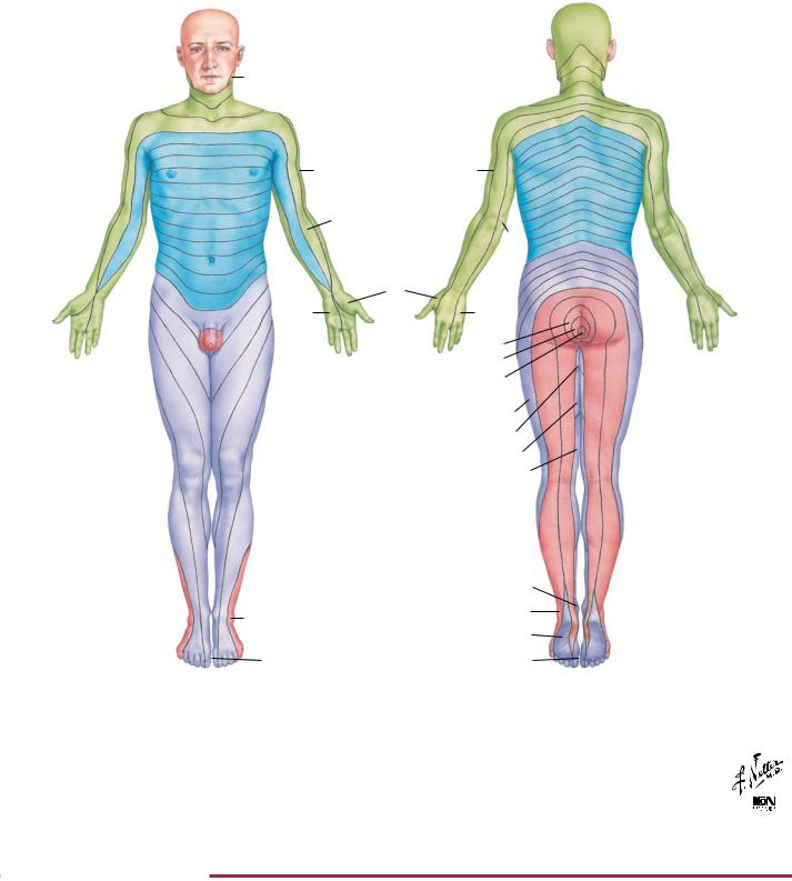
Levels of principal dermatomes
C5 |
Clavicles |
C5, 6, 7 |
Lateral parts of upper limbs |
C8, T1 |
Medial sides of upper limbs |
C6 |
Thumb |
C6, 7, 8 |
Hand |
C8 |
Ring and little fingers |
T4 |
Level of nipples |
|
L3 |
|
|
S1 S2 |
|
|
L4 |
|
|
S1 |
|
|
L5 |
|
|
L4 |
|
T10 |
Level of umbilicus |
|
T12 |
Inguinal or groin regions |
|
L1, 2, 3, 4 |
Anterior and inner surfaces of lower limbs |
|
L4, 5, S1 |
Foot |
|
L4 |
Medial side of great toe |
|
S1, 2, L5 |
Posterior and outer surfaces of lower limbs |
|
S1 |
Lateral margin of foot and little toe |
|
S2, 3, 4 |
Perineum |
© |
FIGURE 2.31 DERMATOMES•
Sensory information below the head is localized to specific areas of the body, which reflect the distribution of peripheral sensory fibers that convey sensations to the spinal cord through the dorsal roots (sensory nerve cell bodies reside in the corresponding dorsal root ganglion). The area of skin subserved by afferent fibers of one
dorsal root is called a dermatome. This figure shows the dermatome segments and lists key dermatome levels used by clinicians. Variability and overlap occur, so all dermatome segments are only approximations.
82

Visual System: Receptors |
NEUROPHYSIOLOGY |
A. Eyeball
Lens
Iris Cornea
Suspensory
ligament Ciliary body
Anterior Posterior chamber chamber
Ora containing serrata aqueous humor
Vitreous humor
Retina
Choroid
Sclera
Fovea
Optic nerve
Synaptic ending depolarized
C. Rod in dark
|
Rhodopsin |
|
Metabolic |
|
energy |
Current |
Retinene |
flow |
+ |
|
Opsin |
Na+ |
Vitamin A |
permeability |
|
increased |
|
|
Circulation |
FIGURE 2.32 VISUAL RECEPTORS•
B. Section through retina
D. Rod in light
Photons of light
 Lumirhodopsin
Metarhodopsin
Retinene
+
Opsin
Vitamin A
Inner limiting membrane
Axons at surface of retina passing via optic nerve, chiasm, and tract to lateral geniculate body
Ganglion cell
Müller cell (supporting glial cell)
Amacrine cell Bipolar cell Horizontal cell Rod
Cone Pigment cells of choroid
Synaptic ending fully polarized
Synaptic bar
Nucleus
Centriole (basal body)
 Na+ permeability decreased
©

The rods and cones of the retina transduce light into electrical signals. As illustrated for the rod, light is absorbed by rhodopsin, and through the second messenger cGMP (not shown), Na channels in the membrane close and the cell hyperpolarizes. Thus, in the
dark the cell is depolarized, but it is hyperpolarized in the light. This electrical response to light is distinct from other receptor responses, in which the response to a stimulus results in a depolarization of the receptor cell membrane.
83