


50
Upper Urinary Tract Trauma
Richard A. Santucci; Mang L. Chen
Questions
1.What method is used to perform "one-shot" intraoperative intravenous pyelography (IVP) in a 50-kg woman?
a.Inject a 50-mL bolus of intravenous contrast agent followed by a full IVP series including abdominal compression to evaluate the ureters.
b.Inject a 100-mL bolus of intravenous contrast agent followed in exactly 10 minutes by a flat plate of the abdomen on the operating room table.
c.Inject a 50-mL bolus of intravenous contrast agent followed in exactly 10 minutes by a flat plate of the abdomen on the operating room table.
d.Determine the patient's serum creatinine level before administration of intravenous contrast agent to make sure that he or she will not experience renal failure as a result of reaction to the contrast agent.
e.Inject 100 mL of intravenous contrast and obtain a kidney, ureter, bladder (KUB) film 20 minutes following injection.
2.What is the best option for repair of midureteral transection after a stab wound?
a.Ureteroureterostomy
b.Transureteroureterostomy
c.Boari flap
d.Nephrectomy
e.Cutaneous ureterostomy
3.When ureteroureterostomy is performed, which of the following is required?
a.Postoperative retroperitoneal Penrose drain
b.Postoperative nephrostomy drain
c.Spatulated, watertight repair
d.Nonabsorbable sutures
e.Intraperitonealization of the ureteral anastomosis
4.Which maneuver is cited as a cause of ureteral injury during stone basketing?
a.Ureteroscopy without dilating the ureteral orifice first
b.Ureteroscopy in nondilated systems
c.Use of the holmium laser
d.Pulsatile saline irrigation to assist visualization
e.Persistence in stone basketing attempts in the face of a ureteral tear
5.Which of the following is a contraindication to transureteroureterostomy for repair of significant lower ureter injury?
a.History of urolithiasis
b.History of ureteral trauma
c.Obesity
d.Neurogenic bladder
e.Spinal fracture
6.What is the treatment of choice for a ureteral contusion caused by a highvelocity bullet?
a.Observation
b.Ureteral stent placement
c.Transureteroureterostomy
d.Ureteroureterostomy
e.Oversewing the contusion with healthy ureteral tissue
7.Which imaging technique is most useful for detecting ureteral injuries after trauma?
a.Computed tomography (CT) without use of contrast material
b.CT with use of contrast agent, obtained immediately after injection of the contrast agent
c.CT with the use of contrast material, obtained 20 minutes after injection of the contrast agent
d.Intravenous pyelography
e.Furosemide (Lasix) renography
8.Which of the following statements is TRUE about ureteral injuries during laparoscopy?
a.The total number of injuries has stayed steady over the years.
b.Surgery for endometriosis greatly increases the risk.
c.Bipolar cautery use during tubal ligation eliminates risk.
d.Most ureteral injuries are recognized immediately.
e. Indigo carmine dye eliminates the risk of injury.
Imaging
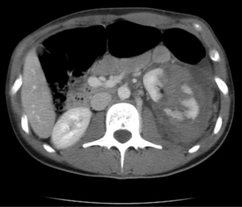
1.A 22-year-old woman has a CT scan (Figure 50-1) after a motor vehicle accident. Her vital signs are stable, and there are no other significant injuries.
FIGURE 50-1
The next step is:
a.open surgical repair of the kidney.
b.delayed imaging to evaluate the collecting system.
c.left nephrectomy to avoid future complications.
d.renal artery embolization.
e.intravenous urogram.
2.A 24-year-old man had a CT scan (Figure 50-2) 36 hours after a motor vehicle accident. The most likely diagnosis is:
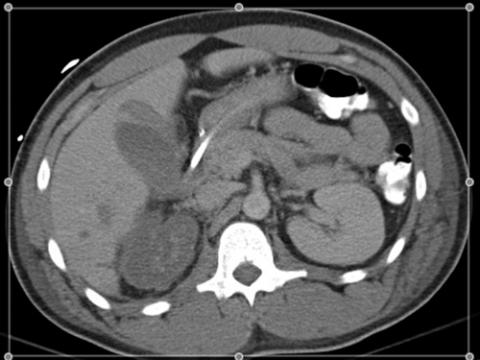
FIGURE 50-2
a.ureteral injury.
b.segmental renal artery transection.
c.main renal artery occlusion.
d.ureteropelvic junction disruption.
e.traumatic renal vein occlusion.
Answers
1.b. Inject a 100-mL bolus of intravenous contrast agent followed in exactly 10 minutes by a flat plate of the abdomen on the operating room table. Only a single film is taken 10 minutes after intravenous injection (IV push) of 2 mL/kg of contrast material.
2.a. Ureteroureterostomy. Ureteroureterostomy, so-called end-to-end repair in injuries to the upper two thirds of the ureter, is common (up to 32% of one large series) and has a reported success rate as high as 90%.
3.c. Spatulated, watertight repair. Repair ureters under magnification with spatulated, tension-free, stented, watertight anastomosis and place a retroperitoneal closed suction drain.
4.e. Persistence in stone basketing attempts in the face of a ureteral tear.

One factor cited as a cause of injury was the persistence in stone basketing attempts after recognition of a ureteral tear. Current recommendations are to stop and place a ureteral stent.
5.a. History of urolithiasis. This operation is contraindicated in patients with a history of urothelial calculi.
6.d. Ureteroureterostomy. Ureteral contusions, although the most "minor" of ureteral injuries, often heal with stricture or break down later if microvascular injury results in ureteral necrosis. Severe or large areas of contusion should be treated with excision and ureteroureterostomy.
7.c. CT with the use of contrast material, obtained 20 minutes after injection of the contrast agent. Because modern helical CT scanners can obtain images before intravenous contrast dye is excreted in the urine, delayed images must be obtained (5 to 20 minutes after contrast material injection) to allow contrast material to extravasate from the injured collecting system, renal pelvis, or ureter.
8.b. Surgery for endometriosis greatly increases the risk. A large percentage of ureteral injuries after gynecologic laparoscopy occur during electrosurgical or laser-assisted lysis of endometriosis.
Imaging
1.b. Delayed imaging to evaluate the collecting system. When blunt renal trauma results in deep parenchymal lacerations, delayed imaging at the time of the CT is essential to evaluate the collecting system for injury. In a stable patient with this type of injury, neither surgical repair nor angiographic embolization is indicated. Most renal injuries heal well with conservative and expectant management, making options a and c incorrect. Intravenous urography no longer plays a role in the acute evaluation of blunt renal trauma.
2.c. Main renal artery occlusion. There is complete absence of enhancement of the entire right kidney, which is caused only by main renal artery occlusion. Segmental occlusion would cause abnormality only in a portion of the right renal nephrogram. Ureteral injury and UPJ disruption may cause delay in the nephrogram but not complete absence of enhancement.
Chapter review
1. The best indication of significant urinary system injury is gross or

microscopic hematuria.
2.Evaluation of urologic trauma in children differs from adults in that children: (1) are at greater risk for renal trauma, (2) often do not become hypotensive with major blood loss, and (3) have a higher propensity for renal anomalies.
3.Rapid deceleration involving high-velocity impact may result in injuries at points of fixation such as the ureteral pelvic junction and the renal hilum (renal artery intimal disruption).
4.The degree of hematuria and the severity of renal injury are not consistently correlated.
5.Criteria for radiologic imaging include (1) all penetrating trauma, (2) high-impact rapid deceleration trauma, (3) all blunt trauma with gross hematuria, (4) all blunt trauma with microhematuria and hypotension, and (5) all pediatric patients with microscopic hematuria.
6.Adult patients with microscopic hematuria without shock may be observed without imaging studies.
7.Findings suggestive of a major renal injury on CT include medial hematoma, medial urinary extravasation, and lack of contrast enhancement of the entire kidney.
8.Nonoperative management for renal injuries is preferred in the hemodynamically stable patient, particularly with grades I to III renal injuries.
9.Exploration of renal gunshot wounds is no longer mandatory in selected cases. Those patients with grade I to IV parenchymal lacerations with well-contained hematomas who are hemodynamically stable may be observed expectantly.
10.Absolute indications for renal exploration are (1) hemodynamic instability with shock, (2) expanding pulsatile hematoma, (3) suspected renal pedicle avulsion, and (4) ureteral pelvic junction disruption.
11.Surgical exploration of the acutely injured kidney is best performed through a transabdominal incision with early isolation of the renal artery to the affected kidney.
12."Damage control" involves packing the wound around the injured kidney with laparotomy pads to control bleeding and a planned return to the operating room in 24 hours. This is rarely indicated but is a management strategy in the multiply injured patient in whom an attempt to salvage the kidney is necessary when the additional time required to do this is

inadvisable during initial post-trauma exploration.
13.Hypertension as a result of renal trauma is due to renal vascular injury or compression of the renal parenchyma by a circumferential hematoma (Page kidney).
14.When repairing ureteral injuries, the ureteral tissue should be debrided back to bleeding tissue to remove all traumatized microvascular damaged tissue.
15.Placement of a vascular graft anterior to the ureter and aneurysms may cause a periureteral inflammatory reaction and ureteral injury/stenosis.
16.In emergent situations when a ureteral repair in the multiply traumatized patient cannot be adequately performed, the ureter may be tied off and the kidney drained postoperatively through a percutaneous nephrostomy with planned reconstruction at a later date, or a single J stent may be placed up the ureter to the renal pelvis, secured to the ureter, and brought out through the flank.
17.Intravenous methylene blue and indigo carmine should be avoided in pregnant patients or those taking a serotonin reuptake inhibitor.
18.When anastomosing the ureter to the bladder for a traumatic distal ureteral injury, the ureter should be spatulated and sewn to the bladder in a nontunneled (refluxing) fashion.
19.Ureteral transections should be repaired within 3 days of the injury, or the repair should be delayed for 6 weeks.
20.It is prudent to isolate the ureteral repair from other organs with omentum or peritoneum.
21.The uterine artery may be a significant source of the blood supply to the distal ureter in females and, when ligated during a hysterectomy, may lead to ureteral ischemia, resulting in ureteral stenosis, urinoma, or ureterovaginal fistula.
22.Ureteral injury occurring during vascular surgery should be repaired and isolated from the graft with normal tissue such as omentum.

PART IX
Urinary Lithiasis and Endourology