

80 |
4 Abnormal Lung Patterns |
Mechanism
Vascular process can be evident with increased or decreased perfusion, and/ or altering diameter of pulmonary vessels. Additionally, distortion from masses or bullae is possible.
In review, the vascular pattern includes processes that effect the visualization of vasculature, however, may not be primarily vascular etiology.
Vascular Examples
There are a number of conditions altering vasculature, hence qualifying for the vascular pattern. Three conditions affecting the CXR are presented, including:
1.Pulmonary Arterial Hypertension: Large central arteries with peripheral tapering
2.Congestion: Engorged veins, especially upper lungs
3.Emphysema: Diminished or compresses or distorted vasculature Other conditions include:
•Lymphangitic Carcinoma: Irregular infiltration around vessels may resemble vessel enlargement.
•Thromboembolism: Locally diminished vessels with possible vessel mass centrally located.
•Bronchial Circulation: Irregular vessels in unusual directions.
•Shunt Vascularity: All vessels enlarged.
Pulmonary Arterial Hypertension (PAH)
PAH presents large central arteries with peripheral tapering. Think about conditions such as chronic thrombophlebitis and, secondarily, obesity (Pickwickianism) and sleep apnea.
Pulmonary arterial hypertension (PAH) is continuous high blood pressure in the pulmonary artery. The average blood pressure in a normal pulmonary artery is about 15 mmHg when the person is resting. In PAH, the average is usually greater than 25 mmHg [8].
In PAH, three types of changes may occur in the pulmonary arteries.
1. The muscles within the walls of the arteries may tighten up. This makes the inside of the arteries narrower.
2. The walls of the pulmonary arteries may thicken as the amount of muscle increases in some arteries. Scar tissue may form in the walls of arteries. As the walls thicken and scar, the arteries become increasingly narrow.
3. Tiny blood clots may form within the smaller arteries, causing blockages [8].
Vascular Pattern |
81 |
Case 4.20
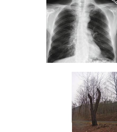
Figure 4.60 illustrates PAH with tapering vessels (often referred to as “pruning”) using an analogy with a tree that was pruned.
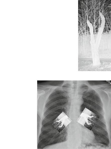
The tree in Fig. 4.60b was pruned and now has small branches growing from the stumps, not dissimilar to how enlarged pulmonary arteries abruptly taper with small branches emanating after the elasticity changes to the intima.
Figures 4.61a and 4.62a demonstrate an enlarged pulmonary trunk, sometimes known as a middle mogul.
Findings: Enlarged pulmonary arteries proximally with tapering peripherally. Pattern: Vascular.
Differential Diagnosis: PAH, COPD, cor pulmonale.
Fig. 4.60a This CXR shows enlarged pulmonary arteries bilaterally. Note how quickly the arteries taper to thinner branches
Fig. 4.60b This photo of a pruned tree shows some smaller branches that have grown since the pruning (Photos by Dr. Les Folio)
82 |
4 Abnormal Lung Patterns |
Fig. 4.60c Same photo, only reversed in black and white to create the following collage
Fig. 4.60d The CXR of PAH with manipulated pruned trees to demonstrate pruning model
“Hypoxic vasoconstriction, obliteration of the pulmonary vascular bed, and volume overload are the three core pathophysiologic processes that lead to pulmonary artery hypertension.”
“Hypoxic vasoconstriction occurs most commonly in the setting of chronic obstructive pulmonary disease. Chronic hypoxia leads to vasoconstriction of the pulmonary vasculature, which leads to pulmonary artery hypertension. Similarly, if there is a 60% or greater loss of the total pulmonary vasculature (obliteration of the

Vascular Pattern |
83 |
pulmonary vascular bed), pulmonary artery hypertension will ensue. This is common in patients with chronic pulmonary emboli and collagen vascular disorders.”
“A multitude of cardiac disorders can lead to elevated pulmonary arterial pressures. Several stem from disorders affecting the left heart. Mitral and aortic valvular disease along with cardiomyopathies increase the pulmonary pressure gradient over time and lead to pulmonary artery hypertension. Pulmonary volume overload occurs with congenital heart diseases such as Atrial Septal Defect (ASD) or Ventricular Septal Defect (VSD) causing left to right shunts [9].”
Figure 4.62c is a 37-year-old female (different patient from above) with large MPA on CXR, suspected of having a pulmonary embolus.
Fig. 4.61a PA of a 34-year-old male patient with sickle cell anemia and PAH. Note the large main pulmonary trunk (arrows). The cardiac silhouette is also enlarged
Fig. 4.61b Lateral of same patient showing large left (posterior circle) and right (anterior circle) pulmonary arteries. Also note the typical H-shaped vertebral bodies of sickle cell disease
84 |
4 Abnormal Lung Patterns |
Fig. 4.61c HRCT (done prone) demonstrating mosaic attenuation (previously known as mosaic “perfusion” or mosaicism). The two arrows contrast the whiter versus darker lung in this pattern often seen with PAH due to perfusion variation. Also note the large segmental and subsegmental pulmonary arteries
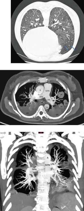
Fig. 4.62a Enlarged pulmonary trunk on axial CT thick slab MIP (Maximum Intensity Projection) in a patient with lung metastasis. Note the larger Main Pulmonary Artery (trunk, or MPA) over the Aorta (A). Note the masses (M) and effusions (eff)
Fig. 4.62b Enlarged pulmonary arteries on coronal CT thick slab MIP in the same patient. Note the truncated branching. Note the atelectasis (atel) LLL adjacent to the effusion (eff)
Vascular Pattern |
85 |
Fig. 4.62c PA CXR demonstrating large MPA mogul (large arrows) and large right interlobar pulmonary artery (small arrows).
Fig. 4.62d Axial CT reformatted 20 mm slab of same patient showing a wall adherent thrombus (arrows) of the right interlobar pulmonary artery (RILPA). Also note the large left lower lobe pulmonary artery (LLLPA) and corkscrew peripheral pulmonary arteries (dotted circle) in LLL. Also noted is the right atrium (RA), right ventricle (RV), left atrium (LA), left ventricle (LV), right main pulmonary artery (RPA), and left pulmonary vein (LPV)
Fig. 4.62e Axial CT reformatted 20 mm slab at level of enlarged MPA and enlarged central pulmonary arteries (RPA, LPA). Also note large pulmonary artery braches bilaterally, and corkscrewing again on the left. Ascending aorta (AA) and descending aorta and superior vena cava (SVC) also seen