

- •Foreword
- •Preface
- •Contents
- •About the Editors
- •Contributors
- •1: Tracheobronchial Anatomy
- •Trachea
- •Introduction
- •External Morphology
- •Internal Morphology
- •Mucous Layer
- •Blood Supply
- •Anatomo-Clinical Relationships
- •Bronchi
- •Main Bronchi
- •Bronchial Division
- •Left Main Bronchus (LMB)
- •Right Main Bronchus (RMB)
- •Blood Supply
- •References
- •2: Flexible Bronchoscopy
- •Introduction
- •History
- •Description
- •Indications and Contraindications
- •Absolute Contraindications
- •Procedure Preparation
- •Technique of FB Procedure
- •Complications of FB Procedure
- •Basic Diagnostic Procedures
- •Bronchoalveolar Lavage (BAL)
- •Transbronchial Lung Biopsy (TBLB)
- •Transbronchial Needle Aspiration (TBNA)
- •Bronchial Brushings
- •Advanced Diagnostic Bronchoscopy
- •EBUS-TBNA
- •Ultrathin Bronchoscopy
- •Transbronchial Lung Cryobiobsy (TBLC)
- •Therapeutic Procedures Via FB
- •LASER Bronchoscopy
- •Electrocautery
- •Argon Plasma Coagulation (APC)
- •Cryotherapy
- •Photodynamic Therapy
- •Airway Stent Placement
- •Endobronchial Valve Placement
- •Conclusion
- •References
- •History and Historical Perspective
- •Indications and Contraindications
- •Procedure Description
- •Procedure Planning
- •Target Approximation
- •Sampling
- •Complications
- •Future Directions
- •Summary and Recommendations
- •References
- •4: Rigid Broncoscopy
- •Innovations
- •Ancillary Equipment
- •Rigid Bronchoscopy Applications
- •Laser Bronchoscopy
- •Tracheobronchial Prosthesis
- •Transbronchial Needle Aspiration (TBNA)
- •Rigid Bronchoscope in Other Treatments for Bronchial Obstruction
- •Mechanical Debridement
- •Pediatric Rigid Bronchoscopy
- •Tracheobronchial Dilatation
- •Foreign Bodies Removal
- •Other Indications
- •Complications
- •The Procedure
- •Some Conclusions
- •References
- •History and Historical Perspective
- •Indications and Contraindications
- •Preprocedural Evaluation and Preparation
- •Physical Examination
- •Procedure-Related Indications
- •Application of the Technique
- •Topical Anesthesia
- •Anesthesia of the Nasal Mucosa and Nasopharynx
- •Anesthesia of the Mouth and Oropharynx
- •Superior Laryngeal Nerve Block
- •Recurrent Laryngeal Nerve Block (RLN)
- •Conscious Sedation
- •Monitored Anesthesia Care (MAC)
- •General Anesthesia
- •Monitoring the Depth of Anesthesia
- •Interventional Bronchoscopy Suites
- •Airway Devices
- •Laryngeal Mask Airway (LMA)
- •Endotracheal Tube (ETT)
- •Rigid Bronchoscope
- •Modes of Ventilation
- •Spontaneous Ventilation
- •Assisted Ventilation
- •Noninvasive Positive Pressure Ventilation (NIV)
- •Positive Pressure Controlled Mechanical Ventilation
- •Jet Ventilation
- •Electronic Mechanical Jet Ventilation
- •Postprocedure Care
- •Special Consideration
- •Anesthesia for Peripheral Diagnostic and Therapeutic Bronchoscopy
- •Anesthesia for Interventional Bronchoscopic Procedures During the COVID-19 Pandemic
- •Summary and Recommendations
- •Conclusion
- •References
- •Background
- •Curricular Structure and Delivery
- •What Is a Bronchoscopy Curriculum?
- •Tradition, Teaching Styles, and Beliefs
- •Using Assessment Tools to Guide the Educational Process
- •The Ethics of Teaching
- •When Learners Teach: The Journey from Novice to Mastery and Back Again
- •The Future Is Now
- •References
- •Interventional Procedure
- •Assessment of Flow–Volume Curve
- •Dyspnea
- •Analysis of Pressure–Pressure Curve
- •Conclusions
- •References
- •Introduction
- •Adaptations of the IP Department
- •Environmental Control
- •Personal Protective Equipment
- •Procedure Performance
- •Bronchoscopy in Intubated Patients
- •Other Procedures in IP Unit
- •References
- •Introduction
- •Safety
- •Patient Safety
- •Provider Safety
- •Patient Selection and Screening
- •Lung Cancer Diagnosis and Staging
- •Inpatients
- •COVID-19 Clearance
- •COVID Clearance: A Role for Bronchoscopy
- •Long COVID: A Role for Bronchoscopy
- •Preparing for the Next Pandemic
- •References
- •Historical Perspective
- •Indications and Contraindications
- •Evidence-Based Review
- •Summary and Recommendations
- •References
- •Introduction
- •Clinical Presentation
- •Diagnosis
- •Treatment
- •History and Historical Perspectives
- •Indications and Contraindications
- •Benign and Malignant Tumors
- •Tumors with Uncertain Prognosis
- •Application of the Technique
- •Evidence Based Review
- •Summary and Recommendations
- •References
- •12: Cryotherapy and Cryospray
- •Introduction
- •Historical Perspective
- •Equipment
- •Cryoadhesion
- •Indications
- •Cryorecanalization
- •Cryoadhesion and Foreign Body Removal
- •Cryoadhesion and Mucus Plugs/Blood Clot Retrieval
- •Endobronchial Cryobiopsy
- •Transbronchial Cryobiopsy for Lung Cancer
- •Safety Concerns and Contraindications
- •Cryoablation
- •Indications
- •Evidence
- •Safety Concerns and Contraindications
- •Cryospray
- •Indications
- •Evidence
- •Safety Concerns and Contraindications
- •Advantages of Cryotherapy
- •Limitations
- •Future Research Directions
- •References
- •13: Brachytherapy
- •History and Historical Perspective
- •Indications and Contraindications
- •Application of the Technique
- •Evidence-Based Review
- •Adjuvant Treatment
- •Palliative Treatment
- •Complications
- •Summary and Recommendations
- •References
- •14: Photodynamic Therapy
- •Introduction
- •Photosensitizers
- •First-Generation Photosensitizers
- •M-Tetrahidroxofenil Cloro (mTHPC) (Foscan®)
- •PDT Reaction
- •Tumor Damage Process
- •Procedure
- •Indications
- •Curative PDT Indications
- •Palliative PDT Indications
- •Contraindications
- •Rationale for Use in Early-Stage Lung Cancer
- •Rationale
- •PDT in Combination with Other Techniques for Advanced-Stage Non-small Cell Lung Cancer
- •Commentary
- •Complementary Endoscopic Methods for PDT Applications
- •New Perspectives
- •Other PDT Applications
- •Conclusions
- •References
- •15: Benign Airways Stenosis
- •Etiology
- •Congenital Tracheal Stenosis
- •Iatrogenic
- •Infectious
- •Idiopathic Tracheal Stenosis
- •Distal Bronchial Stenosis
- •Diagnosis Methods
- •Patient History
- •Imaging Techniques
- •Bronchoscopy
- •Pulmonary Function Test
- •Treatment
- •Endoscopic Treatment
- •Dilatation
- •Laser Therapy
- •Stents
- •How to Proceed
- •Stent Placement
- •Placing a Montgomery T Tube
- •The Rule of Twos for Benign Tracheal Stenosis (Fig. 15.23)
- •Surgery
- •Summary and Recommendations
- •References
- •16: Endobronchial Prostheses
- •Introduction
- •Indications
- •Extrinsic Compression
- •Intraluminal Obstruction
- •Stump Fistulas
- •Esophago-respiratory Fistulas (ERF)
- •Expiratory Central Airway Collapse
- •Physiologic Rationale for Airway Stent Insertion
- •Stent Selection Criteria
- •Stent-Related Complications
- •Granulation Tissue
- •Stent Fracture
- •Migration
- •Contraindications
- •Follow-Up and Patient Education
- •References
- •Introduction
- •Overdiagnosis
- •False Positives
- •Radiation
- •Risk of Complications
- •Lung Cancer Screening Around the World
- •Incidental Lung Nodules
- •Management of Lung Nodules
- •References
- •Introduction
- •Minimally Invasive Procedures
- •Mediastinoscopy
- •CT-Guided Transthoracic Biopsy
- •Fluoroscopy-Guided Transthoracic Biopsies
- •US-Guided Transthoracic Biopsy
- •Thoracentesis and Pleural Biopsy
- •Thoracentesis
- •Pleural Biopsy
- •Surgical or Medical Thoracoscopy
- •Image-Guided Pleural Biopsy
- •Closed Pleural Biopsy
- •Image-Guided Biopsies for Extrathoracic Metastases
- •Tissue Acquisition, Handling and Processing
- •Implications of Tissue Acquisition
- •Guideline Recommendations for Tissue Acquisition in Mediastinal Staging
- •Methods to Overcome Challenges in Tissue Acquisition and Genotyping
- •Rapid on-Site Evaluation (ROSE)
- •Sensitive Genotyping Assays
- •Liquid Biopsy
- •Summary, Recommendations and Highlights
- •References
- •History
- •Data Source and Methodology
- •Tumor Size
- •Involvement of the Main Bronchus
- •Atelectasis/Pneumonitis
- •Nodal Staging
- •Proposal for the Revision of Stage Groupings
- •Small Cell Lung Cancer (SCLC)
- •Discussion
- •Methodology
- •T Descriptors
- •N Descriptors
- •M Descriptors
- •Summary
- •References
- •Introduction
- •Historical Perspective
- •Fluoroscopy
- •Radial EBUS Mini Probe (rEBUS)
- •Ultrasound Bronchoscope (EBUS)
- •Virtual Bronchoscopy
- •Trans-Parenchymal Access
- •Cone Beam CT (CBCT)
- •Lung Vision
- •Sampling Instruments
- •Conclusions
- •References
- •History and Historical Perspective
- •Narrow Band Imaging (NBI)
- •Dual Red Imaging (DRI)
- •Endobronchial Ultrasound (EBUS)
- •Optical Coherence Tomography (OCT)
- •Indications and Contraindications
- •Confocal Laser Endomicroscopy and Endocytoscopy
- •Raman Spectrophotometry
- •Application of the Technique
- •Supplemental Technology for Diagnostic Bronchoscopy
- •Evidence-Based Review
- •Summary and Recommendations, Highlight of the Developments During the Last Three Years (2013 on)
- •References
- •Introduction
- •History and Historical Perspective
- •Endoscopic AF-OCT System
- •Preclinical Studies
- •Clinical Studies
- •Lung Cancer
- •Asthma
- •Airway and Lumen Calibration
- •Obstructive Sleep Apnea
- •Future Applications
- •Summary
- •References
- •23: Endobronchial Ultrasound
- •History and Historical Perspective
- •Equipment
- •Technique
- •Indication, Application, and Evidence
- •Convex Probe Ultrasound
- •Equipment
- •Technique
- •Indication, Application, and Evidence
- •CP-EBUS for Malignant Mediastinal or Hilar Adenopathy
- •CP-EBUS for the Staging of Non-small Cell Lung Cancer
- •CP-EBUS for Restaging NSCLC After Neoadjuvant Chemotherapy
- •Complications
- •Summary
- •References
- •Introduction
- •What Is Electromagnetic Navigation?
- •SuperDimension Navigation System (EMN-SD)
- •Computerized Tomography
- •Computer Interphase
- •The Edge Catheter: Extended Working Channel (EWC)
- •Procedural Steps
- •Planning
- •Detecting Anatomical Landmarks
- •Pathway Planning
- •Saving the Plan and Exiting
- •Registration
- •Real-Time Navigation
- •SPiN System Veran Medical Technologies (EMN-VM)
- •Procedure
- •Planning
- •Navigation
- •Biopsy
- •Complications
- •Limitations
- •Summary
- •References
- •Introduction
- •Image Acquisition
- •Hardware
- •Practical Considerations
- •Radiation Dose
- •Mobile CT Studies
- •Future Directions
- •Conclusion
- •References
- •26: Robotic Assisted Bronchoscopy
- •Historical Perspective
- •Evidence-Based Review
- •Diagnostic Yield
- •Monarch RAB
- •Ion Endoluminal Robotic System
- •Summary
- •References
- •History and Historical Perspective
- •Indications and Contraindications
- •General
- •Application of the Technique
- •Preoperative Care
- •Patient’s Position and Operative Field
- •Incision and Initial Dissection
- •Palpation
- •Biopsy
- •Control of Haemostasis and Closure
- •Postoperative Care
- •Complications
- •Technical Variants
- •Extended Cervical Mediastinoscopy
- •Mediastinoscopic Biopsy of Scalene Lymph Nodes
- •Inferior Mediastinoscopy
- •Mediastino-Thoracoscopy
- •Video-Assisted Mediastinoscopic Lymphadenectomy
- •Transcervical Extended Mediastinal Lymphadenectomy
- •Evidence-Based Review
- •Summary and Recommendations
- •References
- •Introduction
- •Case 1
- •Adrenal and Hepatic Metastases
- •Brain
- •Bone
- •Case 1 Continued
- •Biomarkers
- •Case 1 Concluded
- •Case 2
- •Chest X-Ray
- •Computerized Tomography
- •Positive Emission Tomography
- •Magnetic Resonance Imaging
- •Endobronchial Ultrasound with Transbronchial Needle Aspiration
- •Transthoracic Needle Aspiration
- •Transbronchial Needle Aspiration
- •Endoscopic Ultrasound with Needle Aspiration
- •Combined EUS-FNA and EBUS-TBNA
- •Case 2 Concluded
- •Case 3
- •Standard Cervical Mediastinoscopy
- •Extended Cervical Mediastinoscopy
- •Anterior Mediastinoscopy
- •Video-Assisted Thoracic Surgery
- •Case 3 Concluded
- •Case 4
- •Summary
- •References
- •29: Pleural Anatomy
- •Pleural Embryonic Development
- •Pleural Histology
- •Cytological Characteristics
- •Mesothelial Cells Functions
- •Pleural Space Defense Mechanism
- •Pleura Macroscopic Anatomy
- •Visceral Pleura (Pleura Visceralis or Pulmonalis)
- •Parietal Pleura (Pleura Parietalis)
- •Costal Parietal Pleura (Costalis)
- •Pleural Cavity (Cavitas Thoracis)
- •Pleural Apex or Superior Pleural Sinus [12–15]
- •Anterior Costal-Phrenic Sinus or Cardio-Phrenic Sinus
- •Posterior Costal-Phrenic Sinus
- •Cost-Diaphragmatic Sinus or Lateral Cost-Phrenic Sinus
- •Fissures18
- •Pleural Vascularization
- •Parietal Pleura Lymphatic Drainage
- •Visceral Pleura Lymphatic Drainage
- •Pleural Innervation
- •References
- •30: Chest Ultrasound
- •Introduction
- •The Technique
- •The Normal Thorax
- •Chest Wall Pathology
- •Pleural Pathology
- •Pleural Thickening
- •Pneumothorax
- •Pulmonary Pathology
- •Extrathoracic Lymph Nodes
- •COVID and Chest Ultrasound
- •Conclusions
- •References
- •Introduction
- •History of Chest Tubes
- •Overview of Chest Tubes
- •Contraindications for Chest Tube Placement
- •Chest Tube Procedural Technique
- •Special Considerations
- •Pneumothorax
- •Empyema
- •Hemothorax
- •Chest Tube Size Considerations
- •Pleural Drainage Systems
- •History of and Introduction to Indwelling Pleural Catheters
- •Indications and Contraindications for IPC Placement
- •Special Considerations
- •Non-expandable Lung
- •Chylothorax
- •Pleurodesis
- •Follow-Up and IPC Removal
- •IPC-Related Complications and Management
- •Competency and Training
- •Summary
- •References
- •32: Empyema Thoracis
- •Historical Perspectives
- •Incidence
- •Epidemiology
- •Pathogenesis
- •Clinical Presentation
- •Radiologic Evaluation
- •Biochemical Analysis
- •Microbiology
- •Non-operative Management
- •Prognostication
- •Surgical Management
- •Survivorship
- •Summary and Recommendations
- •References
- •Evaluation
- •Initial Intervention
- •Pleural Interventions for Recurrent Symptomatic MPE
- •Especial Circumstances
- •References
- •34: Medical Thoracoscopy
- •Introduction
- •Diagnostic Indications for Medical Thoracoscopy
- •Lung Cancer
- •Mesothelioma
- •Other Tumors
- •Tuberculosis
- •Therapeutic Indications
- •Pleurodesis of Pneumothorax
- •Thoracoscopic Drainage
- •Drug Delivery
- •Procedural Safety and Contraindications
- •Equipment
- •Procedure
- •Pre-procedural Preparations and Considerations
- •Procedural Technique [32]
- •Medical Thoracoscopy Versus VATS
- •Conclusion
- •References
- •Historical Perspective
- •Indications and Contraindications
- •Evidence-Based Review
- •Endobronchial Valves
- •Airway Bypass Tracts
- •Coils
- •Other Methods of ELVR
- •Summary and Recommendations
- •References
- •36: Bronchial Thermoplasty
- •Introduction
- •Mechanism of Action
- •Trials
- •Long Term: Ten-Year Study
- •Patient Selection
- •Bronchial Thermoplasty Procedure
- •Equipment
- •Pre-procedure
- •Bronchoscopy
- •Post-procedure
- •Conclusion
- •References
- •Introduction
- •Bronchoalveolar Lavage (BAL)
- •Technical Aspects of BAL Procedure
- •ILD Cell Patterns and Diagnosis from BAL
- •Technical Advises for Conventional TLB and TLB-C in ILD
- •Future Directions
- •References
- •Introduction
- •The Pediatric Airway
- •Advanced Diagnostic Procedures
- •Endobronchial Ultrasound
- •Virtual Navigational Bronchoscopy
- •Cryobiopsy
- •Therapeutic Procedures
- •Dilation Procedures
- •Thermal Techniques
- •Mechanical Debridement
- •Endobronchial Airway Stents
- •Metallic Stents
- •Silastic Stents
- •Novel Stents
- •Endobronchial Valves
- •Bronchial Thermoplasty
- •Discussion
- •References
- •Introduction
- •Etiology
- •Congenital ADF
- •Malignant ADF
- •Cancer Treatment-Related ADF
- •Benign ADF
- •Iatrogenic ADF
- •Diagnosis
- •Treatment Options
- •Endoscopic Techniques
- •Stents
- •Clinical Results
- •Stent Complications
- •Other Available Stents
- •Other Endoscopic Methods
- •References
- •Introduction
- •Anatomy and Physiology of Swallowing
- •Functional Physiology of Swallowing
- •Epidemiology and Risk Factors
- •Types of Foreign Bodies
- •Organic
- •Inorganic
- •Mineral
- •Miscellaneous
- •Clinical Presentation
- •Acute FB
- •Retained FB
- •Radiologic Findings
- •Bronchoscopy
- •Airway Management
- •Rigid Vs. Flexible Bronchoscopy
- •Retrieval Procedure
- •Instruments
- •Grasping Forceps
- •Baskets
- •Balloons
- •Suction Instruments
- •Ablative Therapies
- •Cryotherapy
- •Laser Therapy
- •Electrocautery and APC
- •Surgical Management
- •Complications
- •Bleeding and Hemoptysis
- •Distal Airway Impaction
- •Iron Pill Aspiration
- •Follow-Up and Sequelae
- •Conclusion
- •References
- •Vascular Origin of Hemoptysis
- •History and Historical Perspective
- •Diagnostic Bronchoscopy
- •Therapeutic Bronchoscopy
- •General Measures
- •Therapeutic Bronchoscopy
- •Evidence-Based Review
- •Summary
- •Recommendations
- •References
- •History
- •“The Glottiscope” (1807)
- •“The Esophagoscope” (1895)
- •The Rigid Bronchoscope (1897–)
- •The Flexible Bronchoscope (1968–)
- •Transbronchial Lung Biopsy (1972) (Fig. 42.7)
- •Laser Therapy (1981–)
- •Endobronchial Stents (1990–)
- •Electromagnetic Navigation (2003–)
- •Bronchial Thermoplasty (2006–)
- •Endobronchial Microwave Therapy (2004–)
- •American Association for Bronchology and Interventional Pulmonology (AABIP) and Journal of Bronchology and Interventional Pulmonology (JOBIP) (1992–)
- •References
- •Index
702 |
M. Simof et al. |
|
|
argon plasma coagulation, cryotherapy, and electrocautery). Before using laser, argon plasma coagulation (APC), or electrocautery, one must consider if the involved FB has combustible properties and if the patient can tolerate an inspired fraction of oxygen less than 40%.
The choice of which instrument to use for retrieval will depend on whether retrieval is being achieved with rigid and/or exible bronchoscopy, instrument availability, location of FB (i.e. central or distal), and FB qualities (i.e. hard, soft, smooth, rough, sharp, size, etc.). FB repositioning may introduce different options for retrieval and instrument selection, but this always increases the risk of the FB moving more distally and out of reach or further injuring the airway. Retrieval instruments and their respective advantages and disadvantages are discussed in the next section. After the FB has been secured with the selected instrument(s), care must be taken while pulling out to ensure that the path of least resistance is taken not only in the airways, but also through a rigid scope or artifcial airway. Request to hold ventilation from Anesthesia can help the FB to not be displaced distally while retrieving. If the FB is too large for the rigid scope or artifcial airway, the FB attached to the retrieval instrument and airway device (i.e. rigid scope or endotracheal tube) may need to be removed in an en bloc fashion. This also applies to cases where the FB was lost in the airway device. Plans to re- establish airway immediately post-FB retrieval should be established prior to retrieval. Mucosal and vocal cord injury can occur during this process and bleeding may occur.
Once the FB is removed, distal airways must be assessed adequately to ensure there are not any additional foreign bodies or remnants that need removal. Also, assess the physical properties of the removed FB to identify if the FB was retrieved in a complete manner or if there is evidence of FB fracture suggesting the presence of retained pieces. In retained FB cases there may be evidence of secretion retention and/or post- obstructive pneumonia, with expulsion of purulent secretions from the distal airways. Our practice is to perform airway washings, using 10 mL aliquots of normal saline instilled sequen-
tially to remove small plugs which might have formed, assist with drainage of secretions, and ensure that no retained debris is left behind.
After completion of FB retrieval, a detailed airway exam in the region of the FB to assess for in ammation, granulation tissue, mucosal tear, and/or airway stenosis should be performed. Some of these injuries may need subsequent fol- low-up examinations to assess for progression.
Instruments
Grasping Forceps
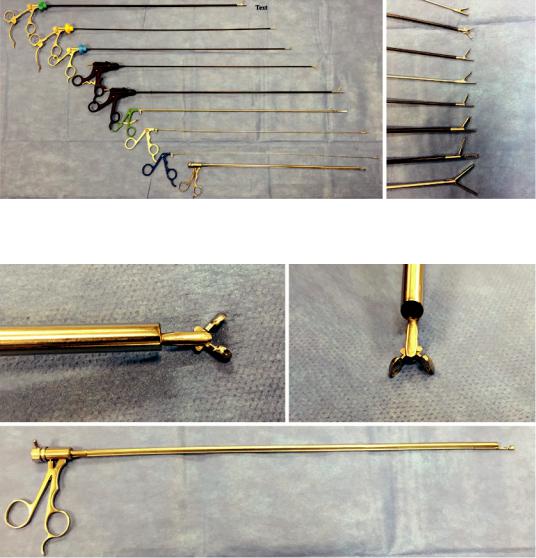
Grasping forceps represent the mainstay of FB retrieval because of their versatility, ease of use, and variety. Forceps are available for both rigid and exible approaches. There are two primary forceps mechanism designs: single and dual action. In single-action forceps, one jaw of the forceps is stationary and in a fxed position, while the other jaw is movable. In dual-action forceps, there are two movable jaws that open symmetrically away from each other (i.e. alligator jaw forceps). There are a variety of jaw surfaces available with variances in serration size, number, and arrangement. Additionally, there are available variances in surface area, shapes, presence of a needle, and fenestrations. Some examples of different available forceps designs include curved, needle-forceps, rat-tooth, V-shape, and shark- tooth. Forceps can also have different coatings, such as latex and rubber, which can help with enhancing grip of specifc FBs. Some rigid forceps have the capability to rotate upon their axis, which can allow for an easier approach (Fig. 40.17a, b). Rigid optical forceps are also available (Fig. 40.18). These forceps can be used in conjunction with a Hopkins telescope, which allows direct visualization when grasping FBs.
FB properties and location should determine the selection of forceps. The use of rigid forceps requires the FB to be directly in the pathway of the rigid tracheoscope/bronchoscope barrel. Distal FBs will often require the addition of aexible bronchoscope and exible forceps. When a FB is located in a confned space or is up against an airway wall, single-action forceps are consid-
40 Foreign Bodies in the Airway: Endoscopic Methods |
703 |
|
|
a |
b |
Fig. 40.17 Rigid forceps and graspers. (a) Variety of rigid forceps and graspers. (b) Close-up of jaws of different forceps that can be used for removal of foreign bodies
Fig. 40.18 Rigid optical forceps. These forceps work in conjunction with the Hopkins telescope and allow for direct visualization during use
ered initially because the space required for jaw opening is less than dual-action forceps. Rat- tooth forceps have a confguration where the teeth of the jaws interlace with each other when closing, which make these useful in retrieval of softer FBs. Objects with at surfaces such as coins and certain dental appliances are better grasped with V-shape and shark-tooth forceps (known for having a frm grip).
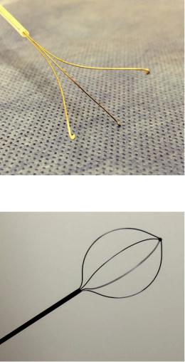
While technically not forceps, multi-pronged graspers serve FB retrieval in a similar manner
(Fig. 40.19). These graspers can have anywhere from three to fve arms that open wide apart, usually to accommodate larger FBs. The tips of these arms come in different shapes, such as rings, to avoid mucosal trauma when opening them.
Baskets
Baskets are essentially complex snares that contain two or more wires that are in a loop formation and can be tightened in order to “capture” a FB (Fig. 40.20). Use of retrieval baskets origi-
Данная книга находится в списке для перевода на русский язык сайта https://meduniver.com/
704 |
M. Simof et al. |
|
|
Fig. 40.19 Flexible 3-pronged grasper
Fig. 40.20 Boston Scientifc Zero Tip™ Airway Retrieval Basket (Spencer, Indiana, USA)
nated in the feld of gastroenterology, with it being the primary tool for removal of resected polyps. However, the use of baskets has spread to many other endoscopic and laparoscopic specialties for removal of various tissues and materials. While baskets are generally used in conjunction with exible bronchoscopes, there are rigid varieties available. Baskets come in different sizes, number of wire loops, stiffness, and different tips. Some baskets have nets built into them to assist in retrieval of smaller material. Baskets are especially useful for airway FBs that are round or spherical in shape, as well as those that are smooth. Caution should be used in FBs that are
less solid or soft in nature, as retraction of wire loops can lead to unintended cutting of the FB into multiple smaller pieces.
Balloons
Balloons used for airway dilation, embolectomies, and bronchial blockage purposes can also be used to assist in the removal of FBs indirectly. For distal FBs that are “wedged” or in a confned space that will not allow for instrumentation, the use of a balloon can be considered. The technique involves passing a balloon distal to the FB and then in ating it. By slowly retracting the balloon proximally, the FB can sometimes be dislodged and/or moved. The purpose of this maneuver is to allow for improved positioning of the FB giving the operator more retrieval options. Another use of a balloon is placing it distal to the FB to prevent migration into smaller airways during instrumentation or to have it available for in ation to facilitate rapid tamponade if removal of FB results in brisk bleeding. This technique usually requires use of rigid bronchoscopy to control multiple tools within the airways. Keep in mind that balloons should be avoided with sharp FBs as the pulling/dislodgement can lead to mucosal injury in addition to balloon rupture. Caution must be maintained when deciding to pass the balloon distal to the airway FB, as this maneuver does pose the risk of pushing the FB further into the airway.
Suction Instruments
Particularly when a FB is sitting more freely within an airway, there are various methods to utilize suction during FB retrieval. Most simply, this can be accomplished directly with the exible bronchoscope’s working channel and/or with the plastic or metal suction catheters used with rigid bronchoscopy. It is very important to acknowledge that despite its ease, this method does not allow for secure procurement of the FB during retrieval. Precautions must be taken during retrieval, especially, when the bronchoscope is used trans-orally without an artifcial airway. It is possible for the FB to become loose and detached from the bronchoscope at any number of levels (i.e. vocal cords, hypopharynx, pharynx,

40 Foreign Bodies in the Airway: Endoscopic Methods |
705 |
|
|
etc.). This may lead to the FB becoming obstructive in a more proximal or central location, such as the trachea, glottis, or supraglottic region. To prevent losing a FB during retrieval, a suction catheter can be utilized to retrieve the FB into proximal large airways, where forceps or basket can be used for removal from the airway. If loss of FB occurs in the upper airway and proximal to the vocal cords, direct laryngoscopy can be performed with the use of Magill forceps (Fig. 40.21) for retrieval of the lost FB. Loss of FB in the trachea can lead to cardiopulmonary decompensation and may require endotracheal intubation. In such cases, bronchoscopically advancing the FB distally into a mainstem may allow for improved ventilation and further stabilization of the patient.
Rigid bronchoscopy allows for the use of large caliber suction catheters. These catheters are specifcally useful in suction of macerated and/or fragmented FBs such as medication pills and chewed edibles. Also, large-bore catheters are excellent for suctioning large volumes of blood, if massive hemoptysis occurs associated with the FB aspiration or its removal.
Ablative Therapies
While ablative therapies are routinely used for their tissue coagulative and destructive effects, they have a unique role in management of FBs. Granulation tissue is a common consequence of retained FBs and can make retrieval challenging. Ablative therapies such as laser, argon plasma coagulation (APC), and electrocautery are all
effective tools in destroying granulation tissue and allowing easier access to the FB. Cryotherapy is unique in that in addition to its ablative effects, it can directly be used for FB retrieval of organic material. Below, we discuss in-depth aspects of cryotherapy, laser therapy, and electrocautery/ APC.
Cryotherapy
Cryotherapy is traditionally used for its destructive properties in the treatment of obstructive airway lesions, but it is also a very useful tool in specifc cases of FB retrieval. The extremely cold temperatures produced by cryotherapy allow organic FBs to freeze and attach to the probe. Often these FBs can be removed without much diffculty adhered to the probe. Once the probe is in contact with the FB a freezing cycle is started. Once the ice ball is forming (Fig. 40.22), the FB should be attached to the probe (approximately 10–20). The operator must be very careful not to touch the airway wall while attempting to remove the FB, as the probe will stick to the wall also.
Cryotherapy can also be used to remove blood clots and mucus plugs from obstructed airways. Because of their water content, blood and mucus are excellent candidates for cryotherapy and are effciently removed by placing the probe into the clot or plug, begin a freezing cycle, and slowly pull out with the bronchoscope. Mucus and blood casts of the tracheobronchial tree can be removed in an en bloc manner. Again, ensure that the probe does not touch the airway wall during this maneuver.
Fig. 40.21 Magill forceps |
Fig. 40.22 Iron pill aspiration-induced bronchostenosis |
of the right lower lobe bronchus |
Данная книга находится в списке для перевода на русский язык сайта https://meduniver.com/