

Color Atlas of Neurology
.pdfVisual Field Defects
Visual field
Directions tested
Blind spot
Test object
Macular region
Confrontation test
Homonymous inferior quadrantanopsia
Homonymous hemianopsia (macular sparing)
Incongruent homonymous quadrantanopsia
Homonymous hemianopsia
Binasal homonymous defect
Monocular defect
Right visual field
Meridian
Left visual field
Line of fixation of eye
Patient ca. 50 cm from examiner
|
|
|
Homonymous |
|
|
|
hemianopic central |
||
|
|
visual field defect |
||
|
|
|
(occipital pole |
|
|
|
|
lesion) |
|
|
|
Sparing of contralat- |
||
Homonymous superior |
eral ”sickle” and |
|||
macula (lesion of |
||||
quadrantanopsia |
||||
calcarine cortex on |
||||
|
|
|||
|
|
medial surface of |
||
|
|
|
hemisphere) |
|
|
|
|||
Bitemporal hemianopsia
 ”Junction scotoma”
Tunnel vision
Cortical blindness
Inferior altitudinal hemianopsia
|
Bilateral |
|
Types and localization of visual field defect |
homonymous |
|
visual field defect |
||
|
Cranial Nerves
83
Rohkamm, Color Atlas of Neurology © 2004 Thieme
All rights reserved. Usage subject to terms and conditions of license.

Cranial Nerves
84
Oculomotor Function
The visual axes of the eyes are directed straight ahead on primary gaze (i.e., 23° inward from the more lateral axes of the orbits). Movements of the eyes are mediated by six extraocular muscles on each side. The lateral and medial rectus muscles are responsible for horizontal eye movements. Vertical eye movements are subserved by the superior and inferior rectus as well as superior and inferior oblique muscles. The rectus muscles elevate and depress the eye when it is abducted, the oblique muscles when it is adducted. The two muscles of each synergistic pair (e. g., the left lateral rectus and right medial rectus muscles) receive equal degrees of innervation (Hering’s law).
Vestibulo-ocular reflex (VOR). Impulses arising in the semicircular canals in response to rapid movement of the head induce reflex movement of the eyes in such a way as to stabilize the visual image (p. 26). For example stimulation of the horizontal semicircular canal activates the ipsilateral medial rectus and contralateral lateral rectus muscles, while inhibiting the ipsilateral lateral rectus and contralateral medial rectus muscles. The VOR makes the eyes move in the direction opposite to the head movements, at the same angular velocity.
Optokinetic reflex. Optokinetic nystagmus (OKN) is triggered by large-scale, moving visual stimuli and serves to stabilize the visual image during slow head movement. OKN is characterized by slow, gliding conjugate movement of the eyes in the direction of an object moving horizontally or vertically across the visual field, in alternation with rapid return movements in the opposite direction (saccades). OKN is intact in psychogenic (pseudo) blindness.
Fixation. Fixation is active adjustment of the gaze (with or without the aid of eye movement) to keep a visualized object in focus.
Saccades. Saccades are rapid, jerky conjugate movements of the eyes that serve to adjust or set the point of fixation of an object on the fovea. Saccades may be spontaneous, reflexive (in response to acoustic, visual, or tactile stimuli), or voluntary; the rapid phase of nystagmus is a saccade. The speed, direction, and amplitude of a saccadic movement are determined before it is carried out and cannot be influenced voluntarily during its execution. Shifts of visual fixation by more than 10° are accompanied by head move-
ments.
Slow ocular pursuit. Voluntary ocular pursuit can occur only when triggered by a moving visual stimulus (e. g., a passing car). Conversely, fixation of the gaze on a resting object while the head is moving leads to gliding eye movements. Fixation-independent ocular pursuit also occurs during somnolence and the early stages of sleep (“floating” eye movements).
Vergence movements (convergence and divergence) are mirror-image movements of the two eyes toward or away from the midline, evoked by movement of an object toward or away from the head in the sagittal plane. They serve to center the visual image on both foveae and are accompanied by an adjustment of the curvature of the lens (accommodation) to keep the object in focus.
Neural pathways. The medial longitudinal fasciculus (MLF) interconnects the nuclei of cranial nerves III, IV, and VI. The MLF also connects with fibers conveying information to and from the cervical musculature, vestibular nuclei, cerebellum, and cerebral cortex and thus mediates the coordination of eye movements with movements of the body and head. Saccades are produced by two parallel systems: Voluntary eye movements are subserved by the frontal system, which consists of the frontal eye fields (areas 4, 6, 8, 9), the supplementary eye field (area 6), the dorsolateral prefrontal cortex (area 46), and a portion of the parietal cortex (area 7). It projects to the contralateral paramedian pontine reticular formation (PPRF), which coordinates vertical and horizontal saccades. Vertical and torsional eye movements are controlled by the rostral interstitial nucleus of the MLF and by the interstitial nucleus of Cajal. Reflex eye movements are initiated in the visual cortex (area 17) and temporal lobe (areas 19, 37, 39) and modulated in the superior colliculus (collicular system). Vergence and accommodation are mediated by the pretectal area in the vicinity of the oculomotor nucleus.
Rohkamm, Color Atlas of Neurology © 2004 Thieme
All rights reserved. Usage subject to terms and conditions of license.

Oculomotor Function
Inferior oblique m. (III)
Secondary position
Primary position
Superior oblique m. (IV)
MLF
Orbital axis |
|
|
|
Superior rectus m. (III) |
|||||
|
|
||||||||
|
|
|
|
|
|
|
|
Visual axis |
|
|
|
23o |
|
|
|
|
|
|
Medial |
|
|
|
|
|
|
|
|
||
|
|
|
|
|
|
|
rectus m. (III) |
||
|
|
|
|
|
|
|
|
Lateral rectus |
|
|
|
|
|
|
|
|
|
|
m. (VI) |
|
|
|
|
|
|
|
Tertiary position |
||
|
|
|
|
|
|
|
|||
|
|
|
|
|
Inferior rectus m. (III) |
||||
|
|
|
|
|
|||||
Conjugate eye movements
(arrows indicate direction of gaze, red = active muscle)
Nu- |
|
|
Pathway for reflex eye movement |
|
|
||||||||||||||||||||||||
|
|
||||||||||||||||||||||||||||
clear |
|
|
Pathway for voluntary eye movement |
||||||||||||||||||||||||||
|
|
||||||||||||||||||||||||||||
region |
|
Nucleus of Darkschewitsch |
|
|
|
|
|
|
|
|
|
|
|
|
|
||||||||||||||
III |
|
|
|
|
|
|
|
|
|
|
|
|
|
|
|
||||||||||||||
|
|
|
|
|
|
|
|
|
|
|
|
|
|
|
|||||||||||||||
|
|
|
|
|
|
|
|
|
|
|
|
|
|
|
Rostral interstitial nucleus of MLF |
||||||||||||||
PPRF |
|
||||||||||||||||||||||||||||
|
Interstitial nucleus |
|
|
|
|
|
|
|
|
|
|
|
|
|
|||||||||||||||
Nu- |
|
|
|
|
|
|
|
|
|
|
|
|
|
|
|
|
|
||||||||||||
|
Trochlear nucleus |
Area 46 |
|
|
|
|
|
|
|
|
|
|
|
|
|||||||||||||||
clear |
|
|
|
|
|
|
|
|
|
|
|
|
|
||||||||||||||||
|
|
|
|
|
|
|
|
|
|
|
|
|
|
|
|||||||||||||||
region |
|
|
To vestibulo- |
|
|
|
|
|
|
|
|
|
|
|
|
|
|||||||||||||
|
|
|
|
|
|
|
|
|
|
|
|
|
|
|
|||||||||||||||
VI |
|
cerebellum |
|
|
|
|
|
|
|
|
|
|
|
|
|
||||||||||||||
Nucleus |
|
|
Vestibular nuclei |
|
|
|
|
|
|
|
|
|
|
|
|
|
|||||||||||||
|
|
|
|
|
|
|
|
|
|
|
|
|
|
|
|||||||||||||||
prepositus |
|
Vestibulospinal |
Pathway for voluntary eye movement |
||||||||||||||||||||||||||
|
|
|
|
|
|
|
|
|
|
|
|
|
|
|
|
|
|
|
|
|
|
|
|
|
|
|
|
||
|
|
|
|
|
Nerve pathways |
|
tract |
|
|
|
|
|
|
|
|
|
|
|
|
|
|||||||||
|
|
|
|
|
|
|
|
|
|
|
|
|
|
|
|
|
|
|
|
|
|||||||||
Rostral interstitial |
|
|
|
|
|
|
|
|
|
|
|
|
|
|
|
|
|
|
|
|
|
||||||||
|
|
|
|
|
|
|
|
|
|
|
|
|
|
|
|
|
|
|
|
|
|||||||||
|
|
|
|
|
|
|
|
|
|
|
|
|
|
|
|
|
|
|
|
|
|||||||||
nucleus of MLF |
IV |
|
|
|
|
|
|
|
|
|
|
|
|
|
|||||||||||||||
Interstitial |
|
|
|
|
|
|
|
|
|
|
|
|
|
|
|
|
|
|
|
|
|||||||||
|
|
|
|
|
|
|
|
|
|
|
|
|
|
|
|
|
|
|
|
|
|
||||||||
|
|
|
|
|
|
|
|
|
|
|
|
|
|
|
|
|
|
||||||||||||
nucleus |
|
|
|
|
|
|
|
|
|
|
|
|
|
|
|
|
|||||||||||||
MLF |
|
|
|
|
|
|
|
|
|
|
|
|
|
|
|
|
|
|
|
|
|
|
|
|
|||||
|
|
|
|
|
|
|
|
|
|
|
|
|
|
|
|
|
|
|
|
|
|
|
|
|
|||||
|
|
|
|
|
|
|
|
|
|
|
|
|
|
|
|
|
|
|
|
||||||||||
Nucleus |
|
|
|
|
|
|
|
|
|
|
|
|
|
|
|
|
|
|
|
|
|
||||||||
|
|
|
|
|
|
|
|
|
|
|
|
|
|
|
|
|
|||||||||||||
prepositus |
|
|
|
|
|
|
|
|
|
|
|
|
|
|
|
|
|||||||||||||
VI |
|
|
|
|
|
|
|
|
|
|
Vestibular |
|
|
|
|
|
|
|
|||||||||||
|
|
|
|
|
|
|
|
|
|
|
|
|
|||||||||||||||||
|
|
|
|
|
|
|
|
|
|
|
|
|
|
|
|
|
|
|
|
|
|||||||||
|
|
|
|
|
|
|
|
|
|
|
|
|
|
|
|
|
|
|
|||||||||||
|
|
|
|
|
|
|
|
|
|
|
|
|
|
|
|
|
nuclei |
||||||||||||
VIII |
|
|
|
|
|
|
|
|
|
|
|
|
|
|
|
|
|
|
|
|
|
|
|
||||||
|
|
|
|
|
|
|
|
|
|
|
|
|
|
|
|
|
|
||||||||||||
(vestibular n.) |
|
|
|
|
|
|
|
|
|
|
|
|
|
|
|
|
|||||||||||||
Extraocular muscles, cranial nerves and nuclei
(anterior view)
Cranial Nerves
85
Rohkamm, Color Atlas of Neurology © 2004 Thieme
All rights reserved. Usage subject to terms and conditions of license.

Oculomotor Disturbances
Peripheral Oculomotor Disturbances
|
Weakness of an extraocular muscle results in di- |
|
|
plopia, which is most pronounced in the direc- |
|
|
tion of action of the affected muscle (p. 85). The |
|
|
cause may be a lesion in the muscle itself, in the |
|
|
cranial nerve that supplies it, or in the cranial |
|
|
nerve nucleus. |
|
|
Examination. The more peripheral of the two |
|
|
images seen by the patient is always derived |
|
|
from the affected eye. The impaired eye move- |
|
|
ment may be seen directly by observation of |
|
|
conjugate eye movements in the nine cardinal |
|
|
directions of gaze (p. 85). Next, the examiner has |
|
|
the patient look in the direction of greatest |
|
Nerves |
image displacement, covers first one eye and |
|
then the other, and asks the patient each time |
||
more peripheral image disappears when the af- |
||
|
which of the two images has disappeared. The |
|
Cranial |
fected eye is covered. Alternatively, the patient |
|
can be asked to look at a point of light while a |
||
|
||
|
red glass is held in front of one eye; if the more |
|
|
peripheral image is red, then the eye with the |
|
|
glass is the affected eye. Another test is to |
|
|
rapidly cover and uncover one eye and then the |
|
|
other while the patient looks in the cardinal |
|
|
directions of gaze. The greatest ocular deviation |
|
|
and the greatest adjustment of the unaffected |
|
|
eye (secondary angle of deviation) occur when |
|
|
the patient looks in the direction of the paretic |
|
|
muscle. As a rule, these tests are helpful when a |
|
|
single muscle is acutely weak; more sophisti- |
|
|
cated ophthalmological tests are needed if the |
|
|
weakness is chronic or affects more than one |
|
|
muscle. |
|
|
Oculomotor nerve palsy. When a compressive |
|
|
lesion causes complete oculomotor nerve palsy, |
|
|
the patient complains of diplopia (with oblique |
|
|
image displacement) only when the ptotic eye- |
|
|
lid is passively elevated. The affected eye is |
|
|
turned downward (action of the intact superior |
|
|
oblique muscle) and outward (intact lateral rec- |
|
|
tus muscle) on primary gaze, and the pupil is |
|
|
fixed, dilated, and irregularly shaped. The in- |
|
|
volved eye can still be abducted (intact CN VI), |
|
|
and looking down causes intorsion (intact CN |
|
|
IV). Incomplete oculomotor nerve palsy because |
|
|
of nuclear or myopathic lesions may differen- |
86tially affect the intraocular and extraocular muscles supplied by CN III and cause different
types of diplopia and pupillary disorders (p. 90).
Trochlear nerve palsy. The affected eye points upward and toward the nose on primary gaze. Diplopia is worst when the affected eye looks toward the nose and downward.
Abducens nerve palsy. The affected eye deviates toward the nose on primary gaze. Horizontal diplopia is worst on looking toward the side of the affected eye.
Supranuclear and Internuclear Oculomo-
tor Disturbances
Internuclear ophthalmoplegia (INO) is characterized by inability to adduct one eye, combined with nystagmus of the other, abducted eye (dissociated nystagmus), on attempted lateral gaze. It is due to a lesion of the medial longitudinal fasciculus (MLF) on the side of the nonadducting eye and at a level between the nuclei of CN III and CN VI. Bilateral MLF lesions cause bilateral INO. Both eyes can adduct normally during convergence. More rostral lesions lead to convergence paresis without nystagmus; more caudal lesions lead to paresis of the lateral rectus muscle. Multiple sclerosis and vascular disorders are the most common causes of INO.
Unilateral pontine lesions cause ipsilateral gaze palsy (the gaze points away from the side of the lesion) but leave vertical eye movement largely intact. Co-involvement of the MLF leads to one- and-a-half syndrome (ipsilateral pontine gaze palsy + INO), e. g., paresis of conjugate gaze to the left and impaired adduction of the left eye on looking to the right.
Supratentorial lesions. Extensive cortical or subcortical hemispheric lesions produce contralateral gaze palsy (patient gazes toward the side of the lesion). Slow reflex movements of the eyes in all directions are still possible because the optokinetic reflex is not affected. In occipital lesions, the optokinetic reflex is absent; voluntary eye movements are preserved, but the eyes can no longer follow slowly moving objects. Abnormal, diffuse elevation of activity within a hemisphere (e. g., because of an epileptic seizure) causes contralateral gaze deviation.
For further information on horizontal and vertical gaze palsy, see page 70.
Rohkamm, Color Atlas of Neurology © 2004 Thieme
All rights reserved. Usage subject to terms and conditions of license.
Oculomotor Disturbances
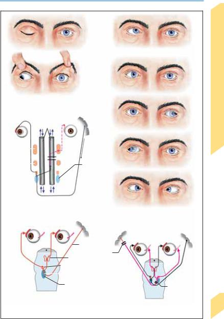
Right trochlear palsy (looking straight ahead)
Abducens palsy (looking straight ahead)
Complete right oculomotor palsy
(looking straight ahead)
|
|
|
|
|
|
MLF |
|
|
|||||
III |
|
|
|
|
|
|
|
|
|
|
|
||
|
|
|
|
|
|
||||||||
IV |
|
|
|
|
|
|
|
|
Lesion |
Nucleus |
|
||
|
|
|
|
|
|
|
|
|
|||||
|
|
|
|
|
|
|
|
||||||
|
|
|
|||||||||||
VI |
|
|
|
|
|
|
|
|
|
praepositus |
|
||
|
|
|
|
|
hypoglossi |
|
|||||||
|
|
|
|
|
|
|
|
|
|
|
|
|
|
|
|
|
|
|
|
|
|
|
|
|
|
(lateral gaze) |
|
Neuroanatomy of internuclear ophthalmoplegia |
|
||||||||||||
(INO) (shown: left INO on rightward gaze) |
|
Bilateral INO |
|||||||||||
|
|
|
|
|
|
|
|
|
|
|
|
|
(leftward, rightward, and downward gaze) |
|
|
|
|
|
|
|
|
|
|
|
|
Irritative lesion |
|
|
|
|
|
|
|
|
|
|
|
|
|
Supra- |
|
|
|
|
|
|
|
|
|
|
|
|
III |
tentorial |
|
|
|
|
|
|
|
|
|
|
|
|
VI |
lesion |
|
|
|
|
|
|
|
|
|
|
|
|
|
|
|
|
|
|
|
|
|
|
|
|
|
|
Irritative lesion |
|
|
|
|
|
|
|
|
|
|
|
|
|
|
||
|
|
|
|
|
|
|
|
|
|
|
Hypoglossal nucleus |
Pontine lesion |
|
|
|
|
|
|
|
|
|
|
|
|
|
|
|
Rightward gaze deviation |
Conjugate supranuclear paresis of leftward gaze |
||||||||||||
(irritative lesion: left, supratentorial; right, pontine) (right supratentorial lesion or left pontine destructive lesion)
Cranial Nerves
87
Rohkamm, Color Atlas of Neurology © 2004 Thieme
All rights reserved. Usage subject to terms and conditions of license.

Cranial Nerves
88
Nystagmus
Nystagmus is involuntary rhythmic movement of the eyes consisting of slow movement in one direction and rapid return movement in the other. The slow component is caused by disturbances of the motor and stabilizing systems of the eye (p. 84) or because of ocular muscle paresis; the fast component represents the rapid return movement of pontine generators. Although the slow component is the actual pathological component of nystagmus, the direction of nystagmus is conventionally said to be that of its fast component, which is easier to detect. The intensity of nystagmus increases when the patient gazes in the direction of the fast component. Nystagmus can be further classified according to the type of movement as pendular, circular, or torsional (rotatory).
Examination. The examiner first observes the eyes on primary gaze, then during horizontal and vertical pursuit (fixation of gaze on a slowly moving object) and vergence. Nystagmus of labyrinthine origin is observed best with Frenzel spectacles (preventing visual fixation and giving the examiner a magnified view of the eyes). The following features of nystagmus are assessed: positional-dependence, coordination (conjugate, dissociated), direction (horizontal, vertical, rotatory, retracting, pendular), amplitude (fine, medium, coarse), and frequency (slow, moderate, fast).
Physiological Nystagmus
Physiological nystagmus serves to stabilize the visual image while the head and body are moving or when the individual looks at a moving object. The different types include congenital nystagmus (often X-linked recessive; fixation nystagmus is most pronounced when gazing fixedly on an object; the direction of nystagmus is usually horizontal), spasmus nutans (pendular nystagmus beginning in the first year of life; often accompanied by nodding of the head and torticollis; disappears spontaneously), end-position nystagmus (occurs during rapid movement; extreme lateral gaze; usually only a few beats), and optokinetic nystagmus (its absence is pathological; see p. 84).
Pathological Nystagmus
Gaze-evoked nystagmus occurs only in certain direction(s) of gaze. The main causes are drug
intoxication and brain stem or cerebellar disturbances. A slower and coarser gaze-paretic nystagmus may be seen in association with supranuclear or peripheral gaze palsy, beating in the direction of the paretic gaze. Peripheral palsy of an eye muscle may cause unilateral nystagmus of the affected eye.
Spontaneous nystagmus is that which occurs when the eyes are in the primary position; it is usually caused by vestibular dysfunction and is rarely congenital.
Peripheral vestibular nystagmus (cf. p. 58) can be seen in patients with benign paroxysmal positional vertigo, vestibular neuritis, Ménière disease, vascular compression of the vestibular nerve, and labyrinthine fistula. Nystagmus decreases on fixation and increases when fixation is blocked (lid closure, Frenzel spectacles). Most patients exhibit rotatory nystagmus that either beats continually toward the nonaffected ear, or else begins a short time after a change of position (positional nystagmus toward the lower ear, see p. 58).
Central vestibular nystagmus (p. 58) is caused by lesions of the brain stem (vestibular nuclei, vestibulocerebellum) or of the thalamocortical projections. It is usually accompanied by other brain stem or cerebellar signs, does not decrease on fixation, depends on the direction of gaze, and usually persists. Central positional nystagmus does not exhibit latency, is not affected by the rate of positional change, occurs with changes of position to either side, beats toward the higher ear, and is not exhaustible, stopping only when the patient is returned to the neutral position. Because positional information from vestibular, visual, and somatosensory systems is integrated in the vestibulo-ocular reflex (VOR; see pp. 26, 84), the phenomena associated with nystagmus can be explained as functional disturbances in one of the major three spatial planes of action of the VOR. Lesions cause an imbalance between the neural inputs to the VOR concerning the two sides of the affected plane. Depending on which plane is affected, the resulting nystagmus may be horizontal (horizontal plane; lesion of the vestibular nuclei), vertical (sagittal plane; pontomesencephalic, pontomedullary, or floccular lesion), or torsional (coronal plane; pontomesencephalic or pontomedullary lesion). Vertical nystagmus (upbeat or downbeat) is always due to a central lesion.
Rohkamm, Color Atlas of Neurology © 2004 Thieme
All rights reserved. Usage subject to terms and conditions of license.

Nystagmus
Direction of nystagmus
Primary gaze
Gaze-evoked nystagmus
(no nystagmus on primary gaze)
Spontaneous nystagmus
Primary gaze
Horizontal plane = yaw
(tendency to fall to ipsilateral side; diminished response to caloric testing in ipsilateral ear)
Retraction nystagmus
(bilateral dorsal midbrain lesion)
Sagittal plane = pitch
(tendency to fall forward/backwards; ”elevator” sensation)
Peripheral vestibular nystagmus
(no nystagmus on primary gaze)
|
Frontal plane = roll |
|
(tendency to fall sideways, lateropulsion) |
Skew deviation (vertical disconjugate gaze) |
Central vestibular nystagmus, |
Vertical upand downbeat nystagmus (brain stem lesion) |
spatial planes |
Cranial Nerves
89
Rohkamm, Color Atlas of Neurology © 2004 Thieme
All rights reserved. Usage subject to terms and conditions of license.

Pupillomotor Function
The colored part of the eye, or iris (Greek “rainbow”), is the posterior wall of the anterior ocular chamber. Its inner edge forms the margin of the pupil. The sphincter pupillae muscle contracts the pupil, and the dilator pupillae muscle dilates it. The upper eyelid contains two muscles: the superior tarsal muscle receives sympathetic innervation, and the levator palpebrae superioris muscle is innervated by the oculomotor nerve.
Nerve Pathways
|
Parasympathetic fibers. The preganglionic |
|||
|
fibers arise in the accessory oculomotor nucleus |
|||
Nerves |
(Edinger–Westphal nucleus), travel in the oculo- |
|||
motor nerve along its outer edge, and enter the |
||||
ciliary ganglion. The postganglionic fibers travel |
||||
to the ciliary and sphincter pupillae muscles in |
||||
Cranial |
the short ciliary nerves (of which there are up to |
|||
20). The parasympathetic fibers and all others |
||||
|
||||
|
on the outer aspect of CN III receive their blood |
|||
|
supply from the pial vessels, while fibers in the |
|||
|
interior of the nerve are supplied by the vasa |
|||
|
nervorum. |
|
|
|
|
Sympathetic fibers. The central sympathetic |
|||
|
fibers exit from the posterolateral portion of the |
|||
|
hypothalamus (first |
preganglionic |
neurons), |
|
|
then pass ipsilaterally through the tegmentum |
|||
|
of the mid brain and pons and through the |
|||
|
lateral medulla to form a synapse onto the sec- |
|||
|
ond preganglionic neurons in the intermedi- |
|||
|
olateral cell column of the spinal cord (ciliospi- |
|||
|
nal center), at levels C8–T2. Most of the fibers |
|||
|
exit the spinal cord with the ventral root of T1 |
|||
|
and join with the sympathetic trunk, which lies |
|||
|
adjacent to the pleural dome at this level. They |
|||
|
travel with the ansa subclavia around the sub- |
|||
|
clavian artery and pass through the inferior |
|||
|
(stellate) and middle cervical ganglia to the su- |
|||
|
perior cervical ganglion, where they form a |
|||
|
(third) synapse onto the postganglionic neu- |
|||
|
rons. Postganglionic fibers to the pupil travel |
|||
|
along the course of the internal carotid artery |
|||
|
(carotid plexus) and the ophthalmic artery, then |
|||
|
in the nasociliary nerve (a branch of CN V) and, |
|||
|
finally, the long ciliary nerves, which innervate |
|||
|
the dilator pupillae |
muscle. Other |
postgan- |
|
90glionic fibers of the sympathetic system pass to the sweat glands, the orbital muscles (bridging the inferior orbital fissure), the superior and inferior tarsal muscles, and the conjunctival ves-
sels. Fibers to the sweat glands arise at the
T3–T4 level and form a synapse with the third neuron in the stellate ganglion; thus, nerve root lesions at C8–T2 do not impair sweating.
Light Reflex
The light reflex regulates the diameter of the pupils according to the amount of light falling on the eye. Each pupil constricts in response to light and dilates in the dark. The afferent arm of the reflex arc consists of fibers of the optic nerve that decussate in the optic chiasm, then pass around the lateral geniculate body and terminate in the mid brain pretectal area, both ipsilaterally and contralaterally. The parasympathetic fibers are the efferent arm. The Edinger– Westphal nuclei of the two sides are connected to each other by interneurons; thus, impulses from each optic nerve arrive at both Edinger– Westphal nuclei, and light falling on one eye leads to contraction of both the ipsilateral pupil (direct light reflex) and the contralateral pupil (consensual light reflex). The pupillary diameter in moderate ambient light is normally 3–4 mm. Excessive pupillary constriction (!2 mm) is referred to as miosis, and excessive dilatation ("5 mm) as mydriasis. Anisocoria (inequality of the diameters of the pupils) often indicates a diseased state (see below); it may be physiological but, if so, is usually mild.
The Near Response: Convergence,
Pupilloconstriction, Accommodation
When a subject watches an approaching object, three things happen: the eyes converge through the action of the medial rectus muscles; the pupils constrict; and the curvature of the lens increases through the action of the ciliary muscle (accommodation). The near response may be initiated voluntarily (by squinting) but is most often the result of a reflex, whose afferent arm consists of the visual pathway to the visual cortex. The efferent arm for convergence consists of descending fibers to the pretectal convergence center (Perlia’s nucleus) and onward to the oculomotor nucleus (nuclear area for the medial rectus muscles); the efferent arm for pupilloconstriction and accommodation is the parasympathetic projection of the Edinger– Westphal nucleus through the oculomotor nerve to the sphincter pupillae and ciliary muscles.
Rohkamm, Color Atlas of Neurology © 2004 Thieme
All rights reserved. Usage subject to terms and conditions of license.

Pupillomotor Function
Dilator muscle |
|
Parasympathetic fibers |
|
Lens |
|
Pial vessels |
|
Sphincter |
|
|
|
muscle |
|
Oculomotor n. |
|
Zonular |
|
Vasa nervorum |
|
fibers |
|
|
|
|
|
|
|
Ciliary |
|
Visual cortex |
|
muscle |
|
|
|
|
(areas 17, 18, 19) |
|
|
(short |
|
|
|
|
|
|
|
ciliary ner- |
|
|
|
ves) |
|
|
|
|
Oculomotor nucleus |
Perlia’s nucleus |
|
|
|
|
|
Pupil |
|
Pretectal area |
|
|
Lateral geniculate |
Nerves |
|
|
|
||
|
|
body |
|
Ciliary |
|
Light reflex |
|
ganglion |
|
Accommodation |
Cranial |
Levator |
|
Convergence |
|
|
|
||
palpebrae |
|
Edinger- |
|
|
|
||
superioris m. (III) |
|
Westphal nuclei |
|
Medial |
|
Sweat glands |
|
rectus muscle |
|
(forehead) |
|
Superior |
|
Orbitalis m. |
|
tarsal m. |
|
Central |
|
|
|
|
|
Dilator |
|
sympathetic pathway |
|
|
Carotid plexus, |
|
|
muscle |
|
|
|
Conjunctival vessels |
|
internal carotid a. |
|
|
|
|
|
|
|
Superior cervical ganglion |
|
|
|
Sudoriparous and vaso- |
|
|
|
motor fibers to skin of |
|
|
|
face traveling along the |
|
Orbicularis |
|
external carotid a. |
|
|
|
|
|
oculi m. (VII) |
|
Middle cervical |
|
|
|
ganglion |
|
|
|
Inferior cervical |
|
Pleural dome |
|
(stellate) ganglion |
|
|
Ciliospinal center |
|
|
|
|
|
|
|
|
Ansa subclavia |
|
|
Subclavian a. |
|
|
|
Pupillomotor Function |
|
91 |
|
|
|
Rohkamm, Color Atlas of Neurology © 2004 Thieme
All rights reserved. Usage subject to terms and conditions of license.

Pupillary Dysfunction
|
Examination. The size and shape of the pupils |
|
|
are first assessed in diffuse light with the |
|
|
patient looking at a distant object to prevent the |
|
|
near response. The room is then darkened and |
|
|
the direct light reflex of each pupil is tested at |
|
|
varying light intensities (by varying the distance |
|
|
of the lamp from the eye). If both pupils con- |
|
|
strict when illuminated, there is no efferent |
|
|
pupillary defect. Next, in the swinging flashlight |
|
|
test, the examiner indirectly illuminates one eye |
|
|
with a bright light for ca. 2 seconds, then quickly |
|
|
switches the light to the other eye, and back |
|
|
again, some 5–7 times. The normal finding is |
|
|
that the two pupils are always of equal diame- |
|
|
ter; an abnormal finding indicates asymmetry of |
|
Nerves |
the afferent arm of the light reflex on the two |
|
sides, e. g., because of an optic nerve lesion |
||
either of these tests is abnormal, or if the pupils |
||
|
(Marcus Gunn pupillary escape phenomenon). If |
|
Cranial |
are significantly unequal, the near response |
|
should be tested and the direct and consensual |
||
|
||
|
light reflexes should be tested separately in each |
|
|
eye. It is easier to identify which pupil is abnor- |
|
|
mal by observing both phases of the light re- |
|
|
sponse (constriction and dilatation): both are |
|
|
slower in the abnormal pupil. In light–near dis- |
|
|
sociation, the pupils constrict as part of the near |
|
|
response, but not in response to light. Phar- |
|
|
macological pupil testing may be necessary in |
|
|
some cases. |
Parasympathetic Denervation
(Unilateral Mydriasis)
|
Oculomotor palsy (p. 86) is accompanied by my- |
|
driasis only when the parasympathetic fibers on |
|
the margin of the oculomotor nerve are affected. |
|
This is usually not the case in ischemic neu- |
|
ropathy of CN III (e. g., in diabetes mellitus), be- |
|
cause the marginal fibers receive their blood |
|
supply from pial vessels (p. 90). A tonic pupil is a |
|
mydriatic pupil with light–near dissociation. |
|
This condition may be due to local causes (infec- |
|
tion, temporal arteritis) or to systemic diseases |
|
such as Adie syndrome (+ reduction/absence of |
|
tendon reflexes in the legs) and Ross syndrome |
|
(+ hyporeflexia + segmental hypohidrosis). The |
|
|
|
use of anticholinergic agents (atropine eye- |
92 |
drops, scopolamine patch) causes iatrogenic |
|
mydriasis. |
|
|
Sympathetic Denervation
(Unilateral Miosis)
Horner syndrome is produced by a lesion at any site along the sympathetic pathway to the eye and is characterized by unilateral miosis (with sluggish dilatation) and ptosis; anhidrosis (absence of sweating) and enophthalmos are part of the syndrome but are of no practical diagnostic value. The affected pupil will fail to dilate in response to the instillation of 5% cocaine eyedrops. Preganglionic lesions (i.e., those proximal to the superior cervical ganglion) can be distinguished from postganglionic lesions by the instillation of 5% pholedrine eyedrops (at least three days after the cocaine test); the miotic pupil dilates more than the normal pupil if the lesion is preganglionic, symmetrically if it is postganglionic. Central Horner syndrome (first preganglionic neuron) may be due to lesions of hypothalamus, brain stem, or cervicothoracic spinal cord; the second preganglionic neuron may be affected by lesions of the brachial plexus, apical thorax, mediastinum, or neck; the postganglionic neuron may be affected by carotid dissection or lesions of the skull base.
Supranuclear Lesions
Lesions above the oculomotor nucleus tend to cause bilateral pupillary dysfunction; the most common cause is dorsal compression of the midbrain (Parinaud syndrome; p. 358). Neurosyphilis produces Argyll–Robertson pupils— unequal, irregularly miotic pupils with a variable degree of iris atrophy, and light–near dissociation.
Coma (see also p. 118)
The cause of coma may be structural, metabolic, or toxic. Pupilloconstriction is produced by opiates, alcohol, and barbiturates, pupillary dilatation by atropine poisoning (mushrooms, belladonna), tricyclic antidepressants, botulinum toxin, cocaine, and other drugs. Focal lesions (clivus, midbrain) may cause unilateral or bilateral pupillary areflexia and mydriasis. Unilateral miosis is seen in central Horner syndrome, and bilateral miosis (pinpoint pupils) in acute pontine dysfunction.
Rohkamm, Color Atlas of Neurology © 2004 Thieme
All rights reserved. Usage subject to terms and conditions of license.