1Paul M Dewick Medicalc Natural / booktext@id88013694placeboie
.pdf
|
|
|
|
|
|
|
|
POLYSACCHARIDES |
|
|
|
|
|
|
|
475 |
||||
|
|
|
|
|
|
|
|
|
|
|
|
|
|
|
|
MannA = mannuronic acid |
||||
|
|
|
|
|
|
|
|
|
|
|
|
|
|
|
|
|
||||
|
O |
CO2H |
|
|
|
|
|
|
|
|
|
|
|
|
||||||
|
|
|
|
|
|
|
|
|
HO2C OH |
β1→4 |
|
|
|
|
|
|
||||
|
|
|
|
O |
|
|
|
|
|
|
|
|
|
|
|
|
||||
HO |
1 |
|
|
|
|
|
O |
O |
|
|
HO |
|
O |
|
|
|
||||
α1→4 |
|
|
|
|
|
1 |
|
|
|
|
|
|||||||||
|
|
|
|
|
|
HO |
|
O |
4 |
|
|
|
|
|
||||||
|
|
|
|
HO |
|
|
|
|
|
O |
|
|
|
|
||||||
|
|
|
|
|
|
|
|
|
|
|
|
|
|
|||||||
|
|
|
|
|
|
|
|
|
|
|
|
|
|
|
|
|
|
|
||
D-GalA |
|
|
|
O |
CO2H |
|
|
|
|
D-MannA |
|
|
|
HO2C OH |
|
|
|
|
||
|
|
|
4 |
O |
|
|
|
|
|
|
|
|
|
|
D-MannA |
|
|
|
||
|
|
|
|
|
|
|
|
|
|
|
|
|
|
|
|
|
||||
|
|
|
|
HO |
|
|
|
|
|
|
|
|
|
|
|
|
|
|
|
|
GalA = galacturonic acid |
HO |
O |
|
|
|
|
|
alginic acid |
|
|
|
|
|
|||||||
|
|
|
|
|
|
|
|
(200−900 residues) |
|
|
|
|
|
|||||||
|
|
|
|
D-GalA |
|
|
|
|
|
|
|
|
||||||||
|
|
|
|
|
|
|
|
|
|
|
|
|
|
|
|
|
|
|
||
|
|
|
|
pectin |
|
|
|
|
|
|
|
|
|
|
|
|
|
|
|
|
|
|
|
|
|
|
|
|
|
|
|
|
|
|
|
|
|
|
|
|
|
|
(400-1000 residues) |
|
|
|
|
|
|
|
|
|
|
|
|
|
|
|
||||
|
|
|
|
|
O α1→4 |
OSO3H |
|
|
|
|
CO2H |
O |
β1→4 |
OSO3H |
||||||
|
|
|
|
|
|
|
|
|
||||||||||||
|
|
|
O |
|
|
O |
|
|||||||||||||
|
|
|
O |
|
|
O |
|
|
|
|
|
|
|
O |
||||||
repeating units of |
|
|
|
HO |
|
|
|
and |
|
|
HO |
|
O |
|
||||||
|
|
|
|
|
|
|
|
|
|
|
|
|
α1→4 |
|||||||
|
|
|
|
HO2C |
HO |
|
|
|
α1→4 |
|
|
|
HO |
HO |
|
|
||||
|
|
|
|
OSO3H |
|
|
HN |
|
|
|
|
|
|
|
HN |
|||||
|
|
|
|
|
|
|
|
|
|
|
|
D-GalA |
|
|
|
|||||
|
|
|
|
L-IdoA |
|
|
|
O |
|
|
|
|
|
D-GlcN |
|
O |
||||
|
|
|
|
|
|
|
|
|
|
|
|
|
||||||||
|
|
|
|
|
|
|
|
|
|
|
|
|
|
|||||||
|
|
|
|
|
|
D-GlcN SO3H note that sulphation is |
|
SO3H |
||||||||||||
|
|
|
|
|
|
|
|
|||||||||||||
|
|
|
|
|
|
|
|
|
|
|||||||||||
IdoA = iduronic acid |
|
|
|
|
|
|
partial and variable |
|
|
|
|
|
||||||||
heparin
(10−30 disaccharide residues)
Figure 8.21
stem from an inhibition of the cross-linking mechanism during the biosynthesis of the bacterial cell wall, and relate to this terminal –D-Ala–D-Ala sequence during biosynthesis. The subdivision of bacteria into Gram-positive or Gram-negative reflects the ability of the peptidoglycan cell wall to take up Gram’s dye stain. In Gram-negative organisms, an additional lipopolysaccharide cell membrane surrounding the peptidoglycan prevents attack of the dye.
Polymers of uronic acids are encountered in pectins, which are essentially chains of galacturonic acid residues linked α1→4 (Figure 8.21), though some of the carboxyl groups are present as methyl esters. These materials are present
in the cell walls of fruit, and the property of aqueous solutions under acid conditions forming gels is the basis of jam making. Alginic acid (Figure 8.21) is formed by β1→4 linkage of mannuronic acid residues, and is the main cell wall constituent of brown algae (seaweeds). Salts of alginic acid are valuable thickening agents in the food industry, and the insoluble calcium salt is the basis of absorbable alginate surgical dressings. The mammalian blood anticoagulant heparin (Figure 8.21) is also a carbohydrate polymer containing uronic acid residues, but these alternate with glucosamine derivatives. Polymers of this kind are known as anionic mucopolysaccharides, or glycosaminoglycans. Heparin consists of two
Polysaccharides
Starch for medicinal and pharmaceutical use may be obtained from a variety of plant sources, including maize (Zea mays; Gramineae), wheat (Triticum aestivum; Gramineae), potato (Solanum tuberosum; Solanaceae), rice (Oryza sativa; Gramineae/Poaceae), and arrowroot (Maranta arudinacea; Marantaceae). Most contain about 25% amylose and 75% amylopectin (Figure 8.19), but these proportions can vary according to the plant tissue.
(Continues)

476 CARBOHYDRATES
(Continued )
Starch is widely used in the food industry, and finds considerable applications in medicine. Its absorbent properties make it ideal for dusting powders, and its ability to swell in water makes it a valuable formulation aid, being the basis for tablet disintegrants. Soluble starch is obtained by partial acid hydrolysis, and is completely soluble in hot water.
Cellulose (Figure 8.19) may be extracted from wood pulp, and is usually partially hydrolysed with acid to give microcrystalline cellulose. These materials are used as tablet diluents. Semi-synthetic derivatives of cellulose, e.g. methylcellulose, hydroxymethylcellulose, and carboxymethylcellulose, are used as emulsifying and suspending agents. Cellulose acetate phthalate is cellulose with about half the hydroxyl groups acetylated, and the remainder esterified with phthalic acid. It is used as an acid-resistant enteric coating for tablets and capsules.
Alginic acid (Figure 8.21) is obtained by alkaline (Na2CO3) extraction of a range of brown seaweeds, chiefly species of Laminaria (Laminariaceae) and Ascophyllum (Phaeophyceae) in Europe, and species of Macrocystis (Lessoniaceae) on the Pacific coast of the USA. The carbohydrate material constitutes 20–40% of the dry weight of the algae. The acid is usually converted into its soluble sodium salt or insoluble calcium salt. Sodium alginate finds many applications as a stabilizing and thickening agent in a variety of industries, particularly food manufacture, and the pharmaceutical industry, where it is of value in the formulation of creams, ointments, and tablets. Calcium alginate is the basis of many absorbable haemostatic surgical dressings. Alginic acid or alginates are incorporated into many aluminiumand magnesium-containing antacid preparations to protect against gastro-oesophageal reflux. Alginic acid released by the action of gastric acid helps to form a barrier over the gastric contents.
Agar is a carbohydrate extracted using hot dilute acid from various species of red algae (seaweeds) including Gelidium (Gelidiaceae) and Gracilaria (Gracilariaceae) from Japan, Spain, Australasia, and the USA. Agar is a heterogeneous polymer, which may be fractionated into two main components, agarose and agaropectin. Agarose yields D- and L-galactose on hydrolysis, and contains alternating β1→3 linked D-galactose and α1→4 linked L-galactose, with the L-sugar in a 3,6-anhydro form. Agaropectin has a similar structure but some of the residues are methylated, sulphated, or in the form of a cyclic ketal with pyruvic acid. Agar’s main application is in bacterial culture media, where its gelling properties are exploited. It is also used to some extent as a suspending agent and a bulk laxative. Agarose is now important as a support in affinity chromatography.
Tragacanth is a dried gummy exudate obtained from Astragalus gummifer (Leguminosae/Fabaceae) and other Astragalus species, small shrubs found in Iran, Syria, Greece, and Turkey. It is usually obtained by deliberate incision of the stems. This material swells in water to give a stiff mucilage with an extremely high viscosity, and provides a useful suspending and binding agent. It is chemically a complex material, and yields D-galacturonic acid, D-galactose, L-fucose, L-arabinose, and D-xylose on hydrolysis. Some of the uronic acid carboxyls are methylated.
Acacia (gum arabic) is a dried gum from the stems and branches of the tree Acacia senegal (Leguminosae/Fabaceae), abundant in the Sudan and Central and West Africa. Trees are tapped by removing a portion of the bark. The gum is used as a suspending agent, and adhesive and binder for tablets. The carbohydrate is a complex branched-chain material, which yields L-arabinose, D-galactose, D-glucuronic acid, and L-rhamnose on hydrolysis. Occluded enzymes (oxidases, peroxidases, and pectinases) can cause problems in some formulations, unless inactivated by heat.
(Continues)

AMINOSUGARS |
477 |
(Continued )
Karaya or Sterculia Gum is a dried gum obtained from the trunks of the tree Sterculia urens (Sterculiaceae) or other Sterculia species found in India. It exudes naturally, or may be obtained by incising through the bark. It contains a branched polysaccharide comprising L-rhamnose, D-galactose, and a high proportion of D-galacturonic acid and D-glucuronic acid residues. The molecule is partially acetylated, and the gum typically has an odour of acetic acid. It is used as a bulk laxative, and as a suspending agent. It has proved particularly effective as an adhesive for stomal appliances, rings of the purified gum being used to provide a non-irritant seal between the stomal bag and the patient’s skin.
Heparin is usually extracted from the mucosa of bovine lung or porcine intestines, where it is present in the mast cells. It is a blood anticoagulant and is used clinically to prevent or treat deep-vein thrombosis. It is administered by injection or intravenous infusion and provides rapid action. It is also active in vitro, and is used to prevent the clotting of blood in research preparations. Heparin acts by complexing with enzymes in the blood which are involved in the clotting process. Although not strictly an enzyme inhibitor, its presence enhances the natural inhibition process between thrombin and antithrombin III by forming a ternary complex. A specific pentasaccharide sequence containing a 3-O-sulphated D-glucosamine residue is essential for functional binding of antithrombin. Natural heparin is a mixture of glycosaminoglycans (Figure 8.21), with only a fraction of the molecules having the required binding sequence, and it has a relatively short duration of action. Partial hydrolysis of natural heparin by chemical or enzymic means has resulted in a range of low molecular weight heparins having similar activity but with a longer duration of action. Certoparin, dalteparin, enoxaparin, and tinzaparin are examples of these currently being used clinically.
Protamine, a basic protein from the testes of fish of the salmon family, e.g. Salmo and Onchorhynchus species (see insulin, page 417), is a heparin antagonist, which may be used to counteract haemorrhage caused by overdosage of heparin.
repeating disaccharide units, in which the amino functions and some of the hydroxyls are sulphated, producing a heterogeneous polymer. The carboxyls and sulphates together make heparin a strongly acidic water-soluble material.
AMINOSUGARS
Aminosugars are readily produced from ketosugars by transamination processes. Whilst many of the natural examples, e.g. glucosamine and galactosamine (Figure 8.10), demonstrate the results of this transamination, there are some further structures where the newly introduced amino group becomes part of a heterocyclic ring system. This arises by using the amino group as a nucleophile to generate an aminohemiacetal linkage, rather than a hydroxyl to produce a hemiacetal. This, of course, is the addition step in the formation of an imine (Schiff base) (see page 18). Should the anomeric hydroxyl then be removed in subsequent
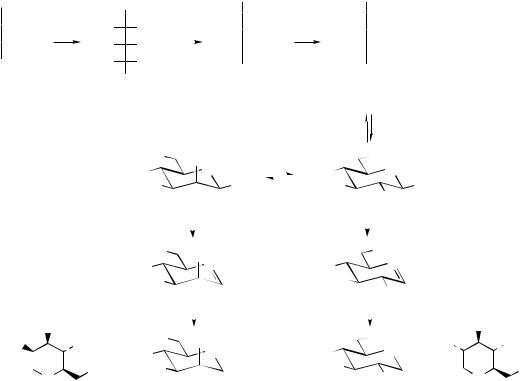
modifications such as imine formation, the product will then be a polyhydroxy-piperidine or pyrrolidine. Any confusion with ornithine/lysine-derived alkaloids (see pages 292, 307) should be dispelled by the characteristic polyhydroxy substitution. The piperidine structures deoxynojirimycin and deoxymannojirimycin (Figure 8.22) from Streptomyces subrutilis are good examples.
The pathways to deoxynojirimycin and deoxymannojirimycin (Figure 8.22) start from the ketosugar fructose, which is aminated and then oxidized to mannojirimycin. This can then form a cyclic aminohemiacetal. Dehydration to the imine can follow, and reduction yields deoxymannojirimycin. Nojirimycin is an epimer of mannojirimycin, and analogous modifications then give deoxynojirimycin. Deoxynojirimycin is found in various strains of Streptomyces and Bacillus, as well as some plants, e.g. Morus spp. (Moraceae), and is attracting considerable attention in the search for anti-HIV agents. This and related structures are inhibitors of glycosidase

478
CHO
H OH HO
OH HO H
H
H OH H
OH H OH CH2OH
D-glucose
CARBOHYDRATES
|
|
|
transamination |
||
|
CH2OH |
|
|
||
|
|
|
O |
|
H2N |
|
|
|
|||
HO |
|
|
H |
|
HO |
H |
|
|
OH |
|
H |
|
|
|
|||
H |
|
|
OH |
|
H |
|
CH2OH |
|
|
||
D-fructose
CH2OH
 H
H
 H O
H O
 OH
OH
 OH
OH
CH2OH
HO |
epimerization |
|||||
HO |
||||||
|
||||||
HO |
NH |
|||||
|
|
|
|
|
||
HO |
OH |
|||||
|
nojirimycin |
|||||
CH2OH
H2N H
H
HO H
H
H OH
OH
H OH
OH
CHO
mannojirimycin
nucleophilic addition of amino on to carbonyl
|
OH |
HO |
NH |
|
|
HO |
OH |
|
HO |
– H2O |
|
– H2O |
|
|
|
|
|
|
HO |
HO |
|
OH |
|
|
|
|
|
|
|
|
N |
|
|
||
|
|
|
HO |
|
N |
HO |
|
|
|
|
|
|
|
|
|
|
|
||
|
|
|
HO |
|
|
HO |
|
|
|
|
|
|
|
|
|
|
HO |
|
|
|
|
|
|
|
reduction |
|
reduction |
|
|
|
OH |
|
HO |
|
|
|
OH |
|
OH |
HO |
OH |
|
HO |
|
HO |
OH |
|||
|
|
|
NH |
||||||
|
|
≡ |
HO |
|
NH |
HO |
|
|
|
|
|
|
|
≡ |
|
|
|||
|
OH |
HO |
|
|
HO |
|
OH |
||
|
|
|
|
HO |
|
||||
|
N |
|
|
|
|
|
|
N |
|
|
H |
|
deoxynojirimycin |
deoxymannojirimycin |
|
H |
|||
Figure 8.22
enzymes (compare indolizidine alkaloids such as castanospermine, page 310). By altering the constitution of glycoproteins on the surface of the virus, such compounds interfere with the binding of the HIV particle to components of the immune system.
AMINOGLYCOSIDES
The aminoglycosides form an important group of antibiotic agents and are immediately recognizable as modified carbohydrate molecules. Typically, they have two or three uncommon sugars attached through glycoside linkages to an aminocyclitol, i.e. an amino-substituted cyclohexane system, which also has carbohydrate origins. The first of these agents to be discovered was streptomycin (see Figure 8.25) from Streptomyces griseus, whose structure contains the aminocyclitol streptamine (Figure 8.23), though both amino
groups are bound as guanidino substituents making streptidine. Other medicinally useful aminoglycoside antibiotics are based on the aminocyclitol
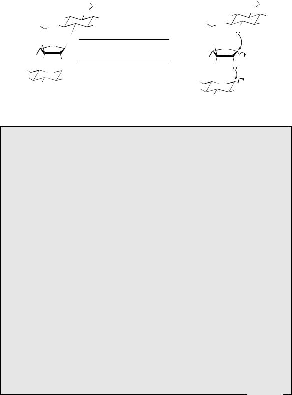
2-deoxystreptamine (Figure 8.24), e.g. gentamicin C1 (see Figure 8.27) from Micromonospora purpurea. Streptamine and 2-deoxystreptamine are both derived from glucose 6-phosphate. The route to the streptamine system can be formulated to involve oxidation (in the acyclic form) of the 5-hydroxyl, allowing removal of a proton from C-6 and generation of an eno-
late |
anion |
(Figure 8.23). |
The |
cyclohexane |
ring |
is then formed by attack |
of |
this enolate |
anion |
||
on |
to the |
C-1 carbonyl. |
Reduction and hydrol- |
||
ysis of the phosphate produces myo-inositol. The amino groups as in streptamine are then introduced by oxidation/transamination reactions. Streptidine incorporates amidino groups from arginine, by nucleophilic attack of the aminocyclitol amino group on to the imino function

|
|
|
|
|
|
AMINOGLYCOSIDES |
|
|
|
|
479 |
||||
|
|
oxidation of |
|
|
formation of |
|
aldol-type |
|
|
||||||
|
OP |
open-chain form |
OP |
enolate anion |
reaction |
|
O |
||||||||
HO |
O |
NAD+ |
|
O |
|
|
|
O |
OP |
|
|||||
HO |
|
|
|
HO |
HO |
OP |
|||||||||
|
|
|
|
|
|
|
|
||||||||
HO |
HO |
OH |
|
|
HO |
|
O |
|
|
HO |
O |
|
|
HO |
OH |
|
|
|
|
|
HO |
|
|
|
HO |
|
|
|
|
HO |
|
|
D-glucose 6-P |
|
|
|
|
|
|
|
|
|
|
|
|
|
|
|
|
|
|
NH |
|
oxidations |
|
|
|||||||
|
|
|
|
|
|
|
transaminations |
|
|
||||||
|
|
|
|
HN NH2 |
|
ATP |
|
|
|||||||
|
|
HO |
|
|
OH |
L-Arg |
HO |
OH |
|||||||
|
|
|
|
|
|
|
|
|
|
|
|||||
|
H2N |
|
HN |
OH |
|
|
|
|
|
|
HO |
OH |
|||
|
|
|
|
|
|||||||||||
|
|
|
|
HO |
|
|
|
|
|
|
|
|
|
HO HO |
|
|
|
|
|
|
|
several steps: amino groups are |
|||||||||
|
|
NH |
streptidine |
|
myo-inositol |
||||||||||
|
|
|
introduced by transamination; |
|
|||||||||||
|
|
|
|
|
|
||||||||||
|
|
|
|
|
|
|
the substrates for N-alkylation |
|
|
||||||
|
|
|
|
|
|
|
are aminocyclitol phosphates, |
|
|
||||||
H2N |
|
|
|
|
|
hence requirement for ATP |
|
|
|||||||
|
|
|
|
|
|
|
|
|
|
|
|
|
|
||
HO |
|
|
|
OH |
|
|
|
|
|
|
|
|
|
|
|
H2N |
|
HO |
OH |
|
|
|
|
|
|
N-alkylation via nucleophilic |
|||||
|
|
|
|
|
|
|
|
|
|
||||||
|
|
|
|
|
|
|
|
|
|
attack of amine on to imine; the |
|||||
|
streptamine |
|
|
|
|
|
|
|
|
||||||
|
|
|
|
|
|
|
|
|
other product is L-ornithine |
||||||
NADH
HO
 OP HO
OP HO


 OH
OH
HO HO myo-inositol 1-P
|
|
NH |
H |
|
|
|
|
|
|
|
CO2H |
H2N N |
|
||||
|
|
||||
|
|
|
H |
|
NH2 |
|
|
|
|
L-Arg |
|
|
|
NH2 |
|
||
|
|
|
|
||
|
|
|
|
||
Figure 8.23
|
|
|
|
oxidation of |
|
|
E2 elimination favoured by |
|
reduction of carbonyl |
|
|
|
||||||||||||
|
|
|
|
4-hydroxyl |
|
|
formation of conjugated system |
|
restoring 4-hydroxyl |
|
|
|
||||||||||||
|
4 |
OP |
|
|
|
|
|
|
O |
OP |
|
|
|
O |
|
|
|
|
|
|
|
|
|
|
HO |
O |
|
NAD+ |
O |
– HOP |
O |
NADH |
HO |
|
|
O |
|||||||||||||
|
|
|
|
|
|
|
||||||||||||||||||
HO |
|
|
|
OH |
|
|
|
|
HO |
|
OH |
|
|
HO |
|
OH |
|
|
|
HO |
|
|
OH |
|
|
|
|
|
|
|
|
|
|
|
|||||||||||||||
|
|
HO |
|
|
|
|
|
|
|
|
HO |
|
|
|
|
HO |
|
|
|
opening of |
HO |
|||
|
D-glucose 6-P |
|
|
|
|
|
|
|
|
|
|
|
|
|
|
|||||||||
|
|
|
|
|
|
|
|
|
|
|
|
hemiacetal ring |
|
|
|
|||||||||
|
|
|
|
|
|
|
|
|
|
|
|
|
|
|
|
|
|
|
|
|
|
|
||
|
|
|
|
oxidation and |
|
|
|
|
aldol-type reaction |
generates enolate |
|
|
||||||||||||
|
|
|
|
|
|
|
|
|
||||||||||||||||
|
|
|
|
|
|
|
|
anion |
|
|
|
|||||||||||||
|
|
|
|
transamination |
|
|
|
|
|
|
|
|||||||||||||
|
|
|
|
|
|
|
|
|
|
|
|
|
|
|
|
|||||||||
H2N 3 |
|
|
O |
|
|
|
|
O |
|
|
|
|
|
|
|
|
||||||||
2 |
|
reactions |
|
|
|
|
|
|
|
|
|
|
|
|
|
O |
||||||||
HO 4 |
|
|
|
|
|
|
|
|
HO |
|
|
|
HO |
|
|
|
|
|
HO |
|
|
|||
|
|
1 |
|
|
|
|
|
|
|
|
|
|
|
|
|
|
|
|
|
|||||
HO |
5 |
6 |
NH2 |
|
|
HO |
OH |
HO |
|
O |
|
HO |
|
|
O |
|||||||||
|
HO |
|
|
|
|
|
|
|
|
|
HO |
|
|
HO |
|
|
|
|
HO |
|||||
|
|
|
|
|
|
|
|
|
|
|
|
|
|
|
|
|
||||||||
2-deoxystreptamine |
|
|
2-deoxy-scyllo-inosose |
|
|
|
|
|
|
|
|
|
|
|||||||||||
Figure 8.24
of arginine (Figure 8.23). However, streptamine itself is not a precursor, and the first guanidino side-chain is built up before the second amino group is introduced. The N -alkylation steps also involve aminocyclitol phosphate substrates. The biosynthesis of 2-deoxystreptamine shares similar features, but the sequence involves loss of the oxygen function from C-6 of glucose 6-phosphate in an elimination reaction (Figure 8.24). The elimination is facilitated by oxidation of the 4-hydroxyl, which thus allows a conjugated enone to develop in the elimination step, but the original hydroxyl is reformed by reduction after the elimination. The cyclohexane ring is then formed by attack of an enolate anion on to the C-1 carbonyl giving a tetrahydroxy-
cyclohexanone, and transamination reactions allow formation of 2-deoxystreptamine. The pathway in Figure 8.24 is remarkably similar to that operating in the biosynthesis of dehydroquinic acid from the seven-carbon sugar DAHP in the early part of the shikimate pathway (see page 122).
The other component parts of streptomycin, namely L-streptose and 2-deoxy-2-methylamino-L- glucose (N -methyl-L-glucosamine) (Figure 8.25), are also derived from D-glucose 6-phosphate, though the detailed features of these pathways will not be considered further. Undoubtedly, these materials are linked to streptidine through stepwise glycosylation reactions via their nucleoside sugars (Figure 8.25).

480 |
|
|
|
|
|
|
|
|
CARBOHYDRATES |
|
|
|
|
|
|
|
|
|
|
|
|
|
|
|
H2N |
|
|
|
|
|
|
|
|
|
H2N |
||||
|
|
|
|
|
|
|
NH |
|
|
|
|
|
|
|
|
|
|
NH |
|
|
|
|
|
|
|
|
|
|
|
|
|
|
|
|
|
|
|
||
|
|
|
|
|
|
|
|
|
|
|
|
|
|
|
|
|
|
||
|
|
|
|
|
HN |
|
|
|
|
|
|
|
|
HN |
|||||
|
|
|
2 |
|
6 |
|
|
|
|
|
|
||||||||
|
|
|
|
|
|
|
|
|
|
|
|||||||||
|
|
|
|
|
|
|
|
HO |
|
|
|
|
|
|
OH |
||||
|
|
|
HO |
|
1 |
|
|
OH |
|
H2N |
|
|
|
|
|
|
|||
|
|
|
|
|
|
|
streptidine |
HN |
|
|
|
|
|
OH |
|||||
H2N |
HN |
4 |
|
|
OH |
HO |
|
|
|
||||||||||
|
|
|
3 |
|
5 |
|
|
|
|
|
|
|
|
|
|
|
|
||
|
|
|
|
|
|
|
|
|
|
|
|
|
|
|
|
||||
|
|
|
|
|
|
|
|
|
|
NH |
|
|
|
|
|
|
|
||
NH |
O |
|
|
|
|
|
|
|
|
|
|
|
|
|
|||||
|
|
|
|
|
|
|
|
|
|
|
|
|
|
|
|
||||
OHC |
|
|
|
|
|
|
|
OHC |
|
|
|
|
|
|
|
|
|||
O |
|
|
|
|
|
|
|
|
|
|
|
|
|
|
|
||||
|
|
1′ |
|
|
|
|
|
L-streptose |
O |
|
|
|
|
|
|
|
|||
Me |
|
|
|
|
|
|
|
|
|
|
|
|
|
|
|
||||
|
|
|
|
|
|
|
|
Me |
|
O |
|
|
UDP |
||||||
HO |
|
O |
|
|
|
|
|
|
|
|
|
||||||||
|
|
|
|
|
|
|
HO |
|
OH |
|
|
|
|
|
|||||
HO |
|
|
|
|
|
|
|
|
|
|
|
|
|
|
|||||
O |
1″ |
|
|
|
|
|
|
|
|
|
|
|
|
|
|
|
|
||
|
|
|
|
|
|
N-methyl-L-glucosamine |
|
|
|
|
|
|
|
|
|
|
|||
HO |
NHMe |
|
|
|
|
|
|
|
|
|
|
|
|
|
|||||
|
|
|
|
HO |
|
|
|
|
|
|
|
|
|||||||
|
3″ |
|
(2-deoxy-2-methylamino-L-glucose) |
|
O |
|
|
UDP |
|||||||||||
HO |
|
|
|
|
|
O |
|
|
|||||||||||
|
|
|
|
|
|
|
|
HO |
|
|
|||||||||
|
|
|
|
|
|
|
|
|
|
|
NHMe |
||||||||
|
|
|
|
|
|
|
|
|
|
|
|
|
|||||||
streptomycin |
HO |
|
Figure 8.25
Aminoglycoside Antibiotics
The aminoglycoside antibiotics have a wide spectrum of activity, including activity against some Gram-positive and many Gram-negative bacteria. They are not absorbed from the gut, and for systemic infections must be administered by injection. However, they can be administered orally to control intestinal flora. The widespread use of aminoglycoside antibiotics is limited by their nephrotoxicity, which results in impaired kidney function, and by their ototoxicity, which is a serious side-effect and can lead to irreversible loss of hearing. They are thus reserved for treatment of serious infections where less toxic antibiotics have proved ineffective. The aminoglycoside antibiotics interfere with protein biosynthesis by acting on the smaller 30S subunit of the bacterial ribosome. Streptomycin is known to interfere with the initiation complex, but most agents block the translocation step as the major mechanism of action. Some antibiotics can also induce a misreading of the genetic code to yield membrane proteins with an incorrect amino acid sequence leading to altered membrane permeability. This actually increases aminoglycoside uptake and leads to rapid cell death.
Bacterial resistance to the aminoglycoside antibiotics has proved to be a problem, and this has also contributed to their decreasing use. Several mechanisms of resistance have been identified. These include changes in the bacterial ribosome so that the affinity for the antibiotic is significantly decreased, reduction in the rate at which the antibiotic passes into the bacterial cell, and plasmid transfer of extrachromosomal R-factors. This latter mechanism is the most common and causes major clinical problems. Bacteria are capable of acquiring genetic material from other bacteria, and in the case of the aminoglycosides this has led to the organisms becoming capable of producing enzymes that inactivate the antibiotic. The modifications encountered are acetylation, adenylylation, and phosphorylation. (Note adenylic acid = adenosine 5 -phosphate). The enzymes are referred to as AAC (aminoglycoside acetyltransferase), ANT (aminoglycoside nucleotidyltransferase) (sometimes AAD (aminoglycoside adenylyltransferase)), and APH (aminoglycoside phosphotransferase). They differ with respect to the reaction catalysed, the position of derivatization (see numbering scheme in gentamicin, Figure 8.26), and the range of substrates attacked. Thus, some clinically significant inactivating enzymes are
(Continues)

AMINOGLYCOSIDES |
481 |
(Continued )
OH
4″ |
5″ |
|
O |
|
|
|
|
2′ |
|
4′ |
R1 |
||
Me |
|
|
|
H2N |
|
3′ |
|
|
|||||
|
2″ |
1″ |
|
|
|
|
|
|
|
||||
MeHN |
|
|
|
|
|
1′ O |
|
6′ |
NHR2 |
||||
3″ |
HO |
6 HO |
|
4 |
5′ |
||||||||
|
5 |
|
|
|
|||||||||
|
O |
|
|
|
|
|
|||||||
|
|
|
|
O |
|
|
|
|
|
|
|
|
|
|
|
|
|
H2N |
|
2 |
|
NH2 |
|
|
|
|
|
|
|
|
|
|
|
|
|
|
|
||||
|
|
|
|
1 |
|
|
3 |
|
|
|
|
|
|
L-garosamine |
2-deoxy- |
|
|
D-purpurosamine |
|||||||||
|
|
||||||||||||
|
|
||||||||||||
streptamine |
|
|
|||||||||||
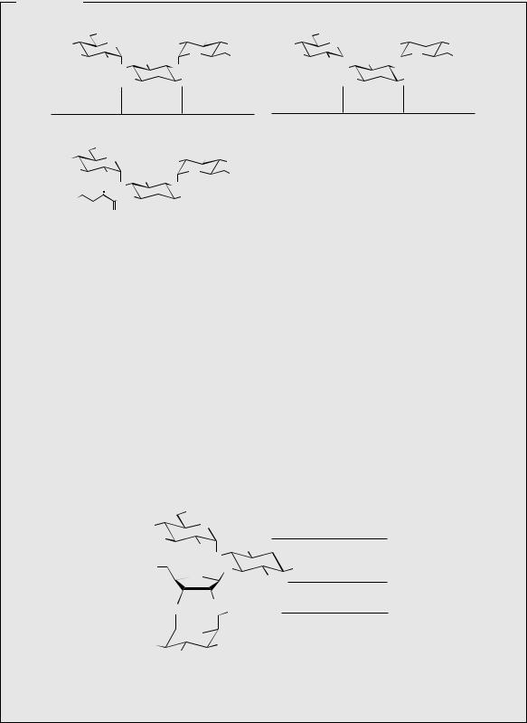
R1 = Me, R2 = Me, gentamicin C1
R1 = Me, R2 = H, gentamicin C2
R1 = H, R2 = H, gentamicin C1a
Figure 8.26
usual sites for derivatization of substituents in aminoglycoside antibiotics by inactivating enzymes
not applicable in case of gentamici
•AAC(3) and AAC(6 ), which acetylate the 3- and 6 -amino functions respectively in gentamicin, tobramycin, kanamycin, neomycin, amikacin, and netilmicin,
•ANT(2 ), which adenylylates the 2 -hydroxy group in gentamicin, tobramycin, and kanamycin,
•APH(3 ), which phosphorylates the 3 -hydroxyl in neomycin and kanamycin, and
•APH(3 ), which phosphorylates the 3 -hydroxyl of streptomycin.
Other changes which may be imparted include acetylation of groups at position 2 , adenylylation of position 4 substituents, and phosphorylation of the position 2 substituent. Position 6 in the streptamine portion of streptomycin is also susceptible to adenylylation and phosphorylation.
Aminoglycoside antibiotics are produced in culture by strains of Streptomyces and Micromonsopora. Compounds obtained from Streptomyces have been given names ending in -mycin, whilst those from Micromonospora have names ending in -micin.
Streptamine-containing Antibiotics
Streptomycin (Figure 8.25) is produced by cultures of a strain of Streptomyces griseus, and is mainly active against Gram-negative organisms. Because of its toxic properties it is rarely used in modern medicine except against resistant strains of Mycobacterium tuberculosis in the treatment of tuberculosis.
Spectinomycin (Figure 8.27) is not strictly an aminoglycoside, but its structure does contain a modified streptamine portion linked by a glycoside bond to a deoxy sugar. It is sometimes written as a ketone at position 4, though this exists as a hydrate as shown in Figure 8.27. Spectinomycin is produced by cultures of Streptomyces spectabilis, and although it displays a broad antibacterial spectrum, it is only used against Neisseria gonorrhoea for the treatment of gonorrhoea where the organism has proved resistant to other antibiotics. It is known to inhibit protein biosynthesis on the 30S ribosomal subunit, but does not appear to cause any misreading of the genetic code.
(Continues)

482 CARBOHYDRATES
(Continued )
HO
NHMe |
|
|
OH |
O |
|
MeHN |
|
|
HO |
|
|
O |
|
OH |
HO |
4 |
|
|
O |
|
|
|
|
spectinomycin Me
Figure 8.27
2-Deoxystreptamine-containing Antibiotics
Gentamicin is a mixture of antibiotics obtained from Micromonospora purpurea. Fermentation yields a mixture of gentamicins A, B, and C, from which gentamicin C is separated for medicinal use. This is also a mixture, the main component being gentamicin C1 (Figure 8.26) (50–60%), with smaller amounts of gentamicin C1a and gentamicin C2. These three components differ in respect to the side-chain in the purpurosamine sugar. Gentamicin is clinically the most important of the aminoglycoside antibiotics, and is widely used for the treatment of serious infections, often in combination with a penicillin when the infectious organism is unknown. It has a broad spectrum of activity, but is inactive against anaerobes. It is active against pathogenic enterobacteria such as Enterobacter, Escherichia, and Klebsiella, and also against Pseudomonas aeruginosa. Compared with other compounds in this group, its component structures contain fewer functionalities that may be attacked by inactivating enzymes, and this means gentamicin may be more effective than some other agents.
Sisomicin (Figure 8.28) is a dehydro analogue of gentamicin C1a, and is produced by cultures of Micromonospora inyoensis. It is used medicinally in the form of the semi-synthetic N-ethyl derivative netilmicin (Figure 8.28), which has a similar activity to gentamicin, but causes less ototoxicity.
The kanamycins (Figure 8.29) are a mixture of aminoglycosides produced by Streptomyces kanamyceticus, but have been superseded by other drugs. Amikacin (Figure 8.29) is a semisynthetic acyl derivative of kanamycin A, the introduction of the 4-amino-2-hydroxybutyryl group helping to protect the antibiotic against enzymic deactivation at several positions, whilst
OH |
|
|
|
Me |
O |
H2N |
|
|
|||
MeHN |
|
HO |
O |
|
HO |
NH2 |
|
|
O |
||
|
|
O |
|
|
|
RHN |
NH2 |
|
|
2-deoxy- |
dehydro- |
L-garosamine |
streptamine |
purpurosamine C |
|
R = H, sisomicin
R = Et, netilmicin
Figure 8.28
(Continues)

AMINOGLYCOSIDES |
483 |
(Continued )
|
OH |
|
OH |
|
OH |
|
3′ |
|
|
|
O |
R |
|
O |
NH2 |
|
|||
HO |
OH |
HO |
OH |
||||||
|
|
|
|||||||
H2N |
|
HO |
O |
H2N |
|
HO |
O |
NH2 |
|
|
HO O |
NH2 |
|
HO O |
|
||||
|
O |
|
|
O |
|
|
|||
|
H2N |
NH2 |
|
H2N |
NH2 |
|
|||
3-amino-3-deoxy- |
|
6-amino-6-deoxy- |
3-amino-3-deoxy- |
2-deoxy- |
|
|
|||
2-deoxy- |
D-glucose |
D-nebrosamine |
|||||||
D-glucose |
streptamine |
(in kanamycin A) |
D-glucose |
streptamine |
|||||
|
R = OH, kanamycin A |
|
|
tobramycin |
|
|
|||
|
R = NH2, kanamycin B |
|
|
|
|
|
|||
|
OH |
|
OH |
|
|
|
|
|
|
HO |
O |
HO |
|
|
|
|
|
||
OH |
|
|
|
|
|
||||
H2N |
|
|
|
|
|
|
|||
HO |
HO |
O |
|
|
|
|
|
||
|
NH2 |
|
|
|
|
|
|||
|
HO O |
O |
NH2 |
|
|
|
|
|
|
H2N |
HN |
|
|
|
|
|
|||
|
|
|
|
|
|
|
|
||
|
O |
amikacin |
|
|
|
|
|
|
|
Figure 8.29
still maintaining the activity of the parent molecule. It is stable to many of the aminoglycoside inactivating enzymes, and is valuable for the treatment of serious infections caused by Gram-negative bacteria which are resistant to gentamicin. Tobramycin (Figure 8.29) (also called nebramycin factor 6) is an analogue of kanamycin B isolated from Streptomyces tenebrarius, and is also less prone to deactivation in that it lacks the susceptible 3 -hydroxyl group. It is slightly more active towards Pseudomonas aeruginosa than gentamicin, but shows less activity against other Gram-negative bacteria.
Neomycin is a mixture of neomycin B (framycetin) (Figure 8.30) and its epimer neomycin C, the latter component accounting for some 5–15% of the mixture. It is produced by cultures of Streptomyces fradiae, and, in contrast to the other clinically useful aminoglycosides described, contains three sugar residues linked to 2-deoxystreptamine. One of these is the common sugar D-ribose. Neomycin has good activity against Gram-positive and Gramnegative bacteria, but is very ototoxic. Its use is thus restricted to oral treatment of intestinal infections (it is poorly absorbed from the digestive tract) and topical applications in eyedrops, eardrops, and ointments.
|
NH2 |
|
|
|
HO |
O |
|
D-neosamine |
|
|
|
|||
HO |
|
NH2 |
|
|
|
H2N |
2-deoxystreptamine |
||
|
O |
|||
HO |
|
NH2 |
||
O |
O |
|||
|
OH |
|
||
|
|
|
||
|
|
OH NH2 |
D-ribose |
|
|
O |
|
||
|
O |
* |
L-neosamine B |
|
H2N |
OH |
|||
HO |
|
|||
|
|
|
||
|
neomycin B (framycetin) |
|
||
(neomycin C is epimer at *)
Figure 8.30

484 |
|
|
|
|
|
|
|
|
|
|
|
|
|
CARBOHYDRATES |
|
|
|
|
|
||
|
|
|
|
|
|
dTDP-D-glucose |
|
|
H2N |
|
O |
|
|
|
|
|
|
||||
|
|
|
|
|
|
|
|
|
|
|
|
|
|
|
|
|
|||||
|
|
|
|
|
|
|
|
|
HO |
|
|
|
Schiff base formation; this |
|
|||||||
|
|
|
|
|
|
|
|
|
|
|
|
|
|
|
|
||||||
|
|
|
|
|
|
|
|
|
|
|
|
|
|
HO |
|
|
|
|
|||
|
|
CH2OH |
|
|
|
|
|
|
|
|
|
|
|
facilitates epimerization at C-2 |
|||||||
|
|
|
|
|
|
|
|
|
|
OPPdT |
|
|
|||||||||
|
|
|
O |
|
|
HO |
CH2OH |
|
dTDP-4-amino- |
HO |
CH2OH |
and 5,6-dehydration |
|
|
|||||||
HO |
|
|
H |
|
|
4,6-dideoxy-D-glucose |
|
|
|
||||||||||||
|
|
|
|
HO |
|
|
|
|
|
|
|
HO |
|
|
|
|
|
||||
H |
|
|
OH |
|
|
|
|
|
|
|
|
|
|
|
5 |
|
|
|
|
|
|
|
|
|
|
|
|
|
|
|
|
|
|
|
2 |
|
|
O |
|
|
|||
|
|
|
|
|
|
|
|
|
|
|
|
|
|
|
|
|
|||||
H |
|
|
OH |
|
|
HO |
|
|
O |
|
|
|
|
N |
|
|
|||||
|
|
|
|
|
|
|
|
|
|
HO |
|
|
|
||||||||
H |
|
|
OH |
|
|
|
OH |
|
|
|
|
|
HO |
|
|
|
|||||
|
|
|
|
|
|
|
|
|
OH |
HO |
|
|
|||||||||
|
CH2OP |
|
|
2-epi-5-epi-valiolone |
|
|
OH |
|
|
OPPdT |
|
|
|||||||||
sedo-heptulose 7-P |
|
|
|
|
|
|
|
|
HO |
|
|
|
maltose |
|
|
||||||
|
|
|
|
|
|
|
|
|
|
|
|
|
|||||||||
|
|
cyclization and other modifications |
|
|
|
|
|
|
|
||||||||||||
|
|
|
|
|
|
|
|
|
|
|
|
||||||||||
|
|
analogous to those in the formation of |
|
|
|
|
|
|
|
|
|
|
|||||||||
|
|
|
|
|
|
|
O |
|
|
|
|||||||||||
|
|
3-dehydroquinic acid from DAHP |
|
|
HO |
N |
|
|
|
||||||||||||
|
|
|
|
|
|
|
|
|
|||||||||||||
|
|
(see Figure 4.1) |
|
|
|
|
|
OH |
H HO |
HO |
OH |
|
|
||||||||
|
CO2H |
|
|
|
|
|
|
|
|
|
O |
|
|
||||||||
|
|
|
|
|
|
|
|
|
|
|
|
|
|
||||||||
|
|
|
|
|
|
|
|
|
|
|
|
O |
|
|
|||||||
|
|
|
|
|
|
|
|
|
|
|
|
|
|
|
|||||||
|
|
|
O |
|
|
HO CO2H |
|
|
OH |
HO |
HO |
OH |
|
||||||||
|
|
|
|
|
|
|
|
|
|
||||||||||||
H |
|
|
H |
|
|
|
|
|
|
O |
|
||||||||||
|
|
|
|
|
|
|
|
|
|
|
|
|
|
|
O |
|
|||||
|
|
|
|
|
|
|
|
|
|
|
|
|
|
|
|
|
|||||
HO |
|
|
H |
|
|
|
|
|
|
|
|
|
|
HO |
|
acarbose |
HO |
HO |
OH |
||
|
|
|
|
|
|
|
|
|
|
|
|
||||||||||
H |
|
|
OH |
|
|
HO |
|
O |
|
|
|
|
|
|
|
|
|
||||
|
|
|
|
|
|
|
HO |
NH2 |
|
|
|
|
|
||||||||
H |
|
|
OH |
|
|
OH |
|
|
|
|
|
|
|
|
|
|
|||||
|
CH2OP |
|
|
3-dehydroquinic |
|
|
OH |
|
|
|
|
|
|
||||||||
|
DAHP |
|
|
acid |
|
|
|
|
|
valienamine |
|
|
|
|
|
||||||
|
|
|
|
|
|
|
|
|
|
|
|
|
|
|
|
|
|||||
Figure 8.31
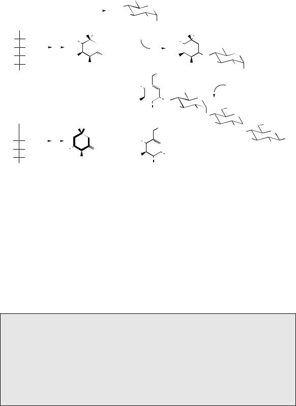
The aminocyclitol found in acarbose (Figure 8.31) is based on valienamine, though this is not a precursor, and the nitrogen is introduced via an imine with the aminosugar, 4-amino-4,6- dideoxyglucose in the form of its deoxyTDP derivative. The cyclitol involved is 2-epi-5-epi- valiolone, and this appears to be produced from the seven-carbon sugar derivative sedo-heptulose 7-phosphate. The reaction sequence is exactly analogous to that seen in the transformation of DAHP into 3-dehydroquinic acid at the beginning of the shikimate pathway (page 122). The valienamine moiety requires subsequent
epimerization and dehydration steps, and these are readily seen to be facilitated by the imine function. Unusually, the two further glucose units are not added sequentially, but via the preformed dimer maltose. Acarbose is produced by strains of Actinoplanes sp., and is of clinical importance in the treatment of diabetes.
The antibiotic lincomycin (Figure 8.32) from
Streptomyces lincolnensis bears a superficial similarity to the aminoglycosides, but has a rather more complex origin. The sugar fragment is termed methyl α-thiolincosaminide, contains a thiomethyl group, and is known to be derived
Acarbose
Acarbose is obtained commercially from fermentation cultures of selected strains of an undefined species of Actinoplanes. It is an inhibitor of α-glucosidase, the enzyme that hydrolyses starch and sucrose. It is employed in the treatment of diabetic patients, allowing better utilization of starchor sucrose-containing diets, by delaying the digestion of such foods and thus slowing down the intestinal release of α-D-glucose. It has a small but significant effect in lowering blood glucose, and is used either on its own, or alongside oral hypoglycaemic agents, in cases where dietary control with or without drugs has proved inadequate. Flatulence is a common side-effect.
