

Reviews in Computational Chemistry
.pdfExample 83
Estimating the Extent of Conformational
Change upon Binding
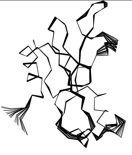
Before applying a certain published method, it is worthwhile to estimate the extent to which large-scale motion will occur for the proteins involved. One approach is to decompose correlated motions of atoms into so-called modes. This terminology stems from normal mode analysis84 of mechanical systems, which is based on evaluation of potential energy derivatives for mechanical systems. Approximate techniques such as essential dynamics85 determine these modes from the positional fluctuations of selected atoms around their mean in an ensemble of conformations by solving an eigenvalue problem. Here, the eigenvectors of the position fluctuation matrix represent independent correlated motions of sets of atoms. As this technique does not require evaluations of potential energy derivatives or extensive energy minimization, it is computationally much more applicable to an approximate prediction of the large-scale motions of a protein than normal mode analysis. Whereas the conformational ensembles in the original essential dynamics technique are generated by MD, the Concoord method developed by de Groot et al.86 perturbs a given protein structure on the basis of experimentally observed positional fluctuations. Thus, a conformational ensemble for use in an essential dynamics study can be generated at negligible computational cost. In Figure 6, the first collective mode of barnase is predicted by analyzing such a Concoord ensemble of conformations. Alternative techniques, such as the detection of motion in a Gaussian network model87 or of independent domain motions,88 differ in how they represent the protein and its potential energy, but represent efficient tools for predicting the large-scale motions of the associating proteins.
In Figure 6, the first essential mode of barnase’s is displayed. One particularly flexible region is located around barnase’s arginine-59 which, from mutational studies,19;89 is known to contribute a large part of the free energy of binding. As this particular region is part of the binding interface, it is not clear whether reasonable predictions for the complex can be obtained by a rigid-body docking approach starting from the unbound structures. Methods to assess qualitatively the extent of large-scale flexibility can help in choosing an appropriate docking approach. In our barnase–barstar demonstration case, the superposition of bound and unbound structures reveals minor overall
˚
backbone changes upon binding; the Ca rmsd is 0.5 A, although local rmsds
˚
of up to 2 A are observed in the barnase–R59 region.
Rigid-Body Docking
When performing rigid-body docking with FTDOCK using the unbound protein structures, without filtering by means of biochemical data, several clusters are proposed by the program as illustrated in Figure 7. The major
84 Protein–Protein Docking
R59
Figure 6 Barnase deformed along the first collective mode computed from perturbed conformations generated by Concoord; see text for details. The location of Arg-59 is indicated.
cluster shown in Figure 7 closely resembles the encounter complex cluster obtained by extensive Brownian dynamics simulation performed by Gabdoulline and Wade.46;63 These diffusional encounter complex structures satisfy the requirement for formation of two native polar contacts at a distance at
˚
ca. 6 A. Formation of these encounter complexes shows that rigid-body docking can provide starting configurations for in-depth studies of the short-range postdiffusional association process.
The best candidate structure from the FTDOCK computations only established 13% of the native contacts, as opposed to 51% when starting

Example 85
Figure 7 The 20 highest ranking putative complexes from the best 100 FTDOCK candidates obtained after fine-grained rigid-body docking of the unbound barnase and barstar structures. Barnase is shown in the middle by dashed lines, surrounded by putative clusters of barstar orientations. The dense cluster to the right contains the correct docking orientation (shown in bold).
from the separated bound conformations (data not shown). Compared to simulations starting from the unbound conformation, the percentage of established native contacts could be improved to 22% when the local loop region around barnase-R59 was deformed to its respective bound conformation while the rest of the structure was held fixed in its unbound conformation.
Although Gabb et al.48 pointed out the shortcomings of their FTDOCK methodology when performing docking experiments with unbound protein structures, the result above shows that backbone structural variability is more important in the interface of the associating proteins. Consequently, a strategy separating backbone and side-chain flexibility might encounter significant difficulties in prediction of a bound complex from the free structures because the relevant small-scale backbone rearrangements should be taken into account explicitly.

86 Protein–Protein Docking
Flexible Docking with Side-Chain Flexibility
To test the impact of localized backbone and side-chain conformational changes when using an interface refinement strategy, we performed constrained MD simulations in dihedral angle space90 (so-called torsion dynamics) of barnase–barstar association from an artificial starting arrangement similar to those resulting from a rigid-body docking approach. In this starting conformation, the two proteins were pulled apart along a line connecting their centers of
˚
mass, leading to an rmsd of the interfacial barstar Ca atoms of 5 A. Using different backbone conformations, only the side chains were allowed to move in this all-atom, molecular dynamics simulation.
Whereas most side-chain optimization techniques described above rely on choosing one of a set of rotamers for a given side chain, the MD-based simulations can sample the full range of side-chain torsional angles. Treating these degrees of freedom as continuous is particularly relevant for so-called hot spot residues88 such as barnase-R59, which contribute a large part to the free energy of binding and do not occupy a standard rotameric state in the bound complex. The favorable energy arising from the nonbonded interactions outweighs the unfavorable torsional energy of the nonstandard rotameric state.
Impact of Side-Chain Conformational Changes
Barnase-R59 has been shown to be involved in an intricate network of water-mediated hydrogen-bonding interactions involving barstar and at least five ordered water molecules buried in the interface. Water molecules are rarely included explicitly in protein–protein docking studies; most approaches utilize implicit solvent approaches. In our torsion dynamics approach, the mechanical properties of the solvent were taken into account by stochastic forces acting on surface protein atoms, and the solvent dielectric properties were represented by a distance-dependent dielectric constant. Thus, we can expect that the use of this approximate model for the solvent will limit the accuracy of predictions of the conformations and interactions of side-chains involved in salt bridges or hydrogen bonds in the solvated interfacial region. As has been pointed out by Bogan and Thorn89 and Xu et al.,33 these residues are often the ones that contribute most to the free energy of binding.
In our simulations, we performed association studies to elucidate their role by selecting pairs of those hot spot residues that established a barnase– barstar contact in the bound complex. In these simulations, we studied what effect the side-chain conformation had on contact formation for certain hot spot residue pairs. A typical result is shown in Figure 8. Starting from the correct rotameric state for certain hot spot residue pairs with the rest of the proteins in their unbound conformations leads to improved contact formation in the initial docking phase. This effect is not observed when performing equivalent simulations with non-hot-spot interfacial residue pairs.

Example 87
Correct contacts [%]
free bn/bs
free bn/bs (bn-Arg59/bs-Glu76 in docked rotamer) free bn/bs (bn-Arg59/bs-Asp35 in docked rotamer) free bn/bs (bn-Arg87/bs-Asp39 in docked rotamer)
80
60
40
20
0
0 |
20 |
40 |
60 |
80 |
100 |
Time [ps]
Figure 8 The impact of hot-spot residue pair conformations on contact formation during docking of barnase (bn) and barstar (bs). Contact formation (as measured by the percentage of native contacts) during the first 100 ps of torsion dynamics simulations is shown; see text for details.
A series of simulations starting from reverse initial conditions (protein structures in bound conformations with hot-spot residue pairs in unbound rotameric states) leads to impaired contact formation during the initial docking phase. These findings indicate, first, that the hot-spot residues can actively hinder or support contact formation; and second, that a side-chain refinement protocol relying on specific side-chain rotameric states may well miss crucial structural detail in the interfacial region.
Impact of Backbone Conformational Changes
From the FTDOCK simulations described above, it is obvious that small localized backbone rearrangements in the protein–protein interface can

88 Protein–Protein Docking
influence the association process. To clarify the extent of this backbone effect, we performed side-chain torsion dynamics simulations using a barnase structure with a deformed loop region surrounding barnase-R59. Starting from the unbound conformation, this loop region was deformed onto the Ca trace of
˚
the respective bound structure. This amounts to a local rmsd of 2 A, compared
˚
to the 0.5-A overall backbone rmsd for the entire structure. Compared to prior simulations with the unbound conformations, an improvement of up to 20% established contacts could be observed for the best in a series of simulations. The difference in docking progress between the bound and unbound starting conformation was almost compensated by introducing the proper backbone conformation in this binding-site loop.
Flexible Docking with Full Flexibility
Although side-chain torsion dynamics simulations can help in understanding the influence of single residues and static backbone conformations on the binding process, they still neglect the effects arising from backbone flexibility. Standard molecular dynamics simulation techniques can be used as a sampling tool to create putative complexes. However, unless simulations are performed with suitable starting configurations close to the docked configuration, a major part of the simulation time will be spent exploring uninteresting regions of conformational space. At the other end of the spectrum, global energy minimization methods can be used to generate complexes while abandoning the physical interpretation of the binding process.
Following an intermediate path, we developed a hybrid simulation method to study the peculiarities of the barnase–barstar binding process. This hybrid method was used to study the short-range association process. Simulations start with orientations resembling encounter complexes, obtained by rigid-body docking governed by long-range electrostatic interactions. This hybrid simulation method is inspired by the genetic algorithm class of algorithms used for global optimization,91,92 in which entire ensembles of candidates are evaluated with respect to an optimality criterion and only promising individuals are propagated to the next generation. In our ‘‘ensemble enriching’’ simulations, multiple copies of barnase interact with the mean field arising from multiple copies of barstar and vice versa. No interaction is simulated between copies of the same protein. In typical simulations, short (10 ps) simulations of 10 barnase and 10 barstar copies are performed with full atomic mobility. After each such simulation, all 100 possible complexes are scored with a heuristic energy function comprising nonbonded interaction energy, pairwise distance potentials, as well as implicit desolvation energy terms. These scores are used to derive an average fitness score for each protein copy in the ensemble. Following this scoring stage, the five lowest scoring conformations in each ensemble are discarded, and the remaining five are duplicated. This effectively leads to an enrichment of the promising candidate

Example 89
conformations while retaining full flexibility and a time interpretation of the binding process. A complete docking simulation consists of several sets of these simulation and enrichment stages.
The results of simulating barnase–barstar association after 100 ps of simple multiple copy MD without ensemble enriching are shown in Figure 9. Whereas contact formation in the simple multiple copy MD simulation has leveled off after the initial 100 ps, ensemble enriching with a linear scoring function is able to shift the ensemble population toward a higher number of established native contacts. Compared to the side-chain torsion dynamics
correct contacts (%)
60
50
40
30
20
10
0 |
|
|
|
|
|
100 |
120 |
140 |
160 |
180 |
200 |
|
|
|
time (ps) |
|
|
Figure 9 Formation of native contacts during a multiple copy MD simulation with and without ensemble enriching after an initial 100 ps multiple copy MD simulation; see text for details. Ensembles that each consist of 10 barnase and 10 barstar copies are simulated, giving rise to 100 putative complexes. The thick lines show the mean and standard deviations, along with the best/worst values for the ensemble enriching method. The superposed thin lines show the corresponding data for the plain multicopy MD simulations without ensemble enriching.

90 Protein–Protein Docking
simulation starting from the unbound conformations of both proteins, an additional 20% of native contacts are gained when treating the simulated proteins as completely flexible. Some barnase copies with large numbers of established contacts exhibit a motion of the barnase–R59 loop region toward its bound conformation.
Although the ensemble enriching multiple copy MD approach is currently too demanding in terms of computational resources to be applied to a large set of protein–protein complexes, it shows that simulations with fully flexible proteins can be useful computational aids when no a priori limitations on flexibility should be made.
FUTURE DIRECTIONS
Computational protein–protein docking can be expected to be a very active field in the near future for several reasons. First, genomics and proteomics studies will provide large amounts of data on protein–protein binding. This binding data will often be of a qualitative kind (protein 1 interacts with protein 2), and computational studies will be necessary for detailed interpretation. Second, large-scale structural genomics studies will mean that 3D structures become available for many more protein targets of therapeutic interest. However, it is anticipated that the structure of a target protein will often be modeled by homology rather than be determined experimentally, and therefore may contain significant structural uncertainties and errors. These errors will be most apparent in loop regions, which can occur at the protein-binding interface (e.g., as in antibody–antigen complexes). Thus, these homology models are a challenge for use in docking studies, and their inaccuracies mean that either low-resolution rigid-body docking strategies or strategies that treat backbone and side-chain flexibility explicitly will be needed. Third, computational advances can be expected in both the sampling and the scoring aspects of docking methods.93
A range of computational docking methods will be required in this context and hierarchical approaches may be adopted so that computations are suited to the type of data available and the questions to be answered. For example, screening of large protein data sets for protein–protein binding requires computationally efficient approaches such as sequence-based methods and low-resolution rigid-body docking. These will be supported by techniques to perform knowledge-based 3D structural searches of protein–protein binding sites.94 When more experimental evidence about a particular complex is available (such as binding free energy differences from mutagenesis studies), the researcher can use these as conditions to filter proposed complexes from a low-resolution rigid-body docking approach. Then, a detailed atomistic model can be constructed and molecular flexibility treated.

Conclusions 91
Computational developments in scoring functions can be expected to focus particularly on solvation. There is presently considerable research effort devoted to the development of new implicit solvation models (see, e.g., Ref. 95) and many of these models should be applicable to docking. Techniques will also be developed to include treatment of explicit water molecules in the docking procedures. Currently, this can only be done during refinement of docked structures. In the small-molecule docking field, ways are being developed to consider interfacial water molecules explicitly during docking.96,97 Related approaches can be expected to be developed for protein– protein docking.
As more computational docking methods are developed, stringent comparisons of docking strategies, as in the Critical Assessment of Structure Prediction (CASP) project (http://predictioncenter.llnl.gov/),98,99 will be needed to evaluate which method is better suited for which problem class. Although CASP does not include a docking section any longer, other forums such as the Critical Assessment of Techniques for Free Energy Evaluation (CATFEE) project (http://uqbar.ncifcrf.gov/ catfee/) may provide objective blind prediction tests for certain aspects of docking strategies. A complementary approach to compare and benchmark docking techniques is to establish a benchmark set of protein–protein docking problems against which new methods can be routinely tested. This benchmark set should contain structurally diverse and energetically well-characterized protein–protein complexes. To test scoring functions, the experimentally determined protein–protein complexes should be distinguished from decoy complexes. Sets of decoy complexes to test scoring functions for rigid-body docking are beginning to be made available.100
CONCLUSIONS
Current protein–protein docking approaches can generate structures
˚
exhibiting rmsds of ca. 1–2 A compared with experimental structures in favorable cases, such as certain enzyme–inhibitor complexes that exhibit only minor structural changes upon binding. Results are usually far less encouraging for cases where structural changes are more prominent, such as in antibody–anti- gen complexes. So far, protein–protein docking programs have not successfully proven themselves able to cope with large-scale structural changes. Intense efforts are underway to integrate flexibility at the backbone level into the current approaches to improve the sampling procedures.
Fueled by the challenges of proteomics and structural genomics, the demand for reliable and fast computational approaches to solve the protein– protein docking problem is likely to increase. With increasing computing power and smarter algorithms, computer docking techniques should be able to meet this demand, although there is much to do.

92 Protein–Protein Docking
REFERENCES
1.ASP atomic solvation parameters, BD Brownian dynamics, DEE dead end elimination, DOF degrees of freedom, EM energy minimization, ES electrostatics, FFT fast Fourier transform, LP linear programming, MCSA Monte Carlo simulated annealing, MM molecular mechanics, PB Poisson–Boltzmann, vdW van der Waals.
2.M. Pellegrini, E. M. Marcotte, M. J. Thompson, D. Eisenberg, and T. O. Yeates, Proc. Natl. Acad. Sci. U.S.A., 96, 4285 (1999). Assigning Protein Functions by Comparative Genome Analysis: Protein Phylogenetic Profiles.
3.E. M. Marcotte, M. Pellegrini, H. L. Ng, D. W. Rice, T. O. Yeates, and D. Eisenberg, Science, 285, 751 (1999). Detecting Protein Function and Protein–Protein Interactions from Genome Sequences.
4.M. Huynen, W. L. B. Snel, and P. Bork, Curr. Opin. Struct. Biol., 10, 366 (2000). Exploitation of Gene Context.
5.P. Uetz, L. Giot, G. Cagney, T. A. Mansfield, R. S. Judson, J. R. Knight, D. Lockshon, V. Narayan, M. Srinivasan, P. Pochart, A. Qureshi-Emili, Y. Li, B. Godwin, D. Conover, T. Kalbfleisch, G. Vijayadamodar, M. Yang, M. Johnston, S. Fields, and J. M. Rothberg, Nature (London), 403, 623 (2000). Comprehensive Analysis of Protein–Protein Interactions in Saccharomyces cerevisiae.
6.B. Ku¨ ster and M. Mann, Curr. Opin. Struct. Biol., 8, 393 (1998). Identifying Proteins and Post-Translational Modifications by Mass Spectrometry.
7.B. Rost, Structure, 6, 259 (1998). Marrying Structure and Genomics.
8.R. Sanchez and A. Sali, Curr. Opin. Struct. Biol., 7, 206 (1997). Advances in Comparative Protein-Structure Modelling.
9.J. Skolnick and J. S. Fetrow, Trends Biotechnol., 18, 34 (2000). From Genes to Protein Structure and Function: Novel Application of Computational Approaches in the Genomic Era.
10.M. J. E. Sternberg, H. A. Gabb, and R. M. Jackson, Curr. Opin. Struct. Biol., 8, 250 (1998). Predictive Docking of Protein–Protein and Protein–DNA Complexes.
11.L. Lo Conte, C. Chothia, and J. Janin, J. Mol. Biol., 285, 2177 (1999). The Atomic Structure of Protein–Protein Recognition Sites.
12.S. Jones and J. M. Thornton, J. Mol. Biol., 272, 133 (1997). Prediction of Protein–Protein Interaction Sites Using Patch Analysis.
13.T. O. Fischmann, G. A. Bentley, T. N. Bhat, G. Boulot, R. A. Mariuzza, S. E. Phillips, D.
Tello, and R. J. Poljak, J. Biol. Chem., 266, 12915 (1991). Crystallographic Refinement
˚
of the Three–Dimensional Structure of the FabD1.3–Lysozyme Complex at 2.5 A Resolution.
14.B. C. Braden, B. A. Fields, and R. J. Poljak, J. Mol. Recognit., 8, 317 (1995). Conservation of Water Molecules in an Antibody–Antigen Interaction.
15.J. Janin, Struct. Fold. Des., 7, R277 (1999). Wet And Dry Interfaces: The Role of Solvent in Protein–Protein and Protein Recognition.
16.D. G. Covell and A. Wallqvist, J. Mol. Biol., 269, 281 (1997). Analysis of Protein–Protein Interactions and the Effects of Amino Acid Mutations on Their Energetics. The Importance of Water Molecules in the Binding Epitope.
17.J. H. Lakey and E. M. Raggett, Curr. Opin. Struct. Biol., 8, 119 (1998). Measuring Protein– Protein Interactions.
18.T. Clackson and J. A. Wells, Science, 267, 383 (1995). Hot Spot of Binding Energy in a Hormone–Receptor Interface.
19.G. Schreiber and A. R. Fersht, J. Mol. Biol., 248, 478 (1995). Energetics of Protein–Protein Interactions: Analysis of the Barnase–Barstar Interface by Single Mutations and Double Mutant Cycles.