

Principles of Enzyme Thermistor Systems
.pdf154 |
S. Grabley · R. Thiericke |
345.Berghöfer-Hochheimer Y, Zurek, C, Langer G, and Munder T (1997) J Cell Biochem 65:184
346.Eigen M, Rigler R (1994) Proc Natl Acad Sci (USA) 91:5740
347.Hogan JC (1997) Nature Biotechnol 15:328
348.Bevan P, Ryder H, Shaw I (1995) TIBTECH 13:115
349.Hurst WJ ed. (1995) Automation in the Laboratory,VCH New York
350.Pharmaceutical Education & Research Institute, Project Management in the Pharmaceutical Industry, 1996
351.Centre for Medicines Research (CMS), Scrip´s Yearbook (several volumes), London
352.Major J (1996) Proceedings of the International Symposium on Laboratory Automation and Robotics, 25
353.Schmid I, Sattler I, Grabley S, Thiericke R (1997) Proceedings International Symposium on Laboratory Automation and Robotics.
354.Beals P (1995) In: Hurst WJ (ed), Automation in the Laboratory,VCH New York, p 109
355.Hurst WJ (1995) In: Hurst WJ (ed), Automation in the Laboratory,VCH New York, p 91
356.Burbaum JJ (1997) Proceedings of the International Symposium on Laboratory Automation and Robotics, in press
357.Elands J (1995) Proceedings of the International Symposium on Laboratory Automation and Robotics, 159
358.Green C (1996) Proceedings of the International Symposium on Laboratory Automation and Robotics, 164
359.Allee C (1996) Laboratory Robotics and Automation 8:307
360.Beese K (1996) Pharmaceutical bioprospecting and synthetic molecular diversity. Draft discussion paper, May 15

Protein Glycosylation: Implications for In Vivo Functions and Therapeutic Applications
Prakash K. Bhatia1, 3 · Asok Mukhopadhyay2
1 National Institute of Immunology, Aruna Asaf Ali Marg, New Delhi-110 067, India 2 National Institute of Immunology, Aruna Asaf Ali Marg, New Delhi-110 067, India.
E-mail: ashok@nii.ernet.in
3 499, Sector 11, Hiranmagri, Udaipur 313001, India
The glycosylation machinery in eukaryotic cells is available to all proteins that enter the secretory pathway. There is a growing interest in diseases caused by defective glycosylation, and in therapeutic glycoproteins produced through recombinant DNA technology route. The choice of a bioprocess for commercial production of recombinant glycoprotein is determined by a variety of factors,such as intrinsic biological properties of the protein being expressed and the purpose for which it is intended, and also the economic target. This review summarizes recent development and understanding related to synthesis of glycans, their functions, diseases, and various expression systems and characterization of glycans. The second section covers processing of N- and O-glycans and the factors that regulate protein glycosylation. The third section deals with in vivo functions of protein glycosylation, which includes protein folding and stability, receptor functioning, cell adhesion and signal transduction. Malfunctioning of glycosylation machinery and the resultant diseases are the subject of the fourth section. The next section covers the various expression systems exploited for the glycoproteins: it includes yeasts, mammalian cells, insect cells, plants and an amoeboid organism. Biopharmaceutical properties of therapeutic proteins are discussed in the sixth section. In vitro protein glycosylation and the characterization of glycan structures are the subject matters for the last two sections, respectively.
Keywords: Glycoprotein, Protein stability, Disease, Expression, Half-life.
1 |
Introduction . . . . . . . . . . . . . . . . . . . . . . . . . . . . . . . |
157 |
2 |
Site and Events of Glycosylation in Eukaryotic Cells . . . . . . . . |
158 |
2.1 |
Entry in the Secretory Route . . . . . . . . . . . . . . . . . . . . . . |
158 |
2.2 |
Early Modifications of Protein . . . . . . . . . . . . . . . . . . . . . |
159 |
2.3 |
Asparagine-Linked Glycosylation . . . . . . . . . . . . . . . . . . . |
160 |
2.3.1 |
Biosynthesis and Processing . . . . . . . . . . . . . . . . . . . . . . |
162 |
2.3.2 |
Factors Regulating Asn-Linked Glycosylation . . . . . . . . . . . . |
166 |
2.4 |
The Biosynthesis of Ser/ Thr-Linked Glycans . . . . . . . . . . . . . |
167 |
2.5 |
Glucosylphosphatidylinositol (GPI) Anchors . . . . . . . . . . . . . |
168 |
3 |
Functions . . . . . . . . . . . . . . . . . . . . . . . . . . . . . . . . |
169 |
3.1 |
Protein Folding and Conformation . . . . . . . . . . . . . . . . . . |
169 |
3.2 |
Protein Stabilization and Structural Integrity . . . . . . . . . . . . |
170 |
3.3 |
Receptor Functioning . . . . . . . . . . . . . . . . . . . . . . . . . . |
172 |
3.4 |
Intracellular Trafficking . . . . . . . . . . . . . . . . . . . . . . . . |
173 |
|
Advances in Biochemical Engineering / |
|
|
Biotechnology,Vol. 64 |
|
|
Managing Editor: Th. Scheper |
|
|
© Springer-Verlag Berlin Heidelberg 1998 |
|

156 |
P.K. Bhatia · A. Mukhopadhyay |
|
3.5 |
Cellular Recognition and Adhesion . . . . . . . . . . . . . . . . . . |
173 |
3.6 |
Receptor Binding and Signal Transduction . . . . . . . . . . . . . . |
174 |
3.7 |
O-Glycosylation Functions . . . . . . . . . . . . . . . . . . . . . . . |
175 |
4 |
Altered Glycosylation Pattern and Diseases . . . . . . . . . . . . . |
176 |
4.1 |
Carbohydrate Deficient Glycoprotein Syndrome (CDGS) . . . . . . |
177 |
4.2 |
Cystic Fibrosis . . . . . . . . . . . . . . . . . . . . . . . . . . . . . . |
177 |
4.3 |
Amyloid Diseases . . . . . . . . . . . . . . . . . . . . . . . . . . . . |
177 |
4.4 |
Lysosomal Storage Diseases . . . . . . . . . . . . . . . . . . . . . . |
178 |
4.5 |
Other Diseases . . . . . . . . . . . . . . . . . . . . . . . . . . . . . . |
178 |
5 |
Choice of Expression Systems . . . . . . . . . . . . . . . . . . . . . |
178 |
5.1 |
Systems Available for Glycoprotein Expression . . . . . . . . . . . . |
178 |
5.1.1 |
Yeasts . . . . . . . . . . . . . . . . . . . . . . . . . . . . . . . . . . . |
180 |
5.1.2 |
Mammalian Cells . . . . . . . . . . . . . . . . . . . . . . . . . . . . |
180 |
5.1.3 |
Insect Cells . . . . . . . . . . . . . . . . . . . . . . . . . . . . . . . . |
182 |
5.1.4 |
Plants . . . . . . . . . . . . . . . . . . . . . . . . . . . . . . . . . . . |
182 |
5.1.5 |
Dictyostelium discoideum . . . . . . . . . . . . . . . . . . . . . . . |
183 |
5.2 |
Protein Glycosylation – Nature of Protein and Host Cells . . . . . . |
183 |
5.3 |
Factors Controlling Glycosylation in Cultured Cells . . . . . . . . . |
184 |
6 |
Biopharmaceutical Properties . . . . . . . . . . . . . . . . . . . . . |
184 |
6.1 |
Antigenicity . . . . . . . . . . . . . . . . . . . . . . . . . . . . . . . |
186 |
6.2 |
Immunogenicity . . . . . . . . . . . . . . . . . . . . . . . . . . . . . |
187 |
6.3 |
Metabolic Clearance and Circulatory Half-Life . . . . . . . . . . . . |
187 |
7 |
Controlled Carbohydrate Remodeling . . . . . . . . . . . . . . . . |
188 |
8 |
Characterization of Sugars . . . . . . . . . . . . . . . . . . . . . . . |
189 |
9 |
Conclusions . . . . . . . . . . . . . . . . . . . . . . . . . . . . . . . |
189 |
10 |
References . . . . . . . . . . . . . . . . . . . . . . . . . . . . . . . . |
196 |
Abbreviations |
|
|
APP |
Acute phase protein |
|
Asn |
Asparagine |
|
ATP |
Adenosine triphosphate |
|
CD2 |
Clusters of diterminant |
|
CG |
Chorionic gonadotropin |
|
CHO |
Chinese hamster ovary |
|
Protein Glycosylation: Implications for In Vivo Functions and Therapeutic Applications |
157 |
|
DolPP |
Dolichol pyrophosphate |
|
Endo F/H/D |
Endopeptidase F/H/D |
|
EPO |
Erythropoietin |
|
ER |
Endoplasmic reticulum |
|
FT |
Fucosyl transferase |
|
Fuc |
Fucose |
|
FSH |
Follicular stimulating hormone |
|
aG |
a-Glucosidase |
|
Glc |
Glucose |
|
Gal |
Galactose |
|
Gnc (GlcNAc) N-acetylglucosamine |
|
|
Glyc T |
Glycosyl transferases |
|
Gn T |
N-acetylglucosaminyl transferase |
|
Gal T |
Galactosyl transferases |
|
GalNAc |
N-acetylgalactosamine |
|
GalNAc T |
N-acetylgalactosaminyl transferase |
|
GPI |
Glucosylphosphatidylinositol |
|
IFNg |
Interferon g |
|
IgG |
Immunoglobulin G |
|
LH |
Luteinizing hormone |
|
Man |
Mannose |
|
a M |
a-Mannosidase |
|
PCR |
Polymerase chain reaction |
|
PNGases |
Peptide N-glycanases |
|
SA |
Sialic acid |
|
ST |
Sialyl transferase |
|
TSH |
Thyroid stimulating hormone |
|
tPA |
Tissue plasminogen activator |
|
UDP |
Uridine diphosphate |
|
Xyl |
Xylose |
|
1 Introduction
To obtain their final structural features and functions, newly formed polypeptides undergo various types of modifications, such as folding, conformation stabilization by disulfide bridges, assembly into homoor hetero-oligomers, acquisition of prosthetic groups, specific proteolytic cleavage and covalent attachment of phosphate, sulfate, fatty acid, complex lipid or sugar groups [1]. The types of modification shown by an individual protein depend on its amino acid sequence, conformation, as well as on cell type and tissue context [2].
Glycoproteins are a conjugated form of proteins containing one or more heterosaccharides covalently bound to the polypeptide chain. These are present in virtually all forms of life and in cell secretions, serum and other body fluids,
158 |
P.K. Bhatia · A. Mukhopadhyay |
connective tissues, and in cell membranes. Glycans make up 10–60% of the molecular weight of glycoproteins. The diverse biological functions that these macromolecules perform include acting as energy sources, as structural components, as key elements in enzymatic catalysis, hormonal control, immunological protection, ion-transport, blood clotting and lubrication, in various molecular recognition processes including bacterial and viral infections, cell adhesion in inflammation and metastasis, differentiation, development and many other intercellular interactions and signal transduction events [3–5].
Oligosaccharides were regarded as compounds completely lacking biological specificity. However, the discovery of the role of protein bound saccharides in biological recognition changed this view. The large structural diversity of protein conjugated glycans, complexity of the biosynthetic pathways, tissue specific and developmentally controlled expression, and plasticity of the structure of complex sugars of glycoproteins in response to pathological conditions lends further support to their importance. Glycans in glycoproteins have diverse biological roles, therefore becoming one of the main topics for discussion. Since the majority of the candidate therapeutic proteins are glycosylated and produced by recombinant DNA technology, alternate systems to express them and characterization of the respective glycan structures have been discussed.
2
Site and Events of Glycosylation in Eukaryotic Cells
The majority of secretory as well as plasma membrane proteins and lysosomal enzymes are glycosylated and have a common biosynthetic origin on the rough ER [6]. Nascent proteins are translocated into the cisternal space of the ER where the signal peptide is cleaved, initial cotranslational folding and formation of disulfide bonds occurs, addition and initial processing of high Man N-linked 14-saccharide core unit (Glc3Man9Gn2) takes place, and finally, for multimeric proteins, oligomerization or subunit assembly is attained before these become competent to be transported out of the ER. The semiprocessed proteins are translocated to the Golgi apparatus, further processed, and then either translocated to the cell surface or packaged into secretory vesicles for secretion [7, 8].
2.1
Entry in the Secretory Route
The majority of proteins to be targeted to the secretory route are initially synthesized as precursors containing a hydrophobic signal being either a cleavable N-terminal peptide [9] or a noncleavable internal sequence located near the N-terminus [10]. Signal peptides of some proteins have been shown to mediate efficient secretion in heterologous environments.
Protein translocation across the ER membrane can occur by two pathways. Most secretory proteins are cotranslationally translocated in contrast to posttranslational translocation destined for other cellular compartments. Transport
Protein Glycosylation: Implications for In Vivo Functions and Therapeutic Applications |
159 |
of precursor proteins across ER membranes occurs most likely through a pro- tein-conducting channel [11]. The signal recognition particle (SRP) and nas- cent-polypeptide-associated complex (NAC), a nonribosomal factor, together provide for fidelity in protein targeting to ER and serve as a versatile targeting chaperone team [12, 13]. Details of the mechanism of protein translocation across membrane are beyond the scope of this review and are not discussed. However readers are advised to see the current literature [14, 15].
2.2
Early Modifications of Protein
The cleavage of the signal peptide by signal peptidase occurs rapidly soon after the translocation of protein across the membrane of rough ER. Prokaryotic signal peptides can be released by eukaryotic signal peptidases and vice versa. Protein folding is facilitated by the resident proteins of ER; some of them are enzymes responsible for disulfide bond formation, isomerization of peptide bonds and glycosylation. Disulfide bond formation is a major rate-limiting step in protein folding,which involves the enzyme protein disulfide isomerase (PDI). The PDI essentially unscrambles intermediates in the protein folding pathway with non-native disulfide bonds [16]. Similarly, cis-trans isomerization of prolylpeptide bonds is catalyzed by peptidyl-prolyl cis-trans isomerase [17]. Other resident proteins are the classes of molecular chaperones, which appear to stabilize protein folding intermediates, prevent competitive aggregation interactions, or promote correct folding [18]. They somehow recognize the kinetically trapped intermediate states of proteins, randomly disrupting and releasing them in less folded states [19]. Their interiors provide a sticky hydrophobic surface that competes with intrachain hydrophobic collapse and helps to pull apart an incorrectly folded protein, allowing opportunities to find pathways leading to the stable native state. Chaperones appear to act sequentially in protein folding pathways.
That folding begins cotranslationally is known for some proteins including IgG, serum albumin and has been suggested for globin molecule [20]. Again, in influenza hemagglutinin, disulfide bond formation and generation of conformational epitopes are cotranslational [21]. After initial modification in ER, some proteins are retained (resident ER proteins) but the majority are exported to final destinations within and outside the cell. Export from ER involves incorporation into transport vesicles that fuse with the next compartment along the secretory pathway. The pathway consists of a series of membrane-bound organelles between which proteins move in a vectorial manner. Specific signals have been identified for the retention/retrieval of ER proteins. Retrieval of soluble ER resident proteins from Golgi is mediated through recognition of amino acid sequence ‘Lys-Asp-Glu-Leu’ by a specific receptor [22], while for transmembrane proteins, the intracytoplasmic dibasic motif plays a similar role [23].
Localization signals of Golgi GlycTs are more complex, and seem conformation dependent. Sequences in the cytoplasmic tails, transmembrane regions and
160 |
P.K. Bhatia · A. Mukhopadhyay |
in luminal regions are important for active Golgi retention [24]. One mechanism for retention is based on protein oligomerization in the environment of a specific Golgi cisterna, as in the case of a-2,6-ST. The oligomer is a disulfide bonded dimer of the enzyme and is catalytically inactive as active site Cys residues participate in dimerization. Under conditions of inadequate supply of donors and/or substrate, the reactive sulfhydryls in the catalytic domain of one molecule may interact with those of another. Only those that are continuously supplied with donor and substrate resist disulfide bond formation and remain active. The disulfide bonded dimer acts primarily as a Gal-specific lectin in the Golgi to retain unsialylated molecules and pass them off to the active ST for sialylation [25].
Proteins that do not reside in ER exit at specialized regions adjacent to the Golgi. Within the Golgi, different subcompartments are distinguishable (cis-, medialand trans-Golgi) on the basis of a distinct content of glycan processing enzymes. Adjacent to the trans-Golgi cisternae is a network of tubules located with coated buds and vesicles that is called the trans-Golgi network (TGN) where sorting of proteins destined to specific intracellular compartments or cell surface domains from constitutively secreted proteins occurs [26]. The secretory pathway involves vesicular transfer to the plasma membrane followed by the secretory event, that is exocytotic discharge of vesicle contents [27].
2.3
Asparagine-Linked Glycosylation
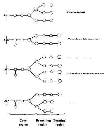
A number of different types of protein glycosylations described in the literature [28, 29] are shown in Table 1. N-Glycans are characterized by a b-glycosidic linkage between a Gn residue and the d amide N of an Asn residue. Though there are many different Asn-glycans, the common feature is the presence of a pentasaccharide (Man3Gn2) core because they all arise from the same biosynthetic precursor lipid-like oligosaccharide that is transferred to the nascent peptide chains. The core can have a Fuc attached to the Gn that is linked to the Asn, and can possess a bisecting Gn residue attached to the central Man of the core. The N-linked glycans fall into three main subgroups as shown in Fig. 1. These are as follows.
1.High-Man structures contain 2–6 additional Man residues at the two terminating Man residues of the core and vary in the number, position and degree of phosphorylation or sulfation of Man residues.
2.Complex-type structures have 2–4 lactosamine (Gal b1,4-Gn) units distributed over the two outer Man residues of the core forming bi-, tri-, or tetraantennary structures. Each of the arms can terminate in sialic acid forming sialyllactosamine. Sulfated lactosamine has been found to substitute sialyllactosamine in some cases, such as the glycoprotein hormones LH, FSH and TSH. Other complex-type structures contain polylactosamine in their outer chains, as in erythroglycan.

Table 1. Classification of glycoproteins and nature of linkage
Type of |
Occurrence |
Glycan - peptide linkage |
Anomeric type |
Stability to acid |
Stability to alkali |
|
glycosylation |
|
|
|
|
|
|
|
Monosaccharide |
Amino acid |
|
|
|
|
|
|
|
|
|
||
|
|
|
|
|
|
|
N-glycosylation |
Widespread |
GlcNAc |
Asn |
b |
+ |
+ |
O-glycosylation |
|
|
|
|
|
|
Mucin |
Mucin, blood group, fetuin, |
GalNAc |
Ser/Thr |
a |
+ |
– |
|
antifreeze glycoproteins |
|
|
|
|
|
Intracellular |
Nucleus and cytosolic |
GlcNAc |
Ser/Thr |
b |
|
|
Proteoglycan |
Proteoglycans |
Xyl core |
Ser |
b |
+ |
– |
Collagen |
Collagens |
Gal |
OH-Lys |
b |
++ |
++ |
Clotting factor |
Factor IX |
Fuc/Glc core |
Ser |
|
|
|
Fungal |
Yeast and Fungal |
Man |
Ser/Thr |
|
|
|
|
glycoproteins |
|
|
|
|
|
Plant |
Plant cell walls, |
Ara |
OH-Pro |
b |
– |
|
|
earthworm, cuticle |
Gal |
Ser |
a |
+ |
– |
GPI anchore |
Cell surface receptors |
|
|
|
|
|
|
|
|
|
|
|
|
Applications Therapeutic and Functions Vivo In for Implications Glycosylation: Protein
161
162 |
P.K. Bhatia · A. Mukhopadhyay |
Oligomannose
Complex : biantennary
Complex : triantennary (solid lines)
Complex : tetraantennary (solid + dotted lines)
Hybrid
Core |
|
Branching |
|
Terminal |
region |
|
region |
|
region |
|
|
|
|
|
|
|
|
|
|
Fig. 1. The basic forms of common N-linked glycans. The N-linked glycans posses three regions of oligosaccharides – core, branching and terminal. All glycans have common core of two N-acetylglucosamine residues and three mannose residues. N-linked glycans are primarily depend on the branching pattern and the type of monosaccharides present in the branching and terminal regions. The branches are distributed over the two terminating core mannose residues. The complex type N-linked glycans can have two to four antennae. In complex and hybrid type N-glycans, the antennae usually terminate in sialic acid or galactose. Sialylation adds the greatest degree of microheterogeneity, however variable core fucosylation also adds heterogeneity to the glycan. ■-GlcNAc (Gn); ▼ -Fuc; ●-Man; ▲ -Gal;  -NueAc (SA)
3.Hybrid-type structures combine the structural features of both the high Man and the complex-type.
2.3.1
Biosynthesis and Processing
Glycosylation begins in the rough ER and proceeds as glycoproteins migrate through the Golgi to their final destination. Asn-glycosylation is a cotranslational event and occurs as the polypeptide is being transferred into the ER, while

Protein Glycosylation: Implications for In Vivo Functions and Therapeutic Applications |
163 |
still in an unfolded state [30]. Initial steps in this pathway, which appear to be generally conserved, involve the synthesis in ER membrane of a dolichol linked precursor glycan and its transfer to an Asn residue of the growing peptide chain. The major steps in phosphodolichol pathway of protein N-glycosylation have been reviewed [31]. The principal pathway for biosynthesis of the dolichollinked oligosaccharides is shown in Fig. 2. The oligosaccharyl transferase (OST) is integral to ER membrane, with active site of the enzyme residing near the membrane on the lumenal side, and transfer only occurs when 12–14 amino acids C-terminal to a sequon have been translocated into the ER lumen [33]. The glycan is added to Asn residue in the tripeptide consensus sequence Asn- X-Ser/Thr (X = any amino acid) and presence of Thr rather than Ser at the hydroxy position favors efficient glycosylation [34]. The OH group of the Thr/Ser has a role in explaining the unique reactivity of this Asn with the DolPP sugar derivative. Secondary structure prediction analysis suggested that these tripeptide consensus sequences have a high probability of occurring in a b-turn or other loop structures, which could serve as recognition sites for enzymes involved in glycosylation [35]. Analysis of occurrence of amino acids at positions around the glycosylated Asn (given the position zero) indicates that Pro is rarely allowed at position +3 and is never observed at position +1 [36].Asp, Glu, Leu and Trp at position +1 are also associated with inefficient glycosylation [37]. This position is preferentially occupied by non-bulky amino acids such as Gly, Val and Ala.
Fig. 2. Pathway of biosynthesis of the dolichol-linked oligosaccharide. ■-UDP: GlcNAc-Uridine diphosphate; ■-PPDol: GlcNAc-Pyrophosphatedolichol; ●-GDP: Man-Guanosine diphosphate; ●-Pdol: Man-Phosphodolichol; ●-Pdol: Glc-Phosphodolichol. (adopted from [32])