

Tools and Applications of Biochemical Engineering Science
.pdf96 |
S. Nath |
6
Molecular Physiological Engineering: a New Frontier
The cell can be considered as a complex network of interacting molecular machines. Traditionally, molecular and cellular processes have been studied by biochemists, microbiologists and molecular biologists, with virtually no input of engineering knowledge. Our research of the past 10 years unambiguously demonstrates that the in vivo dynamic, nonequilibrium mode of operation of biological molecular machines cannot be understood without the application of the principles of thermodynamics, kinetics and transport. The key effects of elastic strain in molecular energy conversion processes cannot be quantitatively described without a sound knowledge of mechanics and dynamics. The application of engineering tools to biology and physiology at the molecular level has led to the development of a new field that I have called “Molecular Physiological Engineering” [15]. Storage of energy within a single molecule (as internal energy) plays a central role in the torsional mechanism of ATP synthesis. Energy storage within single molecules, and its subsequent utilization, via specific mechanisms of the type proposed and detailed in earlier papers and in this work may prove to be one of the great unifying principles of biology: in photoand oxidative phosphorylation, muscle contraction and other related energy transductions. These properties make the above topics very attractive for contemporary interdisciplinary research. Finally, in our view, the aspects dealt with in this work constitute the key elements whose lack of detailed consideration has held back the progress of research in this important field. Thus, it is well known (and generally accepted) that science is indispensable to engineering; however, our research shows that engineering is also indispensable to science, to fundamental science, and this indispensability will, in my opinion, manifest itself even more poignantly in this century as biology becomes increasingly dominated by computation.
Acknowledgement. I am much indebted to my students H. Rohatgi, A. Saha and S. Jain who have greatly contributed to bring our knowledge of the torsional mechanism to this point; their names are to be found in most of our papers on this subject. I thank Dr. N. Pant, in whose classes I sat as a student over many years, for teaching me so much. It is a pleasure to acknowledge Prof. W.-D. Deckwer’s continual encouragement of our approach to bioenergetics. I thank Profs. P. Balaram and M. Saraste for their real help as editors, and Profs. E.H. Battley and L.D. Hansen for their faith in me. I am grateful to the Department of Science and Technology, India, and the All-India Council of Technical Education for funding my research. I am deeply indebted to my father, Prof. M.V. Ranganath for all that he has given me.
References
1.Fiske CH, SubbaRow Y (1929) Science 70:381
2.Lohmann K (1929) Naturwissenschaften 17:624
3.Lipmann F (1941) Adv Enzymol 1:99
4.Penefsky HS, Pullman ME, Datta A, Racker E (1960) J Biol Chem 235:3330
5.Boyer PD (1998) Angew Chem Int Ed Engl 37:2296
6.Slater EC (1953) Nature 172:975
7.Mitchell P (1961) Nature 191:144
The Molecular Mechanism of ATP Synthesis by F1F0-ATP Synthase: A Scrutiny of the Major Possibilities |
97 |
8.Mitchell P (1966) Biol Rev 41:445
9.Williams RJP (1961) J Theor Biol 1:1
10.Williams RJP (1962) J Theor Biol 3:209
11.Boyer PD (1965) In: King TE, Mason HS, Morrison M (eds) Oxidases and related redox systems. Wiley, New York, p 994
12.Boyer PD, Cross RL, Momsen W (1973) Proc Natl Acad Sci USA 70:2837
13.Boyer PD (1993) Biochim Biophys Acta 1140:215
14.Cross RL, Duncan TM (1996) J Bioenerg Biomembr 28:403
15.Nath S (2000) Molecular Physiological Engineering: A New Frontier. 41st Annual Conference of the Association of Microbiologists of India, Jaipur, India, p 3
16.Rohatgi H, Saha A, Nath S (1998) Curr Sci 75:716; erratum (2000) 78:201
17.Nath S, Rohatgi H, Saha A (1999) Curr Sci 77:167
18.Nath S, Rohatgi H, Saha A (2000) Curr Sci 78:23
19.Nath S, Jain S (2000) Biochem Biophys Res Commun 272:629
20.Jain S, Nath S (2000) FEBS Lett 476:113
21.Abrahams JP, Leslie AGW, Lutter R, Walker JE (1994) Nature 370:621
22.Boyer PD (1997) Annu Rev Biochem 66:717
23.Ramasarma T (1998) Curr Sci 74:953
24.Nakamoto RK, Ketchum CJ,Al-Shawi MK (1999) Annu Rev Biophys Biomol Struct 28:205
25.Allison WS (1998) Acc Chem Res 31:819
26.Fillingame RH, Jiang W, Dmitriev OY, Jones PC (2000) Biochim Biophys Acta 1458:387.
27.Bianchet MA, Hullihen J, Pedersen PL,Amzel LM (1998) Proc Natl Acad Sci USA 95:11065.
28.Weber J, Senior AE (1997) Biochim Biophys Acta 1319:19
29.Wilkens S, Capaldi RA (1998) Nature 393:29.
30.Zhou Y, Duncan TM, Cross RL (1997) Proc Natl Acad Sci USA 94:10583
31.Ogilvie I, Aggeler R, Capaldi RA (1997) J Biol Chem 272:16652
32.Sabbert D, Engelbrecht S, Junge W (1996) Nature 381:623
33.Sabbert D, Junge W (1997) Proc Natl Acad Sci USA 94:2312
34.Noji H, Yasuda R, Yoshida M, Kinosita K (1997) Nature 386:299
35.Yasuda R, Noji H, Kinosita K, Yoshida M (1998) Cell 93:1117
36.Shirakihara Y, Leslie AGW,Abrahams JP,Walker JE, Ueda T, Sekimoto Y, Kambara M, Saika K, Kagawa Y, Yoshida M (1997) Structure 5:825
37.Boyer PD (2000) Biochim Biophys Acta 1458:252
38.Löbau S, Weber J, Senior AE (1998) Biochemistry 37:10846
39.Hatefi Y (1993) Eur J Biochem 218:759
39a. Menz RI, Walker JE, Leslie AGW (2001) Cell 106:331
40.Adachi K,Yasuda R, Noji H, Itoh H, Harada Y,Yoshida M, Kinosita K (2000) Proc Natl Acad Sci USA 97:7243
41.Nath S (1994) A fundamental thermodynamic principle for coupling in oxidative phosphorylation. 16th Int Congr Biochemistry and Molecular Biology, New Delhi, India, vol II, p 390
42.Nath S (1998) Pure Appl Chem 70:639
43.Weber J, Senior AE (2000) Biochim Biophys Acta 1458:300
44.Garcia JJ, Capaldi RA (1998) J Biol Chem 273:15940
45.Böttcher B, Gräber P (2000) Biochim Biophys Acta 1458:404
46.Senior AE, Nadanaciva S, Weber J (2000) J Exp Biol 203:35
47.Al-Shawi MK, Ketchum CJ, Nakamoto RK (1997) Biochemistry 36:12961
48.Grubmeyer C, Cross RL, Penefsky HS (1982) J Biol Chem 257:12092
49.Fischer S, Gräber P (1999) FEBS Lett 457:327
50.Hausrath AC, Grüber G, Matthews BW, Capaldi RA (1999) Proc Natl Acad Sci USA 96:13697
51.Possmayer FP, Gräber P (1994) J Biol Chem 269:1896
52.Pänke O, Rumberg B (1996) FEBS Lett 383:196
53.Estabrook W, Holowinski A (1960) J Biophys Biochem Cyt 9:19
54.Mitchell P (1979) Science 206:1148
98 |
S. Nath |
55.Kaim G, Dimroth P (1999) EMBO J 18:4118
56.Jain S, Nath S (2001) Thermochim Acta 378:35
57.Elston T, Wang H, Oster G (1998) Nature 391:510
58.Vik SB, Antonio BJ (1994) J Biol Chem 269:30364
59.Dmitriev OY, Jones PC, Fillingame RH (1999) Proc Natl Acad Sci USA 96:7785
60.Rastogi VK, Girvin ME (1999) Nature 402:263
61.Massari S, Azzone GF (1970) Eur J Biochem 12:301
62.Azzone GF, Massari S (1971) Eur J Biochem 19:97
63.Tedeschi H (1975) FEBS Lett 59:1
64.Tupper JT, Tedeschi H (1969) Science 166:1539
65.Ochoa S (1943) J Biol Chem 151:493
66.Ernster L (1993) FASEB J 7:1520
67.Ferguson SJ (2000) Curr Biol 10:R804
68.Lodish H, Berk A, Zipursky SL, Matsudaira P, Baltimore D, Darnell J (2000) Molecular cell biology, 4th edn. W.H. Freeman, New York, p 647
69.Slater EC, Rosing J, Mol A (1973) Biochim Biophys Acta 292:534
70.Hinkle PC, Kumar MA, Resetar A, Harris DL (1991) Biochemistry 30:3576
71.Lee CP, Gu Q, Xiong Y, Mitchell RA, Ernster L (1996) FASEB J 10:345
72.Zeng AP, Ross A, Deckwer W-D (1990) Biotechnol Bioeng 36:965
73.Stucki JW (1980) Eur J Biochem 109:269
74.Lemasters JJ (1984) J Biol Chem 259:13123
Received: April 2001

Bioreactor Developments for Tissue Engineering Applications by the Example of the Bioartificial Liver
Inka Jasmund1, Augustinus Bader1,2
1Experimental Radiology, Hepatic Tissue Engineering, Medical School Hannover, Carl-Neuberg-Str. 1, 30625 Hannover, Germany
E-mail: jasmund.inka@mh-hannover.de
2National Research Institute for Biotechnology (GBF), Mascheroder Weg 1B, 38124 Braunschweig, Germany
E-mail: Augustinus.Bader@t-online.de, Augustinus.Bader@GBF.de
Dedicated to Prof. Dr. Wolf-Dieter Deckwer on the occasion of his 60th birthday
Tissue engineering is the application of the principles and methods of engineering and the life sciences towards the development of biological substitutes to restore, maintain or improve functions. It is an area which is emerging in importance worldwide. This article is to show the developments in tissue engineering research by the example of the bioartificial liver. As an alternative to liver transplantation, numerous researchers have been working towards the goal of development of a fully functional artificial liver. Liver support systems based on detoxification alone have proven ineffective because they cannot correct biochemical disorders.An effective artificial liver support system should be capable of carrying out the liver’s essential processes, such as synthetic and metabolic functions, detoxification, and excretion. It should be capable of sustaining patients with fulminant hepatic failure and preparing patients for liver transplantation when a donor liver is not readily available. Although several hepatocyte-based liver support systems have been proposed, there is no current consensus on its eventual design configuration.
Keywords. Bioartificial liver, cell culture, hollow fiber bioreactor, flat membrane bioreactor, spheroids
1 |
Introduction . . . . . . . . . . . . . . . . |
. . . . . . . . . . . . . . . . 100 |
2 |
State of the Art . . . . . . . . . . . . . . . . |
. . . . . . . . . . . . . . . 101 |
3 |
Types of Hepatocytes . . . . . . . . . . . . |
. . . . . . . . . . . . . . . 102 |
3.1 |
Cell Lines . . . . . . . . . . . . . . . . . . . |
. . . . . . . . . . . . . . . 102 |
3.2 |
Primary Hepatocytes . . . . . . . . . . . . . |
. . . . . . . . . . . . . . . 103 |
4 |
Bioreactor Design . . . . . . . . . . . . . . |
. . . . . . . . . . . . . . . 103 |
4.1 |
Spheroids . . . . . . . . . . . . . . . . . . . |
. . . . . . . . . . . . . . . 103 |
4.2 |
Hollow Fiber Bioreactors . . . . . . . . . . |
. . . . . . . . . . . . . . . 105 |
4.3 |
Flat Membrane Bioreactor . . . . . . . . . . |
. . . . . . . . . . . . . . . 107 |
5 |
Conclusions . . . . . . . . . . . . . . . . . . |
. . . . . . . . . . . . . . . 107 |
References . . . . . . . . . . . . . . . . . . . . . |
. . . . . . . . . . . . . . . 108 |
|
|
|
Advances in Biochemical Engineering/ |
|
|
Biotechnology, Vol. 74 |
|
|
Managing Editor: Th. Scheper |
|
|
© Springer-Verlag Berlin Heidelberg 2002 |
100 |
I. Jasmund · A. Bader |
1 Introduction
Health care for patients with tissue loss or end-stage organ failure is becoming increasingly expensive. For example the estimated costs in the U.S. for such patients exceed $400 billion p.a. [1]. Transplantation is severely limited by a critical donor shortage, and as a consequence, there has been a rapidly growing interest in the field of tissue engineering - the manufacture of biological substitutes that can restore, maintain or improve tissue function. Tissue engineering offers the possibility of substantial future savings in health care budgets by providing substitutes that are more cost efficient than donor organ transplantations, are life saving, or provide the means of intervention before patients become critically ill.
In the past product development regarding the engineering of hybrid tissues using all cells in an hierarchical and tissue specific arrangement has been plagued by a shortage of bioreactors that could fulfill these expectations. In addition, storage, handling and applications in a clinical environment pose significant hurdles, as live tissues cannot be easily shipped. In contrast, prohibitive costs and time dependent dedifferentiation or infection risks are looming logistical obstacles at present to the shipment of viable artificial organs.
One of the key problems in the in vitro engineering of transplantable bioartificial tissues is the lack of a bioreactor and corresponding bioengineering technology that could solve such limitations [2]. Primary cells need a lead structure supplied in a bioreactor to grow on, no matter what the targeted structure is, it has to provide a unique 3-D matrix structure. The bioreactor should act as a containment, a GMP compatible production unit, and preferably as a transport device with cryostorage capacity. Currently available bioreactor devices cannot fulfill these requirements due to design limitations.
Current technologies do not permit the generation of a full liver in vitro, and full bioartificial organ transplantation still remains an unrealistic dream. As shown previously by Nyberg et al. endogenous retroviruses from animals such as PERV might be transmitted as well [3]. In this context the development of a bioreactor technology to grow organs or parts thereof in vitro represents a clean and working alternative to both, the use of animals as living bioreactors for human embryonal cells and xenotransplantation. Probably, the generated tissue interacts with the human host, i.e. the recipient after transplantation or extracorporeal contact. The extracorporeal liver induces liver regeneration and thus transplantation is not required.
With the goal of mimicking the cellular environment of the native organ, it is fair to assume that pure monolayer cultures are not suitable to reproduce the necessary conditions to induce and maintain differentiation and function of a complex organ [4]. A detailed analysis of the fundamental conditions is therefore necessary to distinguish between a microenvironment that is in a way rather artificial such as the monolayer culture, and a more organotypical 3D situation that contributes to the signal pattern necessary to induce the expression of specific cellular functions. Only with this knowledge, a tissue-like culture device can be constructed and improved to potentially serve as a bioartifi-
Bioreactor Developments for Tissue Engineering Applications by the Example of the Bioartifical Liver |
101 |
cial organ that might be used not only in basic research but also for therapeutic purposes such as artificial organ support.
2
State of the Art
There have already been clinical trials of porcine hepatocyte-based bioartificial livers [5, 6]. However, we believe these systems to represent temporary and short-lived approaches. Compelling evidence from recent experiments show that primary porcine liver cells express and release endogenous retroviral particles that are able to infect human cells. However, long term in vivo investigations of patients previously exposed to porcine tissues over a period of 12 year did not show any porcine endogenous retrovirus (PERV) viremia [7]. Therefore, we consider the further pursuit of porcine bioartificial livers the only solution at present with regard to the cell source. However, as an intermediate term alternative human cell sources are in development [8]. Expansion technologies for human fetal cells may contribute to resolve these limitations in the future.
Hepatocyte-based bioartificial livers will have two major uses: to provide a) short term support for liver-failure patients in the clinic and b) a tool for the pharmaceutical industry to produce drug metabolites for subsequent toxicological studies in vitro.
To be useful to both, clinicians and the pharmaceutical industry, a bioartificial liver will need to maintain a large number of hepatocytes at high cell densities and in a fully differentiated state for prolonged periods of time. Development of such a system has been impeded by three principal problems: a) a requirement for large numbers of cells (>25 · 109); b) loss of liver-specific functions in cultured cells (primary and immortalized); c) nutrient and waste product gradients in high density cultures leading to lowered cell viability and impaired function.
There is a general consensus that human life can be sustained with 10% of normal liver mass [9] or estimated 25 · 109 cells. Therefore, in constructing a bioartificial liver there is an enormous requirement for functional hepatocytes. The use of primary human hepatocytes is limited by the scarcity of human liver tissue and by their poor proliferative potential. Currently available cell lines are limited in that they only display a limited repertoire of liver-specific functions and furthermore, like primary cells, may also lose these during prolonged periods in culture. There is, therefore, a pressing need to develop new human liver cell bioreactor technologies which provide a good proliferative potential and maintain a broad spectrum of liver-specific functions during cultivation time. A new approach is to develop immortalized cell lines by deriving them from human fetal liver cells. These can then be expanded in culture to give the large number of cells required to produce a functional bioartificial liver.
A bioartificial liver will need to maintain a large number of hepatocytes at high cell densities over a prolonged period of time. The metabolic requirements of hepatocytes, particularly the high rates of oxygen consumption, place strin-
102 |
I. Jasmund · A. Bader |
gent demands on the bioreactor system. This problem is exacerbated if the cells are maintained in a 3D matrix, since the matrix material can represent a significant diffusion barrier for nutrients, especially oxygen. Intensive reactor systems are intrinsically inaccessible to conventional monitoring techniques and therefore improvements in design and operation of these systems require new non-invasive methods for assessing reactor performance. A successful bioartificial liver will need to maintain a broad spectrum of liver-specific activities.
3
Types of Hepatocytes
Generally there are two approaches in the incorporation of cells in bioreactors for liver support. One is the use of cell lines [10]. The other is to isolate and use primary hepatocytes from animal livers [11] or human livers [12].
3.1
Cell Lines
Cell lines established from human liver cancer cells can be derived from primary hepatocellular carcinoma or hepatoblastoma cells [10]. For example, the well known HepG2 line was derived from hepatoblastoma cells. Such cells can be employed effectively if the function of normal liver cells has been highly preserved. In practice, however, such cells contain a high proportion of abnormal genetic component, which inhibits their ability to express normal protein synthesis and enzyme activity. Potential problems, such as the loss of liver specific functions and the possibility of metastasis, which could arise from the use of hepatoma cells have not yet been satisfactorily discussed [13].
Artificially immortalized human liver cells would be an ideal source if genetic engineering or other methods could be used to imbue the cells with the highly differentiated functions and proliferative capacity observed in normal cells. At present, cultures of such immortalized cells have not demonstrated longterm viability and the hepatocyte-specific characteristics, such as albumin secretion, disappear quickly. In addition, these cells suffer from a high incidence of malignant change and dedifferentiation. A promising new cell line (HepZ) was created by Werner et al. [14]. The cell line was immortalized through transfection of albumin-promotor-regulated antisense constructions against the negative controlling cell cycle proteins Rb and p53. Furthermore, plasmids including genes coding for the cellular transcription factor E2F and D1 cyclin were cotransfected to overcome the G1-restriction point. The cell line HepZ retains liver-specific functions and is appropriate for a mass cell cultivation. Further investigations have to show whether the biochemical spectrum is sufficient. Research continues to be carried out in this field, however, and promising results are expected in the future [13].
Bioreactor Developments for Tissue Engineering Applications by the Example of the Bioartifical Liver |
103 |
3.2
Primary Hepatocytes
Freshly isolated hepatocytes can be obtained from animal or human liver. Hepatocytes taken from these sources, however, are very vulnerable to contamination and technically difficult to preserve in a fresh state. Although it is not generally possible until now to proliferate human hepatocytes in vitro, the advantages of homology and human-specific functions demand that resources continue to be directed towards the elucidation of the conditions necessary for maintaining viable human hepatocyte cultures.
Methods of collagenase perfusion for hepatocyte isolation were developed on rat livers [15, 16]. Berry and Friend [15] made first experiments with colla- genase-perfusion in rats, essentially comprising three processes: exposure to a medium very low in Ca2+, digestion with collagenase, and gentle mechanical disruption. The two-step procedure [16, 17] was introduced by Seglen as a consequence of his studies of the effects of Ca2+ and collagenase on the isolated perfused liver. The essential feature of the two-step approach is that the liver is first flushed with Ca2+-free medium after which the perfusion medium is changed to one containing Ca2+ and collagenase. This method achieves a good viability and yield in rats.
Because of the need for isolation techniques applicable to other species, Hoogenboom et al [18] investigated the isolation of hepatocytes from porcine livers and developed an enzymatic method with closed-loop and open-loop perfusion stages for the perfusion of liver lobes. We investigated a method to perfuse the whole organ to receive a great amount of liver cells (up to 25 · 109 cells per pig liver) and a viability higher than 90%.
4
Bioreactor Design
The cultivation of hepatocytes in a stationary suspension culture is actually ineffective. The hepatocytes lose their differentiation within hours. An improvement was the attachment culture. Thereby, the cells are cultivated either in selfor microcarrier-induced multicellular aggregates or on membranes. When standard monolayer culture was adequate to maintain the cell viability for 1 to 2 weeks the differentiation was lost after a few days. Different modifications as described beneath allowed the maintenance of differentiation for 2 to 3 weeks.
4.1 Spheroids
It has become increasingly evident that three dimensional rather than monolayer growth is particularly important for maintaining differentiated hepatocyte function in culture. One means of establishing three dimensional hepatocyte growth is the creation of multicellular spheroid aggregates. Sakai et al. have shown that multicellular spheroids can be rapidly created by rotational culture systems [19, 20] or by incubation with a small number of collagen-coated dextran, polystyrene or glass microcarriers as a center for cell aggregation [21].
104 |
I. Jasmund · A. Bader |
Naruse et al. proposed another bioreactor design [22, 23], in which porcine hepatocyte spheroids are immobilized on non-woven polyester fabric. This device allows more direct contact between hepatocytes and perfused medium and improves, therefore, the mass transfer capacity. The non-woven fabric module expressed better metabolic and synthetic functions at 24 hours than a hollow fiber module containing spheroids in suspension culture. Longer term results are not yet available and the immunoexclusion properties of this fabric have not been addressed.
Isolated rat hepatocytes were immobilized in cellulose multiporous microcarriers by Kino et al. [24]. The microcarriers had a pore size of 100 µm and protected the cells from external shear stress. A newly developed stirred tank reactor contained the microcarrier-immobilized hepatocytes. The O2-supply was improved by using an oxygenator. The performance of microcarrier-im- mobilized hepatocytes in the reactor was as good as that in floating culture and they demonstrated good ammonia metabolism.
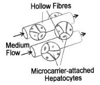
Demetriou et al. [25] described a capillary hollow fiber membrane based bioreactor in which microcarrier-attached hepatocytes are placed in the extracapillary space on the exterior surface of the capillary hollow fiber membranes as shown in Fig. 1. Recent experimental studies with this device have demonstrated its efficacy in animal models. By using cryopreserved microcarrierattached hepatocytes this system offers the convenience of being readily available when needed.
A new concept based on a fluidized bed bioreactor was proposed by Dore et al. [26, 27]. This type of reactor is well known in chemical engineering applications to promote interactions and exchanges between solid and liquid phases.An alginate bead structure provides the hepatocytes with a three-dimensional anchorage framework. Scaling-up only depends on the number of beads employed. The efficacy of this system was tested in vitro and ex vivo in the acute liver failure model of pig [28]. The alginate-embedded hepatocytes maintain most li- ver-specific functions including ammonia removal and urea synthesis in vitro. The animal data suggest that the system arrests the increase of serum ammonia and intracranial pressure significantly in the acute liver failure model of pig.
Fig. 1. Schematic illustration of a hepatocyte bioreactor with microcarrier-attachted hepatocytes. The capillary membranes are perfused with medium. (Modified from Dixit et al. [29])
Bioreactor Developments for Tissue Engineering Applications by the Example of the Bioartifical Liver |
105 |
4.2
Hollow Fiber Bioreactors
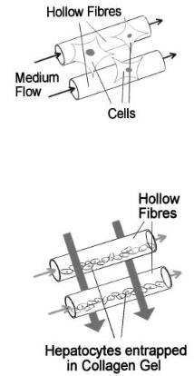
Several innovative membrane-based bioreactor designs have recently been proposed, including that by Sussman et al. [10], which involves the cultivation of hepatoma cells on the exterior surfaces of semipermeable capillary hollow fiber membranes which are bundled together with an enclosing plastic shell (Fig. 2). Required nutrient medium is circulated within the capillaries. After the hepatocytes have attached and formed a mass of liver tissue, the capillary membranes are perfused with the media.
Another modification has been described by Nyberg et al. [30]. In this design primary hepatocytes are entrapped in cylindrical collagen gels inside the lumen of the hollow fibres as shown in Fig. 3. The gel entrapment technique reported by this group enables a large number of hepatocytes to be employed in the bioreactor. Additionally, this technique also provides a three-dimensional frame-
Fig. 2. Hepatoma cells are cultivated on the exterior surfaces of semipermeable capillary hollow fiber membranes. After the hepatocytes have attached and formed a mass of liver tissue, the capillary membranes are perfused with medium. (Modified from Dixit et al. [29])
Blood Flow
Medium
Flow
Fig. 3. The gel-entrapped hepatocytes are located in the intra-luminal capillary space of the device. Culture medium is perfused through and over the gel-entrapped hepatocytes. The host animal’s blood is circulated in the extracapillary compartment between the capillary hollow fibers. (Modified from Dixit et al. [29])