

Tools and Applications of Biochemical Engineering Science
.pdf76 |
S. Nath |
|
Our molecular mechanism for torque generation in the F0 portion of ATP |
synthase [16, 20, 56] considers (and highlights) the roles of both DpH and Dy using two mutually non-colinear half-channels (Fig. 4); torque generation is a result of change in electrostatic potential, D(Dy), brought about by the binding of protons flowing along their concentration gradient to their binding sites in the c-subunit. In our model, entry and exit of protons through the corresponding half-channels are separated by the time it takes the c-rotor to rotate by 15°. We have shown that DpH and Dy are kinetically inequivalent in driving ATP synthesis, and DµH (defined as F Dy – (2.303RT) DpH) is not the true driving force for ATP synthesis [20, 56].
According to the torsional mechanism of ATP synthesis, DpH supplies only half the energy requirement for ATP synthesis; the rest has to be supplied by an independent source of Dy. Further, we have proposed that Dy is created in the vicinity of the ATP synthase enzyme complex, i.e., Dy is localized, and we have postulated a dynamically electrogenic but overall electroneutral ion transport involving membrane-permeable anions (such as succinate) and protons. In order to utilize the energy of the anion gradient, the anion flows through the membrane along its concentration gradient in the vicinity of the enzyme complex and creates a localized Dy (negative inside) of –60 mV. This is followed by the transport of protons (symsequenceport) through the proton half-channel along its concentration gradient. (In general, according to postulates of the torsional mechanism, cations penetrate the inner mitochondrial membrane following a negative internal electrical potential generated by the diffusion of anions.) Hence,the true driving forces for ATP synthesis are DpH and Dy, not DµH . The proton binds to the essential Asp (or Glu) residue of the c-rotor resulting in a change in the overall Dy, D(Dy), of approximately 60 mV (30 mV due to DpH across the entry proton half-channel, and 30 mV due to change in Dy upon proton binding to the Asp residue). This value is estimated based on energy balance and our molecular mechanism which predicts a proton/ATP ratio of 4, and each proton binding and unbinding step to release nearly ~480/4 = ~120 meV of energy in total, i.e., ~60 meV (or equivalently creation of a D(Dy) of ~60 mV) during each elementary step of binding and unbinding. The energy released in this step rotates the c-rotor by 15°. The incoming Asp residue releases its bound proton at the a-c interface, resulting in a D(Dy) of ~60 mV (30 mV due to DpH across the exit proton half-channel, and 30 mV due to change in Dy upon proton unbinding from the Asp residue) and a consequent rotation of the c-rotor by 15°. During each 15° rotation of the c-rotor, the ion-protein interaction energy is transiently stored as a twist in the helices of the ten membrane-bound c subunits. The simultaneous untwisting of the c-subunit helices drives the rotation of the e subunit and the bottom of the g subunit by 15°, and the ion-protein interaction energy is now stored as torsional energy in the g subunit. Hence, the energy transiently stored in DpH and Dy is converted to torsional energy through the process of ion-protein interactions. In this way, through cation binding within the electrostatic potential field created by the transport of anions, the enzyme is able to utilize the energy of both the delocalized DpH and the localized Dy.
Thus, a central feature of the torsional mechanism is the development of a torsional strain in the g subunit due to rotation of the bottom of the central

The Molecular Mechanism of ATP Synthesis by F1F0-ATP Synthase: A Scrutiny of the Major Possibilities |
77 |
Fig. 5. Rotation of the top and bottom of the central stalk as a function of the number of protons as predicted by the torsional mechanism of energy transduction and ATP synthesis [17–20, 56]
stalk of ATP synthase in discrete steps of 30° (considering translocation of a proton across the membrane) or in two discrete steps of 15°, corresponding to each elementary step of proton unbinding from and binding to its binding site on the c subunit in our DpH – Dy two mutually non-colinear half-channel model (Fig. 4), while the top of the g subunit remains stationary due to its interactions with the catalytic sites (Fig. 5). The rotation arises from the change in electrostatic potential D(Dy) caused by ion-protein interactions in
an electrostatic potential field defined by Dyintrinsic , the electrical potential difference due to the charge geometry of the c-rotor–a-stator [16, 20], and the
diffusion potential generated as a result of diffusion of organic anions that are permeable to the membrane (e.g., succinate), the transport of which precede the translocation of cations (e.g., protons). The torsional strain is responsible for storage of torsional energy in the g subunit and causes conformational changes at the catalytic sites [17, 18]. During rotation of the bottom of the central stalk (consisting of the e subunit and the lower portion of the g subunit), the e-bE ester bond gets strained, and Mg2+ interacts with its ligands in the open conformation (bE) and creates a site with the correct conformation for ADP binding. The substrate MgADP binds and the binding energy breaks the strained e-bE interaction, causing the catalytic site to adopt a closed conformation (bC). Rotation of the top of the g subunit by 120° (Fig. 5) causes a change in the conformation of bC to the loose conformation bTP that enables entry and binding of Pi in bTP [18]. Upon another 120° rotation of the top of the g subunit, the g-bTP interactions break, leading to the establishment of the tight conformation, bDP, resulting in synthesis of MgATP by nucleophilic attack involving ADP-O– as the nucleophile and Pi [18]. A further rotation and interaction of the e subunit with the catalytic site leads to opening of the catalytic site (bE) and release of the bound MgATP. The energy for release of preformed MgATP is provided by the formation of the e-bDP ester bond,

78 |
S. Nath |
Fig. 6. One-third of the catalytic cycle according to our proposed torsional mechanism of ATP synthesis by ATP synthase (viewed from the F1 side) [18]. The conformations of the F1 catalytic sites in the diagram on the left are: open, O (bE), tight, T (bDP), and loose, L (bTP). The rotating e subunit has caused bDP to open (see text), thereby releasing the bound MgATP from the previous one-third of the enzyme cycle. MgADP diffuses into this open conformation, but in the absence of a binding site it cannot bind to it. Thus, the open conformation (bE) contains no bound nucleotide. The middle diagram is drawn for the case when the driving force has caused the e subunit to rotate 60° counterclockwise (in two 30° steps, if translocation of one ion through F0 all the way across the membrane is taken as the basis [16, 20], or by four 15° steps, corresponding to each elementary step of proton unbinding from and binding to its binding site on the c subunit, if the ion half-channel is taken as the basis); the top of the g subunit remains stationary in this interval of time because of constraints arising from its interactions with the catalytic sites [17–19]. The substrate has bound and its binding energy has broken the strained e-bE interactions, and the open catalytic site has changed to a closed conformation (bC). bC therefore contains bound MgADP. The tight and the loose catalytic sites contain bound MgATP and bound (MgADP+Pi), respectively. With the passage of four ions through F0 , sufficient torsional energy is accumulated in the g subunit to enable the constraints at the top to be broken, and the top of the g subunit rotates 120° counterclockwise in a single step [17, 18] (i.e., while the bottom of the g subunit, together with e, rotates from 90° to 120°, the top of the g subunit rotates from 0° to 120°), leading to the enzyme conformation depicted in the diagram on the right. This rotation of the top of the g subunit converts the L site (bTP) to the T site (bDP), and the C site (bC) to the L site (bTP). Interaction of the e subunit converts the T site (bDP) to the open site (bE), and an enzyme conformation similar to the diagram on the left appears, but shifted counterclockwise by 120°, and the entire cycle repeats. Thus, the sequence of conformations that a particular catalytic site (b subunit) passes through during ATP synthesis is O to C to L to T
whereas Pi binding and ATP synthesis are driven by rotation of the top of the g subunit by 120° as a consequence of relaxation of the g subunit due to breaking of the constraints at the top of the ‘shaft’ upon accumulation of torsional energy [16–19].
One-third of the catalytic cycle of ATP synthase as proposed by the torsional mechanism of ATP synthesis is shown in Fig. 6. Extension of the sequence of events at a single catalytic site to the enzyme as a whole is shown in Fig. 7. The switching of the enzyme from the two-nucleotide ground state to the threenucleotide catalytic state and back to the ground state is clearly depicted in this figure.

The Molecular Mechanism of ATP Synthesis by F1F0-ATP Synthase: A Scrutiny of the Major Possibilities |
79 |
Fig. 7. Extension of the sequence of events at a single b subunit to the ATP synthase enzyme as a whole during ATP synthesis, as proposed by the torsional mechanism. The diagram is drawn at 60° intervals of the movement of the e subunit. Binding of substrate converts the two-nucleotide state (the resting or ground state) to the three-nucleotide state. Catalysis takes place in the three-nucleotide state, which converts back to the two-nucleotide state with release of product [18]
3.4.1
Some Novel Predictions of the Torsional Mechanism of ATP Synthesis
Some of the predictions made by the torsional mechanism are:
1.Torque generation is a result of a change in electrostatic potential brought about by the ion gradients.
2.DpH and Dy are kinetically inequivalent in driving ATP synthesis, and DµH is not the true driving force for ATP synthesis.
3.Energy storage takes place in the g subunit of ATP synthase.
4.Differential rotation of the top of the g subunit exists with respect to the bottom of the g subunit and the e subunit.
5.The true driving forces for ATP synthesis are DpH and Dy, while for the oxidative phosphorylation process, the overall driving forces are the ion gradients of protons as well as anions or cations, which are transported across the membrane via symsequenceport or antisequenceport, accordingly, followed by ion-protein interactions involving creation of D(Dy) as an intermediate step for rotation of the c-rotor and subsequent storage of torsional energy in the g subunit to be used thereafter for synthesizing ATP.
80 |
S. Nath |
6.K+ ions in the presence of valinomycin do not distribute passively at electrochemical equilibrium; rather, this represents a nonequilibrium state in which K+ creates a diffusion potential following which protons move.
7.Unisite catalysis is a non-physiological mode of operation of the synthase; the two-nucleotide state is the resting (ground) state of the enzyme. Physiological rates of ATP synthesis occur only when all three catalytic sites are filled by bound nucleotide.
8.The sequence of conformations that a particular b subunit cycles through
during ATP synthesis is O (open, bE) to C (closed, bC) to L (loose, bTP) to T (tight, bDP). The order of conformations that a catalytic site passes through during ATP hydrolysis is O to C to T to L.
9.The bound nucleotide occupancies of the catalytic sites during ATP syn-
thesis are: no bound nucleotide in bE (open), MgADP in bC (closed), MgADP+Pi in bTP (loose), and MgATP in bDP (tight). The bound nucleotide occupancies of the catalytic sites during ATP hydrolysis are: no bound
nucleotide in bE (open), MgATP in bC (closed), MgATP in bDP (tight), and MgADP + Pi in bTP (loose).
10.The e subunit plays a crucial role in release of product.
11.The e subunit (in E. coli) is always closest to the catalytic site with the least affinity for ATP (bE, defined only for the two-nucleotide state).
12.Pi binding is not spontaneous but requires energy.
13.A single molecule of ATP synthase catalyzes ATP synthesis by an irreversible mode of catalysis.
14.Product ATP release precedes substrate ADP binding under physiological steady state operating conditions for ATP synthesis.
15.There is no site-site cooperativity in ATP synthase in the physiological steady state mode of operation.
3.4.2
Quantification of the Torsional Mechanism
We focus only on selected results of our quantitative analysis of the torsional mechanism of energy transmission in this section. In the charge geometry of Fig. 4, let l23 be the distance between the two negatively charged rotor residues and d the channel distance between the rotor and the stator. We represent the vertical offset between the leading negatively charged rotor residue and the upper positively charged stator residue by r12. The vertical offset between the trailing negatively charged rotor residue and the lower positively charged stator residue is represented by r34, while the horizontal offset is denoted by l34. Let q be the angle swept by the imaginary line joining the trailing rotor residue and the upper stator residue at any instant of time with respect to the equilibrium position [16, 20]. Thus, we can write the distance x between the trailing rotor residue and the lower stator residue at any instant of time as:
x = [d2 + r234 + {l23 + l34 – r12 tan (tan–1(l23/r12) – q)}2]1/2 |
(1) |
The Molecular Mechanism of ATP Synthesis by F1F0-ATP Synthase: A Scrutiny of the Major Possibilities |
81 |
and the distance y between the trailing rotor residue and the upper stator residue at any time as:
y = [d2 + r122 + {r12 tan (tan–1(l23/r12) – q)}2]1/2 |
(2) |
After a proton hop, the leading rotor residue is neutralized [16, 20] and the electrostatic potential, V, between the two pairs of oppositely charged point charges in the resulting charge configuration can be written as:
V = L[exp (–lx)/x + exp (–ly)/y] |
(3) |
where L is the coupling strength –q1q2/(4pe0 e), and the charges q1 and q2 are taken with the appropriate sign.
The machine electrostatic torque is given by the expression:
tM = –∂V/∂q = –(∂V/∂x)(∂x/∂q) – (∂V/∂y)(∂y/∂q) |
(4) |
Evaluating the partial differentials in Eq. (4) using Eqs. (1)–(3) leads to the angular dependence of the electrostatic torque, i.e.:
tM = –[L{exp(–lx)}{1 + lx}{l23 + l34 – r12 tan (tan–1 (l23/r12) – q)} |
|
· {r12sec2(tan–1(l23/r12) – q)}/x3] + [L{exp (–ly)}{1 + ly} |
|
· {r12 tan(tan–1(l23/r12) – q)}{r12sec2 (tan–1 (l23/r12) – q)}/y3] |
(5) |
Using the equation of motion for a system with differential rotation taking into account the inertial, frictional, torsional and electrostatic forces:
Id2q/dt2 + xdq/dt + kq = tM |
(6) |
with the boundary conditions |
|
w = 0 at x = R2 + r2 – 2Rrcos 30 |
(7) |
and |
|
q = 0 at t = 0, |
(8) |
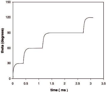
numerical results were obtained. Figure 8 shows the results of this computation in the form of a q vs. time plot for a range of q from 0 to 120° for the bottom of the shaft, which corresponds to the sequential passage of four protons, and represents one-third of the catalytic cycle of ATP synthesis. As seen from Fig. 8, each proton hop leads to a rapid rise in the angular displacement (the transient nonequilibrium state or phase) and subsequently reaches a new local equilibrium, as indicated by the charge configuration shown in Fig. 4. Energy storage in the g subunit takes place rapidly during the transient nonequilibrium phase that minimizes losses due to dissipation and leads to very high efficiencies of energy transfer. Due to the presence of torsion in the shaft with a piecewise linear stress-strain relationship, the time taken by the bottom of the shaft to traverse an angular displacement of 30° increases with the passage of each proton in the 0–90° range. Passage of the fourth proton achieves the break-through stress and causes the constraints at the top of the shaft to be broken, leading to a very rapid movement of the entire shaft (Fig. 8). For the conditions of our
82 |
S. Nath |
Fig. 8. Quantification of the dynamics of the bottom of the g-e shaft as proposed by the torsional mechanism of energy transmission. Angular displacement vs. time in the 0–120° interval obtained by numerical simulation of the equations of motion in the presence of differential rotation is shown. The values of the parameters employed in the simulation are: x = 3.3¥10–25 kg m2 s–1; k = 12.40¥10–20 kg m2 s–2 (0–30°), 6.21¥10–20 kg m2 s–2 (30–60°),
4.14¥10–20 kg m2 s–2 (60–90°), 8.0¥10–20 kg m2 s–2 (90–120°); d = 0.5¥10–9 m; l23 = 1.3¥10–9 m; l34 = 1.8¥10–9 m; r12 = 0.20¥10–9 m; r34 = 0.85¥10–9 m; l = 1¥10–9 m; L = 1.152¥10–28 kg m3 s–2; q = 1.6¥10–19 C; e = 2
simulation, the bottom of the shaft reaches 30° at 0.571 ms, 60° at 1.364 ms, 90° at 3.395 ms, and 120° at 3.859 ms, i.e., it takes 0.571, 0.793, 2.031, and 0.464 ms to traverse successive 30° displacements in the 0–120° range. An average value of angular velocity of approximately 100 revolutions per second is obtained, which is well within the expected range [21, 28].
3.4.3
Resolution of Difficulties Achieved and Experimental Evidence Supporting the Torsional Mechanism
Without exception, none of the specific difficulties discussed in Sects. 3.2 and 3.3 arise in the torsional mechanism. For instance, there is no requirement that nucleotides bind (and stay bound) to the catalytic site in the open conformation in the torsional mechanism; nor does binding of both MgADP and Pi take place in the same catalytic site. In fact, the presence of torsional strain ensures that another conformation (bC) is attained between bE and bTP , where MgADP gets bound. Furthermore, upon rotation and interaction of g with the DELSEED sequence of bC caused by the proton gradient, the conformation of the catalytic
The Molecular Mechanism of ATP Synthesis by F1F0-ATP Synthase: A Scrutiny of the Major Possibilities |
83 |
site is altered (slightly loosened) to bTP and access of inorganic phosphate to the site is created. Binding of Pi takes place in this altered conformation, bTP . Since the torsional mechanism is a tri-site mechanism, substrate enters (and binds) to the same catalytic site from which product had been released earlier; therefore, the problem of leaving a higher-affinity catalytic site unfilled never arises. Each catalytic site serves its particular function in the torsional mechanism, unlike in Boyer’s [37] or Cross’s [14] modification.
3.4.3.1 Optical Probes
The mechanism is in complete agreement with results from recent tryptophan fluorescence experiments (which, due to the inviolability of microscopic reversibility, also hold in the synthesis mode) that establish definitively that (i) Pi cannot simply bind spontaneously, (ii) an enzyme species with all three catalytic sites occupied is the only catalytically competent species, and (iii) release of product and binding of substrate cannot be simultaneous, rather product release must precede substrate binding [38].
3.4.3.2
Electron Microscopy and Image Analysis
In a very recent important development, Böttcher and Gräber carried out a laborious investigation of the structure of chloroplast ATP synthase by electron microscopy and obtained vastly improved interpretation of the data by introducing sophisticated image analysis techniques [45]. 3300 ATP synthase molecules were imaged, classified into 16 classes by difference analysis using multivariate statistical techniques, and the signal-to-noise ratio enhanced by averaging images of molecules within the same class. The class average from electron microscopic data was interpreted based on the Walker structure. The final result (their Fig. 2D) showed the e subunit lying closest to bE and the g subunit interacting with bTP , in complete agreement with the predictions of the torsional mechanism made in 1998 [16, 18]. The small structural detail detected in the form of the tiny third stalk/connection in the electron microscopy data, “formed presumably by a single a helix [45]”, would then correspond to the interaction of the Ser 108 of the e subunit with the DELSEED sequence (b-Glu 381) of the catalytic site proposed in the torsional mechanism [16–20].
3.4.3.3
Single Molecular Spectroscopy
The observed unidirectional (no reversal of rotation) and discrete rotation of the g subunit in steps of 120° during ATP hydrolysis by single molecule spectroscopy [34, 35, 40] supports the irreversible mode of operation of a single enzyme molecule that has been envisaged for the formulation of the thermody-
84 |
S. Nath |
namics and the molecular mechanism right since inception [16, 41, 42]. According to the torsional mechanism, during ATP synthesis, the g subunit rotates counter-clockwise when viewed from the F1 side. Thus, during ATP hydrolysis, the g subunit should rotate clockwise when viewed from the F1 side, which agrees with the direction observed by single molecule epifluorescence microscopy experiments [34]. Moreover, reversing the direction of motion of the g subunit during ATP synthesis, the torsional mechanism predicts that, during ATP hydrolysis, the rotating g subunit will sequentially encounter b subunits in the conformations O followed by L followed by T, in agreement with that determined experimentally [34].
3.4.3.4
Biochemical Nucleotide Site Occupancy Experiments
For ATP hydrolysis, in our view, the bound nucleotide occupancies are: no bound nucleotide in bE (O, open), MgATP in bC (C, closed), MgATP in bDP (T, tight), and MgADP+Pi in bTP (L, loose). The order of conformations that a particular b subunit cycles through during ATP hydrolysis is O to C to T to L. Thus, from the point of view of the order of conformations that a catalytic site passes through, ATP hydrolysis is not simply the reverse of ATP synthesis. Hence, ATP synthesis cannot be understood adequately by merely reversing the reaction arrows in a mechanism for ATP hydrolysis; this point of view has also been expressed by another group recently [43, 46]. This is logical, because the driving force for ATP hydrolysis is the hydrolysis step itself, while, in ATP synthesis, the rotation of g-e is induced by F0 which is driven by the ion gradients. Further, recent studies demonstrate that isolated F1 and F0F1 do not bind Pi to any extent at physiological concentrations (5 mM) in the absence of a proton gradient [38], and the effect of the proton gradient increases the affinity of the catalytic site for Pi by orders of magnitude during ATP synthesis, as discussed earlier, again pointing clearly to irreversibility. This does not conflict with the recent observation that both synthesis and hydrolysis reactions proceed through the same transition state [47]; thus, in our view, the L to T transformation of a b subunit during ATP synthesis and the T to L transformation of the subunit during ATP hydrolysis are the exact reverse of each other and will employ the same reaction pathway and pass through the same transition state.
3.4.3.5
Biochemical Acid Quench/Cold Chase Experiments
In classical acid quench/cold chase experiments [48] with mitochondrial F1 in “unisite” catalysis mode, [g-32P]ATP was used as substrate and the ratio of bound 32Pi/total bound 32P, where total bound 32P includes both bound 32Pi and bound [g-32P]ATP, was measured at different concentrations of F1 and [g-32P] ATP and at different incubation times of the reaction mixture. A kinetic scheme based on a general sequence of events leading to ATP hydrolysis which considers irreversibility of the catalysis steps, as proposed recently by some researchers [16–20, 43, 46, 49], was developed. kr and kr¢ represent the rate con-
The Molecular Mechanism of ATP Synthesis by F1F0-ATP Synthase: A Scrutiny of the Major Possibilities |
85 |
stants for consumption of E.ATP to E.ADP.Pi and of E.ADP.Pi to E.ADP + Pi respectively. We define
f = bound 32Pi/total bound 32P = [E.ADP.Pi]/([E.ATP]+[E.ADP.Pi]) (9)
Unisite catalysis is a non-physiological mode of catalysis in which a very small fraction of the enzyme population contains bound nucleotide in all three catalytic sites.According to the torsional mechanism, only this fraction will contribute to the measured ATPase activity. Based on the site occupancies predicted by the torsional mechanism during ATP hydrolysis (two catalytic sites contain bound MgATP, while one contains bound MgADP + Pi), the fraction f in Eq. (9) is predicted to be 0.33, which is in perfect agreement with experiment [48].
Because of the irreversibility of the catalysis steps, once ATP is bound to the F1-ATPase it has to be hydrolyzed to ADP and Pi and subsequently release the products from the catalytic site. As E.ADP.Pi is an intermediate in ATP hydrolysis, quasi-steady state considerations imply that the rate of formation of E.ADP.Pi is equal to its rate of consumption, i.e.:
vhyd = kr [E.ATP] = kr¢ [E.ADP.Pi] |
(10) |
This implies that
[E.ADP.Pi] = [E.ATP](kr/kr¢) |
(11) |
Substituting Eq. (11) into Eq. (9) leads to
f = kr/(kr + kr¢) |
(12) |
Since f = 0.33 by experiment as well as by theory,
kr/(kr + kr¢) = 0.33, or kr¢ = 2kr |
(13) |
From Eq. (12) we see that f is independent of both the concentrations of the enzyme and the substrate as well as the incubation time, as observed in the experiments. Since kr and kr¢ are rate constants, the fraction f is a characteristic property of the system. Similar to the equilibrium constant for reversible hydrolysis [48], we define a quasi-steady state constant for irreversible hydrolysis as:
Kqss = rate constant for formation of E.ADP.Pi/rate constant |
|
for consumption of E.ADP.Pi = kr/kr¢ = 0.5 |
(14) |
Hence, an irreversible mode of catalysis (where catalysis refers to rotation of the c-rotor in the F0 portion of ATP synthase, and to ADP, Pi binding and ATP synthesis and release in the F1 portion of ATP synthase) may also be employed to explain these experimental observations.
3.4.3.6
Structural Considerations
The Pedersen-Amzel structure revealed a state of the enzyme in which all six a,b subunits adopt a similar closed conformation, and it was argued that this