

Книги фарма 2 / Bertram G. Katzung-Basic & Clinical Pharmacology(9th Edition)
.pdfwater reabsorption will cause the concentration of the solute to rise to a point at which further water reabsorption is prevented. This is the mechanism by which osmotic diuretics act (see below).
Organic acid secretory systems are located in the middle third of the proximal tubule (S2 segment). These systems secrete a variety of organic acids (uric acid, nonsteroidal anti-inflammatory drugs NSAIDs, diuretics, antibiotics, etc) into the luminal fluid from the blood. These systems thus help deliver diuretics to the luminal side of the tubule, where most of them act. Organic base secretory systems (creatinine, choline, etc) are also present, in the early (S1) and middle (S2) segments of the proximal tubule.
Loop of Henle
At the boundary between the inner and outer stripes of the outer medulla, the thin limb of Henle's loop begins. Water is extracted from the thin descending limb of the loop of Henle by osmotic forces created in the hypertonic medullary interstitium. As in the proximal tubule, impermeant luminal solutes such as mannitol oppose water extraction.
The thick ascending limb of the loop of Henle actively reabsorbs NaCl from the lumen (about 35% of the filtered sodium), but unlike the proximal tubule and the thin limb, it is nearly impermeable to water. Salt reabsorption in the thick ascending limb therefore dilutes the tubular fluid, leading to its designation as a "diluting segment." Medullary portions of the thick ascending limb contribute to medullary hypertonicity and thereby also play an important role in concentration of urine.
The NaCl transport system in the luminal membrane of the thick ascending limb is a Na+/K+/2Cl- cotransporter (Figure 15–4). This transporter is selectively blocked by diuretic agents known as "loop" diuretics (see below). Although the Na+/K+/2Cl- transporter is itself electrically neutral (two cations and two anions are cotransported), the action of the transporter contributes to excess K+ accumulation within the cell. This results in back diffusion of K+ into the tubular lumen and development of a lumen-positive electrical potential. This electrical potential provides the driving force for reabsorption of cations—including Mg2+ and Ca2+—via the paracellular pathway (between the cells). Thus, inhibition of salt transport in the thick ascending limb by loop diuretics causes an increase in urinary excretion of divalent cations in addition to NaCl.
Figure 15–4.
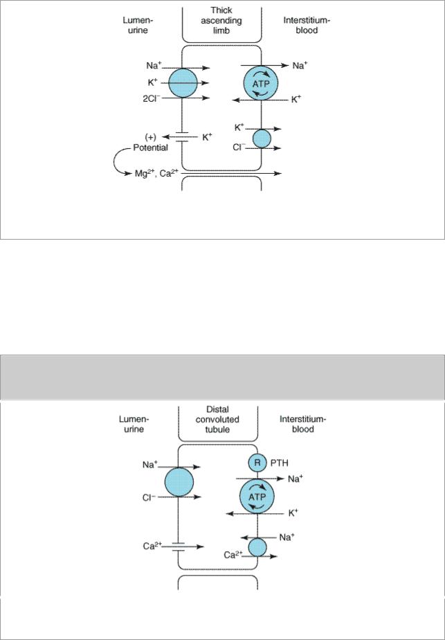
Ion transport pathways across the luminal and basolateral membranes of the thick ascending limb cell. The lumen positive electrical potential created by K+ back diffusion drives divalent cation reabsorption via the paracellular pathway.
Distal Convoluted Tubule
Only about 10% of the filtered NaCl is reabsorbed in the distal convoluted tubule. Like the thick ascending limb, this segment is relatively impermeable to water, and the NaCl reabsorption therefore further dilutes the tubular fluid. The mechanism of NaCl transport in the distal convoluted tubule is electrically neutral Na+ and Cl- cotransport (Figure 15–5). This NaCl transporter is blocked by diuretics of the thiazide class.
Figure 15–5.
Ion transport pathways across the luminal and basolateral membranes of the distal convoluted tubule cell. As in all tubular cells, Na+/K+ ATPase is present in the basolateral membrane. (R, PTH receptor.)

Because K+ does not recycle across the apical membrane of the distal convoluted tubule as it does in the loop of Henle, there is no lumen-positive potential in this segment, and Ca2+ and Mg2+ are not driven out of the tubular lumen by electrical forces. However, Ca2+ is actively reabsorbed by the distal convoluted tubule epithelial cell via an apical Ca2+ channel and basolateral Na+/Ca2+ exchanger (Figure 15–5). This process is regulated by parathyroid hormone. As will be seen below, the differences in the mechanism of Ca2+ transport in the distal convoluted tubule and in the loop of Henle have important implications for the effects of various diuretics on Ca2+ transport.
Collecting Tubule
The collecting tubule is responsible for only 2–5% of NaCl reabsorption by the kidney. Despite this small contribution, the collecting tubule plays an important role in renal physiology and in diuretic action. As the final site of NaCl reabsorption, the collecting tubule is responsible for volume regulation and for determining the final Na+ concentration of the urine. Furthermore, the collecting tubule is a site at which mineralocorticoids exert a significant influence. Lastly, the collecting tubule is the major site of potassium secretion by the kidney and the site at which virtually all diuretic-induced changes in potassium balance occur.
The mechanism of NaCl reabsorption in the collecting tubule is distinct from the mechanisms found in other tubule segments. The principal cells are the major sites of Na+, K+, and H2O transport (Figure 15–6), and the intercalated cells are the primary sites of proton secretion. Unlike cells in other nephron segments, the principal cells do not contain cotransport systems for Na+ and other ions in their apical membranes. Rather, principal cell membranes exhibit separate ion channels for Na+ and K+. Since these channels exclude anions, transport of Na+ or K+ leads to a net movement of charge across the membrane. Because the driving force for Na+ entry into the principal cell greatly exceeds that for K+ exit, Na+ reabsorption predominates, and a 10–50 mV lumen-negative electrical potential develops. Na+ that enters the principal cell from the urine is then transported back to the blood via the basolateral Na+/K+ ATPase (Figure 15–6). The lumen-negative electrical potential drives the transport of Cl- back to the blood via the paracellular pathway and also pulls K+ out of the cell through the apical membrane K+ channel. Thus, there is an important relationship between Na+ delivery to the collecting tubule and the resulting secretion of K+. Diuretics that act upstream of the collecting tubule will increase Na+ delivery to this site and will enhance K+ secretion. If the Na+ is delivered with an anion which cannot be reabsorbed as readily as Cl- (eg, bicarbonate), the lumennegative potential is increased, and K+ secretion will be enhanced. This mechanism, combined with enhanced aldosterone secretion due to volume depletion, is the basis for most diuretic-induced K+ wasting.
Figure 15–6.

Ion and H2O transport pathways across the luminal and basolateral membranes of collecting tubule and collecting duct cells. Inward diffusion of Na+ leaves a lumen-negative potential, which drives reabsorption of Cl- and efflux of K+. (R, aldosterone or ADH receptor.)
Reabsorption of Na+ via the epithelial Na channel (ENaC) and its coupled secretion of K+ is regulated by aldosterone. This steroid hormone, through its actions on gene transcription, increases the activity of both apical membrane channels and the basolateral N+/K+ ATPase. This leads to an increase in the transepithelial electrical potential and a dramatic increase in both Na+ reabsorption and K+ secretion.
A key determinant of the final urine concentration is antidiuretic hormone (ADH; also called vasopressin). In the absence of ADH, the collecting tubule (and duct) is impermeable to water, and dilute urine is produced. However, membrane water permeability of principal cells can be increased by ADH-induced fusion of vesicles containing preformed water channels with the apical membranes (Figure 15–6). ADH secretion is regulated by serum osmolality and by volume status.
Katzung PHARMACOLOGY, 9e > Section III. Cardiovascular-Renal Drugs > Chapter 15. Diuretic Agents >
Basic Pharmacology of Diuretic Agents
Carbonic Anhydrase Inhibitors
Carbonic anhydrase is present in many nephron sites, but the predominant location of this enzyme is the luminal membrane of the proximal tubule cells (Figure 15–3), where it catalyzes the dehydration of H2CO3, a critical step in the reabsorption of bicarbonate. By blocking carbonic

anhydrase, inhibitors block sodium bicarbonate reabsorption and cause diuresis.
The carbonic anhydrase inhibitors were the forerunners of modern diuretics. They are unsubstituted sulfonamide derivatives and were discovered when it was found that bacteriostatic sulfonamides caused an alkaline diuresis and hyperchloremic metabolic acidosis. With the development of newer agents, carbonic anhydrase inhibitors are now rarely used as diuretics, but they still have several specific applications that are discussed below. The prototypical carbonic anhydrase inhibitor is acetazolamide.
Pharmacokinetics
The carbonic anhydrase inhibitors are well absorbed after oral administration. An increase in urine pH from the bicarbonate diuresis is apparent within 30 minutes, maximal at 2 hours, and persists for 12 hours after a single dose. Excretion of the drug is by secretion in the proximal tubule S2 segment. Therefore, dosing must be reduced in renal insufficiency.
Pharmacodynamics
Inhibition of carbonic anhydrase activity profoundly depresses bicarbonate reabsorption in the proximal tubule. At its maximal safely administered dosage, 85% of the bicarbonate reabsorptive capacity of the superficial proximal tubule is inhibited. Some bicarbonate can still be absorbed at other nephron sites by carbonic anhydrase–independent mechanisms, and the overall effect of maximal acetazolamide dosage is about 45% inhibition of whole kidney bicarbonate reabsorption. Nevertheless, carbonic anhydrase inhibition causes significant bicarbonate losses and hyperchloremic metabolic acidosis. Because of this and the fact that HCO3- depletion leads to enhanced NaCl reabsorption by the remainder of the nephron, the diuretic efficacy of acetazolamide decreases significantly with use over several days.
The major clinical applications of acetazolamide involve carbonic anhydrase–dependent bicarbonate transport at sites other than the kidney. The ciliary body of the eye secretes bicarbonate from the blood into the aqueous humor. Likewise, formation of cerebrospinal fluid by the choroid plexus involves bicarbonate secretion into the cerebrospinal fluid. Although these processes remove bicarbonate from the blood (the direction opposite to that in the proximal tubule), they are significantly inhibited by carbonic anhydrase inhibitors, which in both cases dramatically alter the pH and quantity of fluid produced.
Clinical Indications & Dosage
See Table 15–1.
Table 15–1. Carbonic Anhydrase Inhibitors Used Orally in Treatment of Glaucoma.
Drug |
Usual Oral Dose (1–4 Times Daily) |
|
|
Acetazolamide |
250 mg |
|
|
Dichlorphenamide |
50 mg |
|
|
Methazolamide |
50 mg |
|
|
Glaucoma
The reduction of aqueous humor formation by carbonic anhydrase inhibitors decreases the intraocular pressure. This effect is valuable in the management of severe forms of glaucoma, making it the most common indication for use of carbonic anhydrase inhibitors. Topically active carbonic anhydrase inhibitors (dorzolamide, brinzolamide) are also available. These topical compounds reduce intraocular pressure, but plasma levels are undetectable. Thus, diuretic and systemic metabolic effects are eliminated.
Urinary Alkalinization
Uric acid, cystine, and some other weak acids are relatively insoluble in, and easily reabsorbed from, acidic urine. Renal excretion of these compounds can be enhanced by increasing urinary pH with carbonic anhydrase inhibitors. In the absence of continuous bicarbonate administration, these effects of acetazolamide are of relatively short duration (2–3 days). Prolonged therapy requires bicarbonate administration.
Metabolic Alkalosis
Metabolic alkalosis is generally treated by correction of abnormalities in total body K+, intravascular volume, or mineralocorticoid levels.
However, when the alkalosis is due to excessive use of diuretics in patients with severe heart failure, saline administration may be contraindicated. In these cases, acetazolamide can be useful in correcting the alkalosis as well as producing a small additional diuresis for the correction of heart failure. Acetazolamide has also been used to rapidly correct the metabolic alkalosis that may develop in the setting of respiratory acidosis.
Acute Mountain Sickness
Weakness, dizziness, insomnia, headache, and nausea can occur in mountain travelers who rapidly ascend above 3000 m. The symptoms are usually mild and last for a few days. In more serious cases, rapidly progressing pulmonary or cerebral edema can be life-threatening. By decreasing cerebrospinal fluid formation and by decreasing the pH of the cerebrospinal fluid and brain, acetazolamide can enhance performance status and diminish symptoms of mountain sickness.
Other Uses
Carbonic anhydrase inhibitors have been used as adjuvants for the treatment of epilepsy, in some forms of hypokalemic periodic paralysis, and to increase urinary phosphate excretion during severe hyperphosphatemia.
Toxicity
Hyperchloremic Metabolic Acidosis
Acidosis predictably results from chronic reduction of body bicarbonate stores by carbonic anhydrase inhibitors and limits the diuretic efficacy of these drugs to 2 or 3 days.
Renal Stones

Phosphaturia and hypercalciuria occur during the bicarbonaturic response to inhibitors of carbonic anhydrase. Renal excretion of solubilizing factors (eg, citrate) may also decline with chronic use. Calcium salts are relatively insoluble at alkaline pH, which means that the potential for renal stone formation from these salts is enhanced.
Renal Potassium Wasting
Potassium wasting can occur because NaHCO3 presented to the collecting tubule increases the lumen-negative electrical potential in that segment and enhances K+ secretion. This effect can be counteracted by simultaneous administration of KCl.
Other Toxicities
Drowsiness and paresthesias are common following large doses. Carbonic anhydrase inhibitors may accumulate in patients with renal failure, leading to nervous system toxicity. Hypersensitivity reactions (fever, rashes, bone marrow suppression, and interstitial nephritis) may also occur.
Contraindications
Carbonic anhydrase inhibitor-induced alkalinization of the urine will decrease urinary excretion of NH4+ and may contribute to the development of hyperammonemia and hepatic encephalopathy in patients with cirrhosis.
Loop Diuretics
Loop diuretics selectively inhibit NaCl reabsorption in the thick ascending limb of the loop of Henle. Due to the large NaCl absorptive capacity of this segment and the fact that diuresis is not limited by development of acidosis, as it is with the carbonic anhydrase inhibitors, these drugs are the most efficacious diuretic agents available.
Chemistry
The two prototypical drugs of this group are furosemide and ethacrynic acid. The structures of several loop diuretics are shown in Figure 15–7. Like the carbonic anhydrase inhibitors, furosemide, bumetanide, and torsemide are sulfonamide derivatives.
Figure 15–7.

Some loop diuretics. The shaded methylene group on ethacrynic acid is reactive and may combine with free sulfhydryl groups.
Ethacrynic acid—not a sulfonamide derivative—is a phenoxyacetic acid derivative containing an adjacent ketone and methylene group (Figure 15–7). The methylene group (shaded) forms an adduct with the free sulfhydryl group of cysteine. The cysteine adduct appears to be an active form of the drug.
Organic mercurial diuretics also inhibit salt transport in the thick ascending limb but are no longer used because of their high toxicity.
Pharmacokinetics
The loop diuretics are rapidly absorbed. They are eliminated by tubular secretion as well as by glomerular filtration. Absorption of oral torsemide is more rapid (1 hour) than that of furosemide (2–3 hours) and is nearly as complete as with intravenous administration. Diuretic response is extremely rapid following intravenous injection. The duration of effect for furosemide is usually 2– 3 hours and that of torsemide is 4–6 hours. Half-life depends on renal function. Since loop agents act on the luminal side of the tubule, their diuretic activity correlates with their secretion by the proximal tubule. Reduction in the secretion of loop diuretics may result from simultaneous administration of agents such as NSAIDs or probenecid, which compete for weak acid secretion in the proximal tubule. Metabolites of ethacrynic acid and furosemide have been identified, but it is not known if they have any diuretic activity. Torsemide has at least one active metabolite with a

half-life considerably longer than that of the parent compound.
Pharmacodynamics
These drugs inhibit the luminal Na+/K+/2Cl- transporter in the thick ascending limb of Henle's loop. By inhibiting this transporter, the loop diuretics reduce the reabsorption of NaCl and also diminish the lumen-positive potential that derives from K+ recycling (Figure 15–4). This electrical potential normally drives divalent cation reabsorption in the loop, and by reducing this potential, loop diuretics cause an increase in Mg2+ and Ca2+ excretion. Prolonged use can cause significant hypomagnesemia in some patients. Since Ca2+ is actively reabsorbed in the distal convoluted tubule, loop diuretics do not generally cause hypocalcemia. However, in disorders that cause hypercalcemia, Ca2+ excretion can be greatly enhanced by combining loop agents with saline infusions.
Loop diuretics induce renal prostaglandin synthesis, and these prostaglandins participate in the renal actions of these drugs. NSAIDs (eg, indomethacin) can interfere with the actions of the loop diuretics by reducing prostaglandin synthesis in the kidney. This interference is minimal in otherwise normal subjects but may be significant in patients with nephrotic syndrome or hepatic cirrhosis.
In addition to their diuretic activity, loop agents appear to have direct effects on blood flow through several vascular beds. Furosemide increases renal blood flow. Furosemide and ethacrynic acid have also been shown to reduce pulmonary congestion and left ventricular filling pressures in heart failure before a measurable increase in urinary output occurs, and in anephric patients.
Clinical Indications & Dosage
The most important indications for the use of the loop diuretics include acute pulmonary edema, other edematous conditions, and acute hypercalcemia (Table 15–2). The use of loop diuretics in these conditions is discussed in Clinical Pharmacology. Other indications for loop diuretics include hyperkalemia, acute renal failure, and anion disease.
Table 15–2. Loop Diuretics: Dosages.
Drug |
Daily Oral Dose1 |
|
|
|
|
Bumetanide |
0.5–2 mg |
|
|
Ethacrynic acid |
50–200 mg |
|
|
Furosemide |
20–80 mg |
|
|
Torsemide |
2.5–20 mg |
|
|
1As single dose or in two divided doses.
Hyperkalemia
In mild hyperkalemia—or after acute management of severe hyperkalemia by other measures—loop diuretics can significantly enhance urinary excretion of K+. This response is enhanced by
simultaneous NaCl and water administration.
Acute Renal Failure
Loop agents can increase the rate of urine flow and enhance K+ excretion in acute renal failure. However, they do not seem to shorten the duration of renal failure. If a large pigment load has precipitated acute renal failure or threatens to do so, loop agents may help flush out intratubular casts and ameliorate intratubular obstruction. On the other hand, loop agents can theoretically worsen cast formation in myeloma and light chain nephropathy.
Anion Overdose
Loop diuretics are useful in treating toxic ingestions of bromide, fluoride, and iodide, which are reabsorbed in the thick ascending limb. Saline solution must be administered to replace urinary losses of Na+ and to provide Cl-, so as to avoid extracellular fluid volume depletion.
Toxicity
Hypokalemic Metabolic Alkalosis
Loop diuretics increase delivery of salt and water to the collecting duct and thus enhance the renal secretion of K+ and H+, causing hypokalemic metabolic alkalosis. This toxicity is a function of the magnitude of the diuretic effect and can be reversed by K+ replacement and correction of hypovolemia.
Ototoxicity
Loop diuretics can cause dose-related hearing loss that is usually reversible. It is most common in patients who have diminished renal function or who are also receiving other ototoxic agents such as aminoglycoside antibiotics.
Hyperuricemia
Loop diuretics can cause hyperuricemia and precipitate attacks of gout. This is caused by hypovolemia-associated enhancement of uric acid reabsorption in the proximal tubule. It may be avoided by using lower doses.
Hypomagnesemia
Magnesium depletion is a predictable consequence of the chronic use of loop agents and occurs most often in patients with dietary magnesium deficiency. It can be reversed by administration of oral magnesium preparations.
Allergic Reactions
Skin rash, eosinophilia and, less often, interstitial nephritis are occasional side effects of furosemide, bumetanide, and torsemide therapy. These usually resolve rapidly after drug withdrawal. Allergic reactions are much less common with ethacrynic acid.
Other Toxicities