

Книги фарма 2 / Bertram G. Katzung-Basic & Clinical Pharmacology(9th Edition)
.pdfApart from being a vasodilator, nitric oxide is also a potent inhibitor of neutrophil adhesion to the vascular endothelium. This is due to the inhibitory effect of nitric oxide on the expression of adhesion molecules on the endothelial surface. The role of nitric oxide in protecting the endothelium has been demonstrated by studies that showed that treatment with nitric oxide donors protects against ischemiaand reperfusion-mediated endothelial dysfunction.
Respiratory Disorders
Nitric oxide has been shown to improve cardiopulmonary function in adult patients with pulmonary artery hypertension and is approved for this indication (see Preparations Available). It is administered by inhalation. It has also been administered by inhalation to newborns with pulmonary hypertension and acute respiratory distress syndrome. The current treatment for severely defective gas exchange in the newborn is with extracorporeal membrane oxygenation (ECMO), which does not directly affect pulmonary vascular pressures. Nitric oxide inhalation decreases pulmonary arterial pressure and improves blood oxygenation. Thus, when pulmonary resistance is elevated, it is possible to exploit the vasodilator properties of nitric oxide by administering it via inhalation of a few parts per million. Adults with respiratory distress syndrome also appear—in open trials—to benefit from nitric oxide inhalation. Nitric oxide may have an additional role in relaxing airway smooth muscle and thus acting as a bronchodilator. For these reasons, nitric oxide inhalation therapy is being widely tested in both infants and adults with acute respiratory distress syndrome. The adverse effects of this use of nitric oxide are being assessed.
Septic Shock
As mentioned previously, increased urinary excretion of nitrate, the oxidative product of nitric oxide, is reported in gram-negative bacterial infection. Lipopolysaccharide components from the bacterial wall activate the inducible NOS (NOS-2), resulting in exaggerated hypotension, shock, and possible death. This hypotension is reversed by NOS inhibitors such as L-NMMA (Table 19–3) in humans as well as animal models and by compounds such as methylene blue, which prevent the action of nitric oxide, as well as by scavengers of nitric oxide such as hemoglobin. Furthermore, knockout mice lacking a functional NOS-2 gene are more resistant to endotoxin than wild-type mice. However, there has been no correlation between the hemodynamic effects of the nitric oxide inhibitors and survival rate in gram-negative sepsis thus far.
Atherosclerosis
Vascular plaque formation in hypercholesterolemia leads to reduced nitric oxide formation and endothelium-dependent vasodilator responses. In vitro, nitric oxide carriers and donors and cGMP analogs inhibit smooth muscle cell proliferation. In animal models, myointimal proliferation following angioplasty can be blocked by feeding arginine, by using nitric oxide donors, by NOS gene transfer, and by nitric oxide inhalation. In addition, nitric oxide may act as an antioxidant, blocking the oxidation of low-density lipoproteins (LDL) and thus preventing the formation of foam cells in the vascular wall.
Platelets
Abnormal activation of platelets is associated with increased platelet adhesion and aggregation and therefore a higher incidence of thrombotic events. Platelet activation also leads to release of smooth muscle mitogens such as growth factors and thromboxane. Nitric oxide is a potent inhibitor of platelet adhesion and aggregation. Thus, endothelial dysfunction and the associated decrease in nitric oxide generation may result in abnormal platelet function. Platelets also contain both
constitutive and inducible NOS, although to a much lesser extent than endothelial cells. As in vascular smooth muscle, cGMP mediates the protective effect of nitric oxide in platelets. Nitric oxide may have an additional beneficial effect on blood coagulation by enhancing fibrinolysis via an effect on plasminogen.
Organ Transplantation
Accelerated graft atherosclerosis following organ transplantation is a chronic condition and is a major cause of transplant failure, leading to retransplantation or death. Continuous vascular smooth muscle proliferation occurs within the vasculature of most grafts and is a central event in luminal narrowing. Ischemic and reperfusion injuries at the time of organ harvesting, preservation, and revascularization initiate myointimal proliferation, which is also promoted by the continuous immune response to the allogeneic organ graft. By reducing free radical toxicity under these conditions, nitric oxide may act as a cytoprotective agent, inhibiting platelet and neutrophil aggregation and adhesion to the vascular wall. Dietary L-arginine supplementation increases plasma nitrite and nitrate formation and has been shown to attenuate accelerated graft atherosclerosis. However, excessively high concentrations of nitric oxide may be detrimental during acute organ rejection due to up-regulation of inducible NOS by cytokines; under these circumstances, inhibition of nitric oxide synthesis has been shown to prolong graft survival in experimental animals.
The Central Nervous System
Nitric oxide has been proposed to have a major role in the central nervous system—as a neurotransmitter, as a modulator of ligand-gated receptors, or both. In addition, nitric oxide probably plays a role in neuronal degeneration in some conditions. The likely cellular targets of nitric oxide in the central nervous system include presynaptic and postsynaptic nerve terminals. Nitric oxide modifies neurotransmitter release in different areas of the brain. Postsynaptic release of nitric oxide following activation of the NMDA receptor may initiate presynaptic transmitter release of glutamate, ie, nitric oxide may function as a retrograde messenger that is synthesized in postsynaptic sites following opening of the Ca2+ channels and activation of NOS. It is proposed that the nitric oxide thus produced rapidly diffuses to the presynaptic nerve terminal where guanylyl cyclase is activated to yield cGMP and thus facilitate transmitter release. In the cerebellum and in neuroblastoma cells, this effect is blocked by NOS inhibitors such as L-NMMA and is enhanced by L-arginine. It has been suggested that nitric oxide (like many other substances) may have a role in shortand long-term potentiating effects on excitatory amino acids in brain development and learning.
7-Nitroindazole, an inhibitor of NOS-1, and L-NAME, a less selective inhibitor of neuronal NOS, have significant antinociceptive effects in humans and animals and 7-nitroindazol reduces the signs of opioid withdrawal and cocaine action in animal models. This inhibitor also reduces cerebral blood flow. Nevertheless, 7-nitroindazole can reduce the size of cerebral infarcts in animal models. In contrast, NOS-3-deficient mice are more susceptible to ischemic cerebral damage. NOS-1 inhibition by 7-nitroindazole also reduces the neurotoxicity of MPTP and MPP+ (see Chapter 28: Pharmacologic Management of Parkinsonism & Other Movement Disorders) in several animal models.
It is well known that prolonged NMDA glutamate receptor activation results in degeneration of neurons (excitotoxicity). This has been attributed to a large increase in calcium influx, which activates the calmodulin-dependent NOS-1 and leads to sustained elevation of nitric oxide concentrations. The increase in neurodegeneration caused by excitatory amino acids may be due to enhanced oxygen radical formation since superoxide dismutase has a beneficial effect in

Nitric Oxide (INOmax)
Inhalation: 100, 800 ppm gas
Katzung PHARMACOLOGY, 9e > Section IV. Drugs with Important Actions on Smooth Muscle > Chapter 19. Nitric Oxide, Donors, & Inhibitors >
Chapter 20. Drugs Used in Asthma
Katzung PHARMACOLOGY, 9e > Section IV. Drugs with Important Actions on Smooth Muscle > Chapter 20. Drugs Used in Asthma >
Drugs Used in Asthma: Introduction
The clinical hallmarks of asthma are recurrent, episodic bouts of coughing, shortness of breath, chest tightness, and wheezing. In mild asthma, symptoms occur only occasionally, eg, on exposure to allergens or certain pollutants, on exercise, or after a viral upper respiratory infection. More severe forms of asthma are associated with frequent attacks of wheezing dyspnea, especially at night, and even chronic limitation of activity. Asthma is the most common chronic disabling disease of childhood, but it affects all age groups.
Asthma is physiologically characterized by increased responsiveness of the trachea and bronchi to various stimuli and by widespread narrowing of the airways that changes in severity either spontaneously or as a result of therapy. Its pathologic features are contraction of airway smooth muscle, mucosal thickening from edema and cellular infiltration, and inspissation in the airway lumen of abnormally thick, viscid plugs of mucus. Of these causes of airway obstruction, contraction of smooth muscle is most easily reversed by current therapy; reversal of the edema and cellular infiltration requires sustained treatment with anti-inflammatory agents. Asthma therapies are thus sometimes divided into two categories: "short-term relievers" and "long-term controllers."
Short-term relief is most effectively achieved with bronchodilators, agents that increase airway caliber by relaxing airway smooth muscle, and of these the  -adrenoceptor stimulants (see Chapter 9: Adrenoceptor-Activating & Other Sympathomimetic Drugs) are the most widely used. Theophylline, a methylxanthine drug, and antimuscarinic agents (see Chapter 8: CholinoceptorBlocking Drugs) are also used for reversal of airway constriction. Long-term control is most often achieved with an anti-inflammatory agent such as an inhaled corticosteroid, with a leukotriene antagonist, or with an inhibitor of mast cell degranulation, eg, cromolyn or nedocromil. The distinction between "short-term relievers" and "long-term controllers" has become blurred by the finding that theophylline inhibits some lymphocyte functions and modestly reduces airway mucosal inflammation and that budesonide, an inhaled corticosteroid, produces modest immediate bronchodilation. Similarly, two recently released long-acting
  -adrenoceptor stimulants, salmeterol and formoterol, appear to be effective in improving asthma control when taken regularly. Finally, clinical trials are demonstrating the efficacy of specifically targeting a mechanism thought to be fundamental to asthma's pathogenesis by repeated treatment with a humanized monoclonal anti-IgE antibody.
This chapter presents the basic pharmacology of the methylxanthines, cromolyn, leukotriene pathway inhibitors, and monoclonal anti-IgE antibody—agents whose medical use is almost exclusively for pulmonary disease. The other classes of drugs listed above are discussed in relation

to the therapy of asthma.
Pathogenesis of Asthma
A rational approach to the pharmacotherapy of asthma depends on an understanding of the disease's pathogenesis. In the classic immunologic model, asthma is a disease mediated by reaginic (IgE) antibodies bound to mast cells in the airway mucosa (Figure 20–1). On reexposure to an antigen, antigen-antibody interaction on the surface of the mast cells triggers both the release of mediators stored in the cells' granules and the synthesis and release of other mediators. The agents responsible for the early reaction—immediate bronchoconstriction—include histamine, tryptase and other neutral proteases, leukotrienes C4 and D4, and prostaglandins. These agents diffuse throughout the airway wall and cause muscle contraction and vascular leakage. Other mediators are responsible for the more sustained bronchoconstriction, cellular infiltration of the airway mucosa, and mucus hypersecretion of the late asthmatic reaction that occurs 2–8 hours later. These mediators are thought to be cytokines characteristically produced by TH2 lymphocytes, especially GM-CSF and interleukins 4, 5, 9, and 13, which attract and activate eosinophils and stimulate IgE production by B lymphocytes. It is not clear whether lymphocytes or mast cells in the airway mucosa are the primary source of the cytokines and other mediators responsible for the late inflammatory response, but it is now thought that the benefits of corticosteroid therapy may result from their inhibition of cytokine production in the airways.
Figure 20–1.
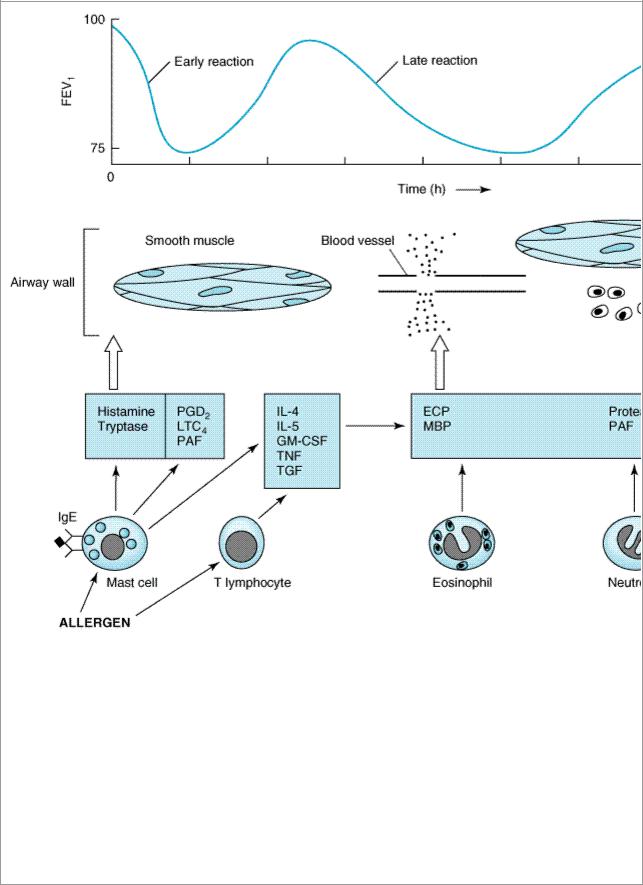
Conceptual model for the immunopathogenesis of asthma. Exposure to allergen causes synthesis of IgE, which binds to mast cells in the airway mucosa. On reexposure to allergen, antigenantibody interaction on mast cell surfaces triggers release of mediators of anaphylaxis: histamine, tryptase, prostaglandin D2 (PGD2), leukotriene C4, and platelet-activating factor (PAF). These agents provoke contraction of airway smooth muscle, causing the immediate fall in FEV1. Reexposure to allergen also causes the synthesis and release of a variety of cytokines: interleukins 4 and 5, granulocyte-macrophage colony stimulating factor (GM-CSF), tumor necrosis factor (TNF), and tissue growth factor (TGF) from T cells and mast cells. These cytokines in turn attract and activate eosinophils and neutrophils, whose products include eosinophil cationic protein (ECP), major basic protein (MBP), proteases, and platelet-activating factor. These mediators cause the edema, mucus hypersecretion, smooth muscle contraction, and increase in bronchial reactivity associated with the late asthmatic response, indicated by a fall in FEV1 2–8 hours after the exposure.
with asthma have no evidence of immediate hypersensitivity to antigens, most severe exacerbations of asthma appear to be provoked by viral respiratory infection, the severity of symptoms correlates poorly with the quantity of antigen in the atmosphere, and in many patients bronchospasm can be provoked by nonantigenic stimuli such as distilled water, exercise, cold air, sulfur dioxide, and rapid respiratory maneuvers.
This tendency to develop bronchospasm upon encountering stimuli that do not affect healthy nonasthmatic airways is characteristic of asthma and is sometimes called "nonspecific bronchial hyperreactivity" to distinguish it from bronchial responsiveness to specific antigens. Bronchial hyperreactivity is quantitated by measuring the fall in forced expiratory volume in 1 second (FEV1) provoked by inhaling serially increasing concentrations of aerosolized histamine or methacholine. This exaggerated sensitivity of the airways appears to be fundamental to asthma's pathogenesis, for it is nearly ubiquitous in patients with asthma and its degree correlates with the symptomatic severity of the disease.
The mechanisms underlying bronchial hyperreactivity are somehow related to inflammation of the airway mucosa. The agents that increase bronchial reactivity, such as ozone exposure, allergen inhalation, and infection with respiratory viruses, also cause airway inflammation. In both dogs and humans, the increase in bronchial reactivity induced by ozone is associated with an increase in the number of polymorphonuclear leukocytes found in fluid obtained by bronchial lavage or from bronchial mucosal biopsies. The increase in reactivity due to allergen inhalation is associated with an increase in both eosinophils and polymorphonuclear leukocytes in bronchial lavage fluid. The increase in reactivity that is associated with the late asthmatic response to allergen inhalation (Figure 20–1) is sustained and, because it is prevented by treatment with inhaled corticosteroids immediately before antigen challenge, is thought to be caused by airway inflammation.
How the increase in airway reactivity is linked to inflammation is uncertain. Much evidence points to the eosinophil. The most consistent difference in bronchial mucosal biopsies obtained from asthmatic and healthy subjects is an increase in the number of eosinophils found beneath the airway epithelium. Immunohistochemical staining shows increased levels of eosinophil cationic protein, indicating activation of the cells. The number of eosinophils in expectorated sputum or in fluid lavaged from the lungs correlates roughly with the degree of bronchial hyperreactivity. Eosinophil products have in turn been shown to cause epithelial sloughing and an increase in contractile responsiveness of airway smooth muscle. The importance of the eosinophil has been challenged, however, by a study showing that treatment with an anti-IL-5 monoclonal antibody effectively blocks airway eosinophilia caused by allergen challenge but does not prevent bronchoconstriction or any further increase in bronchial hyperactivity (Leckie, 2000).
The products of other cells in the airways, such as lymphocytes, macrophages, mast cells, sensory nerves, and epithelial cells, have also been shown to alter airway smooth muscle function, so a specific antagonist to a single mediator or class of mediators might not prove wholly effective as asthma therapy. Other evidence suggests a role for sensitization of sensory nerves in the airways as a mechanism for hyperreactivity (see Pharmacologic Significance of Lung Innervation).
Whatever the mechanisms responsible for bronchial hyperreactivity, bronchoconstriction itself seems to result not simply from the direct effect of the released mediators but also from their activation of neural or humoral pathways. Evidence for the importance of neural pathways stems largely from studies of laboratory animals. Thus, the bronchospasm provoked in dogs by histamine can be greatly reduced by pretreatment with an inhaled topical anesthetic agent, by transection of the vagus nerves, and by pretreatment with atropine. Studies of asthmatic humans, however, have shown that treatment with atropine causes only a reduction in—not abolition of—the


(ACh), which binds to muscarinic receptors on airway smooth muscle. Inhaled materials may provoke bronchoconstriction by several possible mechanisms. First, they may trigger the release of chemical mediators from mast cells. Second, they may stimulate afferent receptors to initiate reflex bronchoconstriction or to release tachykinins (eg, substance P) that directly stimulate smooth muscle contraction.
The hypothesis suggested by these studies—that asthmatic bronchospasm results from a combination of release of mediators and an exaggeration of responsiveness to their effects— predicts that asthma may be effectively treated by drugs with different modes of action. Asthmatic bronchospasm might be reversed or prevented, for example, by drugs that reduce the amount of IgE bound to mast cells (anti-IgE antibody), prevent mast cell degranulation (cromolyn or nedocromil, sympathomimetic agents, calcium channel blockers), block the action of the products released (antihistamines and leukotriene receptor antagonists), inhibit the effect of acetylcholine released from vagal motor nerves (muscarinic antagonists), or directly relax airway smooth muscle (sympathomimetic agents, theophylline).
The second approach to the treatment of asthma is aimed not just at preventing or reversing acute bronchospasm but at reducing the level of bronchial responsiveness. Because increased responsiveness appears to be linked to airway inflammation and because airway inflammation is a feature of late asthmatic responses, this strategy is implemented both by reducing exposure to the allergens that provoke inflammation and by prolonged therapy with anti-inflammatory agents, especially inhaled corticosteroids.
Katzung PHARMACOLOGY, 9e > Section IV. Drugs with Important Actions on Smooth Muscle > Chapter 20. Drugs Used in Asthma >
Pharmacologic Significance of Lung Innervation
As noted previously, the airways are richly supplied with afferent and efferent vagal nerves. The cholinergic motor fibers are clearly responsible in some patients for a portion of the bronchoconstriction characteristic of acute asthma. Such fibers innervate M3 receptors on the smooth muscle and contain modulatory M2 receptors on the nerve terminals. Selective inhibition of M2 receptors can increase bronchoconstrictor responses to a variety of stimuli, while M3 inhibitors can produce dilation of constricted airways.
In contrast, noradrenergic sympathetic innervation of the airways is sparse, and these fibers do not appear to play a major role in controlling airway diameter. Bronchodilation may be brought about by nonadrenergic, noncholinergic nerves releasing nitric oxide since nitric oxide synthase inhibitors have been shown to reduce bronchodilation produced by electrical field stimulation in vitro.
The role of peptidergic neurons is not so clear. Capsaicin, the hot chile pepper chemical that evokes release of peptide transmitters from several types of sensory nerves, has been shown to reproduce some of the signs of bronchial hyperreactivity in animal and human experiments. These findings led to the proposal that sensitization of afferent nerve endings played a major role in chronic airway hyperreactivity. However, peptide transmitter antagonists have not been able to prevent bronchoconstriction in several models. Clearly, much remains to be learned about airway pharmacology.
Katzung PHARMACOLOGY, 9e > Section IV. Drugs with Important Actions on Smooth Muscle > Chapter 20. Drugs Used in Asthma >
Basic Pharmacology of Agents Used in the Treatment of Asthma
The drugs most used for management of asthma are adrenoceptor agonists (used as "relievers" or bronchodilators) and inhaled corticosteroids (used as "controllers" or anti-inflammatory agents). Their basic pharmacology is presented elsewhere (see Chapter 9: Adrenoceptor-Activating & Other Sympathomimetic Drugs and Chapter 39: Adrenocorticosteroids & Adrenocortical Antagonists). In this chapter, we review their pharmacology relevant to asthma.
Sympathomimetic Agents
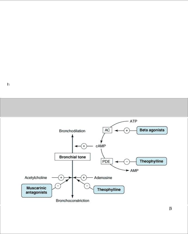
The adrenoceptor agonists have several pharmacologic actions important in the treatment of asthma. They relax airway smooth muscle and inhibit release of some bronchoconstricting substances from mast cells. They may also inhibit microvascular leakage and increase mucociliary transport by increasing ciliary activity or by affecting the composition of mucous secretions. As in other tissues, the  agonists stimulate adenylyl cyclase and increase the formation of cAMP in the airway tissues (Figure 20–3).
Figure 20–3.
Bronchodilation is promoted by cAMP. Intracellular levels of cAMP can be increased by  - adrenoceptor agonists, which increase the rate of its synthesis by adenylyl cyclase (AC); or by phosphodiesterase (PDE) inhibitors such as theophylline, which slow the rate of its degradation. Bronchoconstriction can be inhibited by muscarinic antagonists and possibly by adenosine antagonists.
The best-characterized action of the adrenoceptor agonists on airways is relaxation of airway smooth muscle that results in bronchodilation. Although there is no evidence for significant sympathetic innervation of human airway smooth muscle, there is ample evidence for the presence