

Урология - Urolithiasis
.pdfPossible location of calculi
http://www.npl-rez.ru/litra/pochky.htm

Diagnostics
Gathering of complaints and analysis
Examination and palpation of lumbal area and abdomen
Positive shaking symptom
Laboratory tests – erytrocytes and crystals of salts in urine, urine density
Instrymental methods – chromocystoscopy, ultrasound, X-Ray, CT, angiography

Complaints - differential diagnosis
Although obstructing urinary tract stones are typically associated with symptoms, a definitive diagnosis of urolithiasis cannot be based on symptoms alone. Because of the embryonic development of the kidneys and genital system, as well as the close nerve and vascular supply, pain due to stones may be referred to the gonads or confused with gastrointestinal pathology such as cholecystitis, appendicitis, gastric ulcer, or diverticulitis. Likewise, cystitis and pyelonephritis may mimic acute renalcolic. Musculoskeletal pain, particularly over theflanks, may also be incorrectly attributed to stone pain.

A definitive diagnosis of a stone requires either direct stone retrieval after spontaneous passage or surgical intervention, or identification by radiologic imaging.

X-Ray (KUB) and IVP
Although an abdominal x-ray of the kidneys-ureters-bladder (KUB) is simple and requires no preparation, it can fail to reveal small or adiolucent stones. Excretory urography, also known as intravenous pyelography (IVP), is more sensitive than KUB and provides more anatomicinformation, but IVP can still miss small or radiolucent nonobstructing stones.
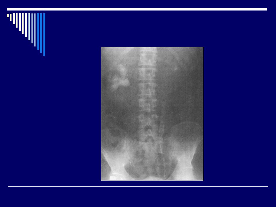
Plain X-ray. Coral-like calculus of
right kidney, calculus of left renal pelvis.

KUB – stone of vesical bladder
by http://www.uroconsultant.ru/index.php?t=28

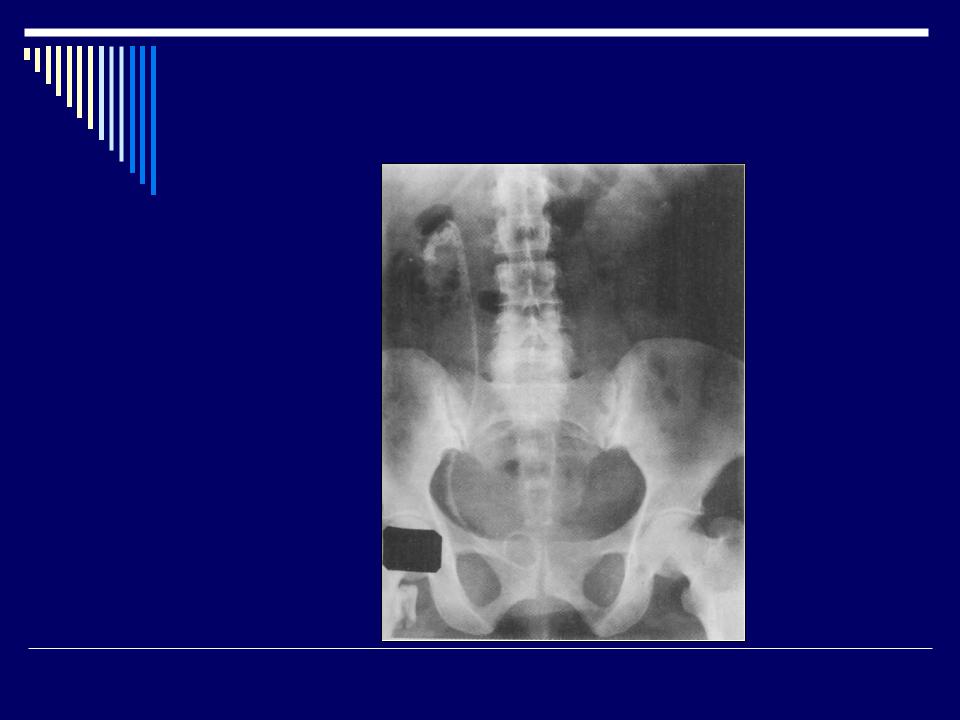
Stone of right renal pelvis
KUB and IVP
Retrograde ureteropyelography – calculus of upper third part of right ureter.
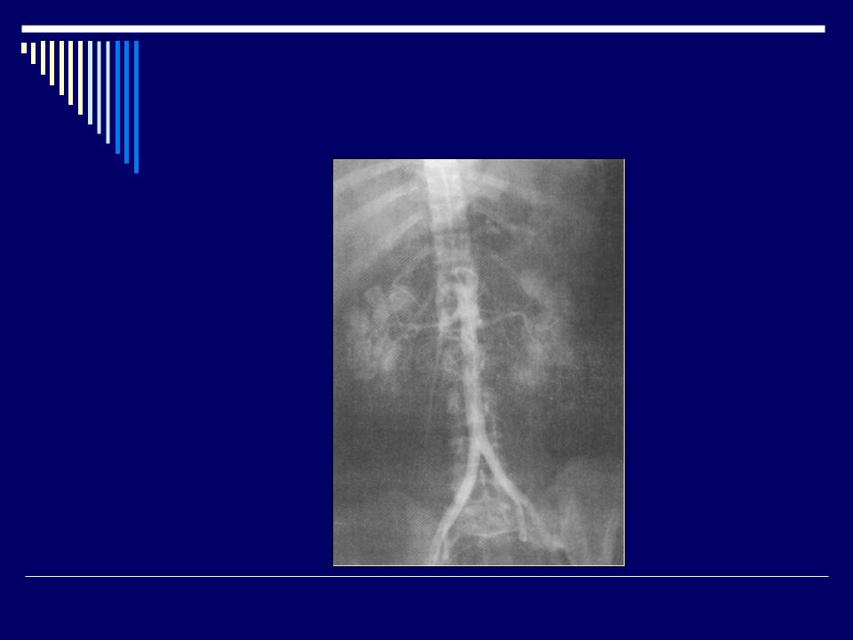
Asngiography of a petient with
coral-like stone of right kidney