

МРТ
.pdfACCF/ACR/AHA/NASCI/SCMR 2010 Expert Consensus Document on Cardiovascular Magnetic Resonance: A Report of the American College of Cardiology Foundation Task Force on Expert Consensus Documents
WRITING COMMITTEE MEMBERS, W. Gregory Hundley, David A. Bluemke, J. Paul Finn, Scott D. Flamm, Mark A. Fogel, Matthias G. Friedrich, Vincent B. Ho, Michael Jerosch-Herold, Christopher M. Kramer, Warren J. Manning, Manesh Patel, Gerald M. Pohost, Arthur E. Stillman, Richard D. White and Pamela K. Woodard
Circulation. 2010;121:2462-2508; originally published online May 17, 2010; doi: 10.1161/CIR.0b013e3181d44a8f
Circulation is published by the American Heart Association, 7272 Greenville Avenue, Dallas, TX 75231 Copyright © 2010 American Heart Association, Inc. All rights reserved.
Print ISSN: 0009-7322. Online ISSN: 1524-4539
The online version of this article, along with updated information and services, is located on the World Wide Web at:
http://circ.ahajournals.org/content/121/22/2462
Permissions: Requests for permissions to reproduce figures, tables, or portions of articles originally published in Circulation can be obtained via RightsLink, a service of the Copyright Clearance Center, not the Editorial Office. Once the online version of the published article for which permission is being requested is located, click Request Permissions in the middle column of the Web page under Services. Further information about this process is available in the Permissions and Rights Question and Answer document.
Reprints: Information about reprints can be found online at:
http://www.lww.com/reprints
Subscriptions: Information about subscribing to Circulation is online at:
http://circ.ahajournals.org//subscriptions/
Downloaded from http://circ.ahajournals.org/ by guest on October 24, 2013

Expert Consensus Document
ACCF/ACR/AHA/NASCI/SCMR 2010 Expert Consensus Document on Cardiovascular Magnetic Resonance
A Report of the American College of Cardiology Foundation Task Force on Expert Consensus Documents
WRITING COMMITTEE MEMBERS
W.Gregory Hundley, MD, FACC, FAHA, Chair*; David A. Bluemke, MD, PhD, FAHA†; J. Paul Finn, MD†; Scott D. Flamm, MD‡; Mark A. Fogel, MD, FACC, FAHA, FAAP§;
Matthias G. Friedrich, MD, FESC‡; Vincent B. Ho, MD, MBA, FAHA **; Michael Jerosch-Herold, PhD; Christopher M. Kramer, MD, FACC, FAHA*;
Warren J. Manning, MD, FACC*; Manesh Patel, MD; Gerald M. Pohost, MD, FACC, FAHA¶; Arthur E. Stillman, MD, PhD, FACR, FAHA#; Richard D. White, MD, FACC, FAHA#; Pamela K. Woodard, MD, FACR, FAHA
ACCF TASK FORCE MEMBERS
Robert A. Harrington, MD, FACC, FAHA, Chair; Jeffrey L. Anderson, MD, FACC, FAHA††;
Eric R. Bates, MD, FACC; Charles R. Bridges, MD, MPH, FACC, FAHA;
Mark J. Eisenberg, MD, MPH, FACC, FAHA; Victor A. Ferrari, MD, FACC, FAHA;
Cindy L. Grines, MD, FACC††; Mark A. Hlatky, MD, FACC, FAHA; Alice K. Jacobs, MD, FACC, FAHA;
Sanjay Kaul, MD, MBBS, FACC, FAHA; Robert C. Lichtenberg, MD, FACC††;
Jonathan R. Lindner, MD, FACC††; David J. Moliterno, MD, FACC; Debabrata Mukherjee, MD, FACC; Gerald M. Pohost, MD, FACC, FAHA††; Robert S. Rosenson, MD, FACC, FAHA;
Richard S. Schofield, MD, FACC, FAHA††; Samuel J. Shubrooks, Jr, MD, FACC, FAHA††;
James H. Stein, MD, FACC, FAHA; Cynthia M. Tracy, MD, FACC, FAHA††; Howard H. Weitz, MD, FACC;
Deborah J. Wesley, RN, BSN, CCA
*American College of Cardiology Foundation Representative.
†North American Society for Cardiovascular Imaging Representative.
‡Society for Cardiovascular Magnetic Resonance Representative. §American Academy of Pediatrics.
American College of Radiology Representative. ¶ACCF Task Force Liaison.
#American Heart Association Representative.
**The findings and conclusions in this expert consensus document reflect ACCF policy and do not necessarily represent the views of the Uniformed Services University of the Health Sciences, the U.S. Department of Defense, or the U.S. Government, by whom Dr. Ho is employed.
††Former Task Force member during the writing effort.
This document was approved by the American College of Cardiology Board of Trustees in January 2009, the American College of Radiology in December 2009, the American Heart Association Science Advisory and Coordinating Committee in September 2009, the North American Society for Cardiovascular Imaging in December 2009, and the Society for Cardiovascular Magnetic Resonance in December 2009.
The American Heart Association requests that this document be cited as follows: Hundley WG, Bluemke DA, Finn JP, Flamm SD, Fogel MA, Friedrich MG, Ho VB, Jerosch-Herold M, Kramer CM, Manning WJ, Patel M, Pohost GM, Stillman AE, White RD, Woodard PK. ACCF/ACR/AHA/NASCI/ SCMR 2010 expert consensus document on cardiovascular magnetic resonance: a report of the American College of Cardiology Foundation Task Force on Expert Consensus Documents. Circulation. 2010;121:2462–2508.
This article has been copublished in the Journal of the American College of Cardiology.
Copies: This document is available on the World Wide Web sites of the American College of Cardiology (www.acc.org) and the American Heart Association (my.americanheart.org). A copy of the document is also available at http://www.americanheart.org/presenter.jhtml?identifier 3003999 by selecting either the “topic list” link or the “chronological list” link (No. KB-0037). To purchase additional reprints, call 843–216-2533 or e-mail kelle.ramsay@wolterskluwer.com.
Expert peer review of AHA Scientific Statements is conducted at the AHA National Center. For more on AHA statements and guidelines development, visit http://www.americanheart.org/presenter.jhtml?identifier 3023366.
Permissions: Multiple copies, modification, alteration, enhancement, and/or distribution of this document are not permitted without the express permission of the American Heart Association. Instructions for obtaining permission are located at http://www.americanheart.org/presenter. jhtml?identifier 4431. A link to the “Permission Request Form” appears on the right side of the page.
(Circulation. 2010;121:2462-2508)
© 2010 by the American College of Cardiology Foundation and the American Heart Association, Inc.
Circulation is available at http://circ.ahajournals.org |
DOI: 10.1161/CIR.0b013e3181d44a8f |
2462
Hundley et al |
Expert Consensus on Cardiovascular Magnetic Resonance |
2463 |
TABLE OF CONTENTS
Preamble . . . . . . . . . . . . . . . . . . . . . . . . . . . . . . . . . . . . . . . . . . . . . . . . . . .2464
1. Introduction . . . . . . . . . . . . . . . . . . . . . . . . . . . . . . . . . . . . . . . . . . . .2465
1.1. Writing Committee Organization. . . . . . . . . . . . . . . . .2465
1.2. Document Development Process . . . . . . . . . . . . . . . . .2465
1.2.1. Relationships With Industry . . . . . . . . . . . . . . .2465
1.2.2. Consensus Development . . . . . . . . . . . . . . . . . . .2465
1.2.3. External Peer Review . . . . . . . . . . . . . . . . . . . . . .2465
1.2.4.Final Writing Committee and Task Force Sign-Off on the
Document . . . . . . . . . . . . . . . . . . . . . . . . . . . . . . . . . . .2465
1.2.5. Document Approval . . . . . . . . . . . . . . . . . . . . . . . .2465
1.3. Purpose of This Expert Consensus
Document . . . . . . . . . . . . . . . . . . . . . . . . . . . . . . . . . . . . . . . . . .2465
1.4. Document Overview . . . . . . . . . . . . . . . . . . . . . . . . . . . . . .2465
1.5. CMR Physics . . . . . . . . . . . . . . . . . . . . . . . . . . . . . . . . . . . . . .2466
1.6. Magnetic Field Strength . . . . . . . . . . . . . . . . . . . . . . . . . .2466
1.7. Configuration and Instrumentation
Within the CMR Suite . . . . . . . . . . . . . . . . . . . . . . . . . . . .2466
1.8. Advantages of CMR . . . . . . . . . . . . . . . . . . . . . . . . . . . . . .2466
2. Assessment of Cardiovascular Structure
and Function With CMR . . . . . . . . . . . . . . . . . . . . . . . . . . . . . .2466
2.1. Dimension and Morphology . . . . . . . . . . . . . . . . . . . . . .2466
2.1.1. Dark Blood Imaging . . . . . . . . . . . . . . . . . . . . . . .2466
2.1.2. Bright Blood Imaging . . . . . . . . . . . . . . . . . . . . . .2466
2.2. Myocardial Function . . . . . . . . . . . . . . . . . . . . . . . . . . . . . .2467
2.3. Metabolism . . . . . . . . . . . . . . . . . . . . . . . . . . . . . . . . . . . . . . . .2467
2.4. Phase-Contrast Blood Flow. . . . . . . . . . . . . . . . . . . . . . .2468
2.5. Myocardial Perfusion . . . . . . . . . . . . . . . . . . . . . . . . . . . . .2468
2.6. Angiography. . . . . . . . . . . . . . . . . . . . . . . . . . . . . . . . . . . . . . .2468
2.7. Tissue Characterization . . . . . . . . . . . . . . . . . . . . . . . . . . .2468
3. Important Applications . . . . . . . . . . . . . . . . . . . . . . . . . . . . . . . .2470
3.1. Heart Failure. . . . . . . . . . . . . . . . . . . . . . . . . . . . . . . . . . . . . . .2470
3.1.1. Potential Advantages of CMR
Relative to Other Imaging
Modalities. . . . . . . . . . . . . . . . . . . . . . . . . . . . . . . . . . .2471
3.1.2. Summary of Existing Guidelines
and Appropriate Use Criteria . . . . . . . . . . . . . .2472
3.2. Coronary Artery Disease. . . . . . . . . . . . . . . . . . . . . . . . . .2472
3.2.1. Anomalous Coronary Artery
Identification . . . . . . . . . . . . . . . . . . . . . . . . . . . . . . . .2472
3.2.2. Potential Advantages of CMR
Relative to Other Imaging
Modalities. . . . . . . . . . . . . . . . . . . . . . . . . . . . . . . . . . .2472
3.2.3. Coronary Artery Aneurysms . . . . . . . . . . . . . . .2472 3.2.4. Coronary CMR for Identification of
Native Vessel Coronary Stenoses . . . . . . . . .2473 3.2.5. Coronary CMR for Coronary
Artery Bypass Graft Assessment . . . . . . . . . .2473
3.2.6.Potential Advantages of CMR Relative to Other Imaging
Modalities. . . . . . . . . . . . . . . . . . . . . . . . . . . . . . . . . . .2474
3.2.7. Summary of Existing Guidelines
and Appropriate Use Criteria . . . . . . . . . . . . . .2474
3.3. Ischemic Heart Disease . . . . . . . . . . . . . . . . . . . . . . . . . . .2474
3.3.1. Myocardial Perfusion Imaging. . . . . . . . . . . . .2475
3.3.2. Stress Imaging of Ventricular
Function. . . . . . . . . . . . . . . . . . . . . . . . . . . . . . . . . . . . .2475
3.3.3. Stress Perfusion and Functional
Imaging for Prognosis Assessment. . . . . . .2476
3.3.4. Magnetic Resonance Spectroscopy. . . . . . .2477
3.3.5.Potential Advantages of CMR Relative to Other Imaging
Modalities . . . . . . . . . . . . . . . . . . . . . . . . . . . . . . . . .2477
3.3.6. Summary of Existing Guidelines
and Appropriate Use Criteria. . . . . . . . . . . . .2477
3.4. Myocardial Infarction/Scar . . . . . . . . . . . . . . . . . . . . . . . .2477
3.4.1. Infarct Imaging . . . . . . . . . . . . . . . . . . . . . . . . . . . .2477
3.4.2. LV Remodeling After Acute
Myocardial Infarction . . . . . . . . . . . . . . . . . . . . .2478
3.4.3.Potential Advantages of CMR Relative to Other Imaging
Modalities . . . . . . . . . . . . . . . . . . . . . . . . . . . . . . . . .2479
3.4.4. Summary of Existing Guidelines
and Appropriate Use Criteria. . . . . . . . . . . . .2479
3.5. Nonischemic Cardiomyopathy/
Myocarditis. . . . . . . . . . . . . . . . . . . . . . . . . . . . . . . . . . . . . . . . .2479
3.5.1. Hypertrophic Cardiomyopathy
(HCM) . . . . . . . . . . . . . . . . . . . . . . . . . . . . . . . . . . . . .2479
3.5.2. Arrhythmogenic Right Ventricular
Cardiomyopathy (ARVC) . . . . . . . . . . . . . . . .2479
3.5.3. Noncompaction Cardiomyopathy . . . . . . . .2480
3.5.4. Dilated Cardiomyopathy . . . . . . . . . . . . . . . . . .2480
3.5.5. Acute Viral Myocarditis . . . . . . . . . . . . . . . . . .2480
3.5.6. Sarcoidosis . . . . . . . . . . . . . . . . . . . . . . . . . . . . . . . .2480
3.5.7. Amyloidosis . . . . . . . . . . . . . . . . . . . . . . . . . . . . . . .2480
3.5.8. Hemochromatosis . . . . . . . . . . . . . . . . . . . . . . . . .2481
3.5.9.Potential Advantages of CMR Relative to Other Imaging
Modalities . . . . . . . . . . . . . . . . . . . . . . . . . . . . . . . . .2482
3.5.10. Summary of Existing Guidelines and
Appropriate Use Criteria. . . . . . . . . . . . . . . . . .2482
3.6. Assessment of Valvular Heart Disease . . . . . . . . . . .2482
3.6.1. Potential Advantages of CMR
Relative to Other Imaging
Modalities . . . . . . . . . . . . . . . . . . . . . . . . . . . . . . . . .2483
3.6.2. Summary of Existing Guidelines
and Appropriate Use Criteria. . . . . . . . . . . . .2483
3.7. Cardiac Masses . . . . . . . . . . . . . . . . . . . . . . . . . . . . . . . . . . . .2483
3.7.1. Characterization of Cardiac Masses. . . . . .2483
3.7.2. Benign Versus Malignant
Cardiac Masses. . . . . . . . . . . . . . . . . . . . . . . . . . . .2483
3.7.3. Potential Advantages of CMR Relative
to Other Imaging Modalities . . . . . . . . . . . . .2484
3.7.4. Summary of Existing Guidelines
and Appropriate Use Criteria. . . . . . . . . . . . .2484
3.8. Pericardial Disease (Constrictive
Pericarditis). . . . . . . . . . . . . . . . . . . . . . . . . . . . . . . . . . . . . . . . .2484
3.8.1.Potential Advantages of CMR Relative to Other Imaging
Modalities . . . . . . . . . . . . . . . . . . . . . . . . . . . . . . . . .2484
3.8.2. Summary of Existing Guidelines
and Appropriate Use Criteria. . . . . . . . . . . . .2484
3.9. Congenital Heart Disease. . . . . . . . . . . . . . . . . . . . . . . . . .2485
3.9.1. Anatomy . . . . . . . . . . . . . . . . . . . . . . . . . . . . . . . . . . .2485
3.9.2. Physiology. . . . . . . . . . . . . . . . . . . . . . . . . . . . . . . . .2485
2464 |
Circulation |
June 8, 2010 |
3.9.3. Biventricular Function . . . . . . . . . . . . . . . . . . .2485
3.9.4. Congenital Aortic Disease . . . . . . . . . . . . . . .2485
3.9.5. Potential Advantages of
CMR Relative to Other
Imaging Modalities. . . . . . . . . . . . . . . . . . . . . . .2486
3.9.6. Summary of Existing Guidelines
and Appropriate Use Criteria . . . . . . . . . . . .2486
3.10. Pulmonary Angiography . . . . . . . . . . . . . . . . . . . . . . . . .2487
3.10.1. Pulmonary Emboli. . . . . . . . . . . . . . . . . . . . . . . .2487
3.10.2. Summary of Existing Guidelines
and Appropriate Use Criteria . . . . . . . . . . . .2487
3.11. Atrial Fibrillation . . . . . . . . . . . . . . . . . . . . . . . . . . . . . . . . .2487
3.11.1. Preablation Planning . . . . . . . . . . . . . . . . . . . . .2487
3.11.2. Potential Advantages of CMR
Relative to Other Imaging
Modalities. . . . . . . . . . . . . . . . . . . . . . . . . . . . . . . . .2488
3.11.3. Summary of Existing Guidelines
and Appropriate Use Criteria . . . . . . . . . . . .2488
3.12. Peripheral Arterial Disease. . . . . . . . . . . . . . . . . . . . . . .2488
3.12.1.Potential Advantages of CMR Relative to Other Imaging
Modalities. . . . . . . . . . . . . . . . . . . . . . . . . . . . . . . . .2489
3.12.2. Summary of Existing Guidelines
and Appropriate Use Criteria . . . . . . . . . . . .2490
3.13. Carotid Arterial Disease. . . . . . . . . . . . . . . . . . . . . . . . . .2490
3.13.1. Potential Advantages of CMR
Relative to Other Imaging
Modalities. . . . . . . . . . . . . . . . . . . . . . . . . . . . . . . . .2490
3.13.2. Summary of Existing Guidelines
and Appropriate Use Criteria . . . . . . . . . . . .2490
3.14. CMR of Thoracic Aortic Disease. . . . . . . . . . . . . . . .2490
3.14.1. Potential Advantages of CMR
Relative to Other Imaging
Modalities. . . . . . . . . . . . . . . . . . . . . . . . . . . . . . . . .2491
3.14.2. Summary of Existing Guidelines
and Appropriate Use Criteria . . . . . . . . . . . .2491
3.15. Renal Arterial Disease . . . . . . . . . . . . . . . . . . . . . . . . . . .2491
3.15.1. Potential Advantages of CMR
Relative to Other Imaging
Modalities. . . . . . . . . . . . . . . . . . . . . . . . . . . . . . . . .2491
|
3.15.2. Summary of Existing |
|
|
Guidelines and Appropriate |
|
|
Use Criteria . . . . . . . . . . . . . . . . . . . . . . . . . . . . . . . |
2491 |
4. CMR Safety. . . . . . . . . . . . . . . . . . . . . . . . . . . . . . . . . . . . . . . . . . . . . |
2491 |
|
4.1. |
Introduction. . . . . . . . . . . . . . . . . . . . . . . . . . . . . . . . . . . . . . . |
2491 |
4.2. General Safety Considerations for |
|
|
|
Implanted Devices. . . . . . . . . . . . . . . . . . . . . . . . . . . . . . . . |
2492 |
4.3. CMR Scanning Post Device |
|
|
|
Implantation . . . . . . . . . . . . . . . . . . . . . . . . . . . . . . . . . . . . . . |
2492 |
4.4. Coronary Artery and Peripheral |
|
|
|
Vascular Stents . . . . . . . . . . . . . . . . . . . . . . . . . . . . . . . . . . . |
2492 |
4.5. |
Aortic Stent Grafts . . . . . . . . . . . . . . . . . . . . . . . . . . . . . . . |
2492 |
4.6. |
Intracardiac Devices. . . . . . . . . . . . . . . . . . . . . . . . . . . . . . |
2493 |
4.7. Inferior Vena Cava Filters . . . . . . . . . . . . . . . . . . . . . . . |
2493 |
|
4.8. |
Embolization Coils . . . . . . . . . . . . . . . . . . . . . . . . . . . . . . . |
2493 |
4.9. |
Hemodynamic Monitoring and |
|
|
Temporary Pacing Devices . . . . . . . . . . . . . . . . . . . . . . |
2493 |
4.10. Permanent Cardiac Pacemakers and
Implantable Cardioverter
Defibrillators . . . . . . . . . . . . . . . . . . . . . . . . . . . . . . . . . . . . . .2493
4.11. Retained Transvenous Pacemaker
and Defibrillator Leads. . . . . . . . . . . . . . . . . . . . . . . . . . .2494
4.12. Hemodynamic Support Devices . . . . . . . . . . . . . . . . .2494
4.13. Gadolinium Contrast Agents. . . . . . . . . . . . . . . . . . . . .2494
5. Summary . . . . . . . . . . . . . . . . . . . . . . . . . . . . . . . . . . . . . . . . . . . . . . . .2494
References . . . . . . . . . . . . . . . . . . . . . . . . . . . . . . . . . . . . . . . . . . . . . . . . .2496
Appendix 1. Author Relationships With Industry
and Other Entities. . . . . . . . . . . . . . . . . . . . . . . . . . .2506
Appendix 2. Peer Reviewer Relationships With
Industry and Other Entities . . . . . . . . . . . . . . . . .2507
Preamble
This document was developed by the American College of Cardiology Foundation (ACCF) Task Force on Clinical Expert Consensus Documents (ECDs) and cosponsored by the American College of Radiology (ACR), American Heart Association (AHA), North American Society for Cardiovascular Imaging (NASCI), and the Society for Cardiovascular Magnetic Resonance (SCMR), to provide a perspective on the current state of cardiovascular magnetic resonance (CMR). ECDs are intended to inform practitioners and other interested parties of the opinion of the ACCF and document cosponsors concerning evolving areas of clinical practice and/or technologies that are widely available or new to the practice community. Topics are chosen for coverage because the evidence base, the experience with technology, and/or the clinical practice are not considered sufficiently well developed to be evaluated by the formal ACCF/AHA practice guidelines process. Often the topic is the subject of ongoing investigation. Thus, the reader should view the ECD as the best attempt of the ACCF and document cosponsors to inform and guide clinical practice in areas where rigorous evidence may not be available or the evidence to date is not widely accepted. When feasible, ECDs include indications or contraindications. Typically, formal recommendations are not provided in ECDs as these documents do not formally grade the quality of evidence, and the provision of “Recommendations” is felt to be more appropriately within the purview of the ACCF/AHA Practice Guidelines. However, recommendations from ACCF/AHA Clinical Practice Guidelines and ACCF Appropriate Use Criteria are presented where pertinent to the discussion. The writing committee is in agreement with these recommendations. Finally, some topics covered by ECDs will be addressed subsequently by the ACCF/AHA Practice Guidelines Committee.
The task force makes every effort to avoid any actual or potential conflicts of interest that might arise as a result of an outside relationship or personal interest of a member of the writing panel. Specifically, all members of the writing panel are asked to provide disclosure statements of all such relationships that might be perceived as real or potential conflicts of interest to inform the writing effort. These statements are reviewed by the parent task force, reported orally to all members of the writing panel at the first
Hundley et al |
Expert Consensus on Cardiovascular Magnetic Resonance |
2465 |
meeting, and updated as changes occur. The relationships and industry information for writing committee members and peer reviewers are published in Appendix 1 and Appendix 2 of the document, respectively.
Robert A. Harrington, MD, FACC, FAHA Chair, ACCF Task Force on Clinical Expert Consensus Documents
1.Introduction
1.1.Writing Committee Organization
The writing committee consisted of acknowledged experts in the field of CMR, as well as a liaison from the ACCF Task Force on Clinical ECDs, the oversight group for this document. In addition to 2 ACCF members, the writing committee included 1 representative from the American Academy of Pediatrics (AAP) and 2 representatives from the ACR, AHA, NASCI, and the SCMR. Representation by an outside organization does not necessarily imply endorsement.
1.2. Document Development Process
1.2.1. Relationships With Industry
At its first meeting, each member of the writing committee reported all relationships with industry and other entities relevant to this document topic. This information was updated, if applicable, at the beginning of all subsequent meetings and full committee conference calls. As noted in the Preamble, relevant relationships with industry and other entities of writing committee members are published in Appendix 1.
1.2.2. Consensus Development
During the first meeting, the writing committee discussed the topics to be covered in the document and assigned lead authors for each section. Authors conducted literature searches and drafted their sections of the document outline. Over a series of meetings and conference calls, the writing committee reviewed each section, discussed document content, and ultimately arrived at a consensus on a document that was sent for external peer review. Following peer review, the writing committee chair engaged authors to address reviewer comments and finalize the document for document approval by participating organizations. Of note, teleconferences were scheduled between the writing committee chair and members who were not present at the meetings to ensure consensus on the document.
1.2.3. External Peer Review
This document was reviewed by 8 official representatives from the ACCF, ACR, AHA, NASCI, and SCMR, as well as 4 content reviewers, resulting in 279 peer review comments. See list of peer reviewers, affiliations for the review process, and corresponding relationships with industry and other entities in Appendix 2. Peer review comments were entered into a table and reviewed in detail by the writing committee chair. The chair engaged writing committee members to respond to the comments, and the document was revised to incorporate reviewer comments where deemed appropriate by the writing committee.
In addition, a member of the ACCF Task Force on Clinical ECDs served as lead reviewer for this document. This person conducted an independent review of the document at the time of peer review. Once the writing committee documented its response to reviewer comments and updated the manuscript, the lead reviewer assessed whether all peer review issues were handled adequately or whether there were gaps that required additional review. The lead reviewer reported to the task force chair that all comments were handled appropriately and recommended that the document go forward to the task force for final review and sign-off.
1.2.4.Final Writing Committee and Task Force Sign-Off on the Document
The writing committee formally signed off on the final document, as well as the relationships with industry that would be published with the document. The ACCF Task Force on Clinical ECDs also reviewed and formally approved the document to be sent for organizational approval.
1.2.5.Document Approval
The final version of the document along with the peer review comments and responses to comments were circulated to the ACCF Board of Trustees for review and approval. Several issues arose during board review that were addressed by the writing committee. The document was approved in November 2009. The document was then sent to the governing boards of the ACR, AHA, NASCI, and SCMR for endorsement consideration, along with the peer review comments/ responses for their respective official peer reviewers. All 4 organizations formally endorsed this document. This document will be considered current until the ACCF Task Force on Clinical ECDs revises or withdraws it from publication.
1.3. Purpose of This Expert Consensus Document
This document is the first ACCF/ACR/AHA/NASCI/SCMR Expert Consensus Document on CMR. It serves the following purposes: 1) it introduces the basic instrumentation, physics, scan techniques, safety parameters, and contraindications associated with CMR acquisitions; 2) it reviews the use of CMR for assessing patients with cardiovascular disease processes; and 3) unique capabilities of image data generated with CMR are provided relative to other imaging techniques. Finally, recommendations from ACCF/AHA clinical practice guidelines and ACCF appropriate use criteria are presented where pertinent. In addition, new recommendations for the use of CMR in clinical practice were developed by this writing committee and are presented for those situations where guidelines are unavailable.
1.4. Document Overview
CMR is an imaging modality that provides a mechanism to assess cardiac or vascular anatomy, function, perfusion, and tissue characteristics in a highly reproducible manner during a single examination. Images can be acquired in patients of various body habitus, in a time-efficient fashion, without an invasive procedure or exposure to ionizing radiation or iodinated intravenous contrast medium.
2466 |
Circulation |
June 8, 2010 |
1.5. CMR Physics
CMR is based on the detection of signals from hydrogen nuclei which are in very high concentration within the body (approximately 100 M).1 Upon a patient entering a scanner, hydrogen nuclei align with and “precess” about the axis of the magnetic field. This precession can be perturbed by application of additional small magnetic field pulses. By applying these pulses in a controlled manner in the form of “pulse sequences,” signals can be received and processed to produce an image of the spatial distribution of the spins or protons within the body. A unique feature of CMR is the availability of multiple types of pulse sequences for imaging that can define cardiac structure, characterize tissue, or measure cardiovascular function.
1.6. Magnetic Field Strength
The strength of the magnetic field within the scanner is measured in Tesla (T).2 Typical commercially available CMR field strengths for use in patients with cardiovascular disease are 1.0-, 1.5-, and 3.0-T. In general, images acquired at higher field strengths exhibit proportionally greater signals, and thus can produce images with higher spatial resolution and more precise delineation of cardiac or vascular structures. On occasion, however, artifacts become more prominent at higher field strengths, which may sometimes negate the advantage provided by the higher spatial resolution.
1.7. Configuration and Instrumentation
Within the CMR Suite
CMR suites are comprised of 5 components: 1) the room housing the scanner; 2) the console room used to direct the scanning process; 3) an image interpretation room; 4) a space allocated for the preparation and recovery of patients; and 5) a technical room for magnet-related equipment. In addition to the magnet, accessory equipment for the scanning procedure is also present in the CMR scanner room. This equipment includes special devices that function in a high magnetic field to monitor heart rate and blood pressure, as well as administer intravenous medications or CMR contrast agents. The operator console for the scanner is located outside of the scanning room. This master console is utilized by the technologist or physician to direct image acquisition, implement pulse sequences, and to display images for immediate review after acquisition. Once images are acquired, they are often transferred to other computer workstations for the purpose of image analysis, storage, and physician review.
1.8. Advantages of CMR
CMR possesses several advantages for the study of patients with cardiovascular disease.3 First, images are acquired without application of ionizing radiation or the administration of radioactive isotopes or iodinated contrast. The noninvasive acquisition of images without the use of ionizing radiation facilitates the diagnosis and subsequent monitoring of medical conditions without incurring the risk of developing conditions related to ionizing radiation exposure. Second, CMR images can be acquired throughout the body in any tomographic plane without limitations imposed by body habitus. This feature can be helpful in patients with acoustic
window limitations during transthoracic echocardiography or attenuation artifacts during radionuclide scintigraphy.
Third, CMR is a flexible imaging modality that allows assessment of multiple different parameters of cardiovascular anatomy and function. As mentioned, CMR can define cardiovascular anatomy and structure, characterize tissue composition (including myocardial viability), measure function in terms of heart wall motion or blood flow, assess metabolism with spectroscopic techniques, visualize and quantify myocardial perfusion, and define the course and orientation of epicardial coronary arteries. Importantly, recent advances allow for the acquisition of this type of information throughout the body; thus, the ability exists to precisely define cardiovascular phenotype in patients with disease processes such as atherosclerosis, cardiomyopathies, diabetes, and hypertension that commonly affect individuals with cardiovascular disease.3
A fourth advantage of CMR imaging is the ability to quantify with relatively high spatial and temporal resolution meaningful measures of cardiovascular structure or performance that discriminate normal from abnormal pathologic conditions or denote adverse cardiovascular prognoses.3 At 1.5-T, voxel sizes of 1 1 3 cm can be acquired with most pulse sequence strategies. When cine sequences are required, frame rates of 20 to 40 ms are routinely available allowing for the characterization of time-dependent processes such as left ventricular (LV) diastolic function. Measurements of myocardial mass; blood flow through vessels or across valves; LV or right ventricular (RV) myocardial thickening, strain, or tissue perfusion; infarct size; or plaque burden can be quantified in absolute terms. Studies have confirmed high reproducibility and low variance of these measures in repeated samples indicating marked precision of CMR for use in clinical or research examinations.4
2. Assessment of Cardiovascular Structure
and Function With CMR
2.1. Dimension and Morphology
2.1.1. Dark Blood Imaging
Dark blood imaging sequences, for example, those acquired with spin echo or inversion recovery techniques, are used to acquire morphologic images of the heart.5– 8 In these techniques, protons in nonmoving or slowly moving structures such as the myocardium provide high signal in the images, while rapidly flowing blood within the heart and great vessels moves out of the imaging slice (and are therefore not exposed to both of the radiofrequency pulses), resulting in a signal void (hence the term “dark blood”).
Dark blood imaging strategies are used throughout the spectrum of cardiovascular diseases, including the assessment of cardiac and great vessel morphology in congenital heart disease and thoracic aortic disease,9 –11 the assessment of myocardial masses, and the evaluation of the pericardium.12–14
2.1.2. Bright Blood Imaging
Bright blood imaging is advantageous for acquiring high temporal resolution cine movies of LV and RV systolic and diastolic function. Imaging strategies include gradient echo (GRE), segmented k-space GRE, GRE hybridized with an
Hundley et al |
Expert Consensus on Cardiovascular Magnetic Resonance |
2467 |
echo-planar readout, and steady-state free precession (SSFP) techniques. These sequences produce images in which the blood pool is bright relative to the adjacent intermediate signal intensity of the myocardium. These techniques can also be used to identify intravoxel dephasing related to turbulent blood flow from valvular stenosis or regurgitation.15
Cine CMR for evaluation of cardiac volumes and systolic function is considered a standard of reference by which other modalities are validated.7 This includes normal physiology such as atrial or right-sided myocardial assessment, as well as pathological conditions with low flow states such as congestive heart failure.
2.2. Myocardial Function
CMR is an accurate and highly reproducible technique for measuring ejection fraction and ventricular volumes in 3 dimensions.16 Unlike 2-dimensional (2D) projection techniques, cine CMR imaging does not rely on geometric assumptions or calculations based on incomplete sampling of the cardiac vol- umes.17–19 Newer SSFP techniques have largely replaced conventional GRE for cine CMR assessment of myocardial volumes, mass, and systolic function.20,21 An offset exists between the older conventional GRE techniques and SSFP cinegenerated CMR measures. The offset between volumes and mass between the 2 CMR methods is linear over the range of interest, so that normal databases for myocardial function may be adapted for the newer SSFP cine CMR method.22
For CMR measurement of myocardial volume and mass, consecutive breath-hold short axis 6- to 10-mm tomographic cine short-axis cross-sections of the heart are obtained; the summation of discs method is then applied to determine the total myocardial mass and volume.3 A series of long-axis views rotated around the anatomical axis of the left ventricle can also be used to assess LV function with comparable accuracy.23–25 In a typical application, the temporal resolution of cine CMR for myocardial function determination is 50 ms or less. Breath-hold time for each cross-sectional slice is approximately 5 to 10 seconds; the lower imaging times are achieved with newer CMR scanners that use parallel imaging techniques. For myocardial mass, the total volume of the myocardial wall at end-diastole is multiplied by the specific gravity of the myocardium (1.05 g/mm3). Myocardial mass and ventricular volumes are commonly adjusted for body size by dividing raw measures by body surface area to derive indexed values. Single acquisition, 3-dimensional (3D) CMR acquisition methods for the heart are available. The temporal resolution in thin, relatively new acquisition is typically lower (100 ms) than the slice-by-slice acquisition methods; spatial resolution is lower as well. The primary advantage is a single breath-hold of 20 to 30 seconds to cover the entire myocardium in this cine 3D mode.
A significant advantage of CMR for evaluation of myocardial mass and volume is its reproducibility and accuracy compared with 2D planar or projection techniques that depend on geometric assumptions in order to define mass and volume determinations. As a result, small changes in myocardial mass and/or volume can be detected over time or as a result of therapy. This is particularly useful for determining the impact of therapy or for research purposes in clinical trials where sample size can
be reduced by an order of magnitude compared with planar or projection techniques using LV geometric assumptions.26,27 CMR LV size and systolic function are precisely determined with standard errors of about 5%.16,19,28 –30
Using CMR, normal LV volumes and mass have been determined to be smaller for women than men even after adjustment for body size.16 In normal individuals, LV mass is relatively constant with increasing age in adults, although LV volumes decrease by about 3% per decade from age 45 years. Asian-American men tend to have slightly smaller body size– adjusted LV mass and volumes (5%) compared with Whites, African-Americans, and Hispanics.
Regional myocardial function may be assessed using CMR tagging.31,32 In this method, specialized radiofrequency pulses are applied prior to the beginning of the cine CMR pulses sequence. These additional pulses result in alteration of the magnetic properties of the heart, typically in a grid stripe pattern. The grids or stripes are dark relative to the remaining myocardium, and the grids are displaced as a result of myocardial motion/contraction. For research purposes, specialized software is available for dynamic analysis of the spacing between the magnetic stripes, allowing regional myocardial strain to be calculated. CMR tagging has allowed precise quantification of regional heterogeneity in myocardial contraction in the setting of coronary artery disease (CAD) and nonischemic cardiomyopa- thy.33–36 In clinical practice, CMR tagging is most commonly interpreted qualitatively rather than quantitatively. New methods (DENSE [displacement encoding with stimulated echoes in CMR]37 and HARP [harmonic phase]38) may offer more automated methods for myocardial strain analysis.
2.3. Metabolism
CMR can be used to assess myocardial metabolism without the need for administration of radioactive tracers; the basis for the assessment of myocardial metabolism is magnetic resonance spectroscopy. For spectroscopy, nuclei other than hydrogen may be studied, but there are substantial scanner hardware modifications and signal-to-noise compromises involved in using other nuclei. At the time of writing, clinical cardiac spectroscopy is not available as a routine tool. Spectroscopic approaches have been applied to evaluate the behavior of the high-energy phosphates; phosphorus-31 provides the basis for such evaluation.39 The spectrum is represented by a series of peaks, each of which represents 1 or more molecular species, including adenosine triphosphate (ATP), phosphocreatine (PCr), and inorganic phosphate. The position of a spectral peak is determined by the phenomenon of chemical shift, which is related to the chemical nature and environment of the molecule. For example, the position of or chemical shift of the inorganic phosphate peak is related to the intracellular pH. With ischemia, the environment becomes acidic, and the inorganic phosphate peak is shifted to the right. Due to the relatively low concentration of 31P, a large volume of myocardium (20 to 30 cm3) must be interrogated to generate a 31P spectrum at 1.5-T. Spectral resolution can be improved by using a higher field strength, for example, 3.0-T, and thus, 3.0-T is often preferred.
2468 |
Circulation |
June 8, 2010 |
2.4. Phase-Contrast Blood Flow
In addition to the magnitude data used to generate cine CMR images of cardiac function, the phase data collected from the image acquisition can be used to measure velocity.40 The use of the phase data, termed the “phase-contrast” (PC) technique, relies on the fact that blood flowing through a magnetic field gradient produces a phase shift that is proportional to the velocity of flow.41 By summing the PC-generated velocities within the area of the lumen throughout the cardiac cycle, blood flow within the vessel can be calculated. PC-CMR measures of blood flow agree strongly with those obtained in phantom models as well as by both noninvasive and other accepted invasive techniques.42,43 Conventional PC magnetic resonance (MR) usually encodes the velocity in a single direction. More recently developed tridirectional PC MR allows velocity encoding in multiple directions, facilitating direct visualization of flow disturbances such as vortices or turbulent flow.44
Clinically, PC-CMR measures of blood flow velocity have been acquired in the aorta,43 the pulmonary arteries,45 coronary artery bypass grafts,46 and across heart valves.47 These data are useful for identifying abnormalities of blood flow in patients with diseases of the aorta (aortic dissection, aneurysms, or coarctation),46 congenital heart disease (either through native vessels or surgically placed conduits),48,49 or stenotic/regurgitant valve lesions.3
2.5. Myocardial Perfusion
Myocardial perfusion imaging by CMR is most commonly achieved with rapid dynamic imaging during the first pass of a tracer or contrast agent.50 Coronary autoregulation provides an efficient mechanism for maintaining adequate myocardial blood flow during resting conditions in the presence of flow-limiting epicardial lesions. However, during stress, myocardial perfusion is inadequate in the setting of flowlimiting epicardial coronary artery stenoses. The myocardial perfusion examination therefore consists of a measurement at baseline (rest) and a comparative measurement during stress. The term stress is used here in a generic form, and in most cases, a vasodilator is administered to induce maximal hyperemia and determine the coronary flow capacitance. The pharmacological agents that are most widely used for myocardial perfusion imaging with CMR include adenosine and dipyridamole. Exercise-induced stress is currently performed in specialized academic centers.
Contrast agents used for CMR generally reduce both the longitudinal (T1) and transverse (T2) relaxation times.51 Pulse sequence techniques sensitive to T1, T2, or both can be employed to detect the transit of contrast agent through a perfusion bed. Currently, myocardial perfusion studies are mostly based on T1-weighted 2D, multislice imaging, with 3 to 5 slices being considered the minimum for coverage of the heart. As an alternative to vasodilator perfusion imaging, dobutamine can be administered for assessment of regional contractile response during rest and stress conditions. Recent data on the prognostic value of CMR perfusion imaging indicate that patients with a normal myocardial vasodilator perfusion reserve and normal dobutamine stress (DS) wall motion have a 3-year event-free survival rate of 99.2%.52
In patients with suspected coronary disease, myocardial perfusion reserve measured by CMR yields high diagnostic accuracy for the detection of flow-limiting lesions.53–55 CMR perfusion imaging has also been used to assess functional improvements after percutaneous coronary interventions.56 –58 Microvascular dysfunction and microvascular obstruction after myocardial infarction are detected by CMR,59,60 and the presence of microvascular obstruction detected by early hypoenhancement carries valuable prognostic information, independent of infarct size.61– 63 The extent and incidence of microvascular obstruction observed with CMR has been associated with the duration of ischemia before coronary intervention.64
An international, multicenter study demonstrated that CMR perfusion imaging exhibits high specificity for detecting coronary disease.65 Other single-center studies have shown similar findings.66 High spatial resolution provides high utility for detecting flow deficits within the subendocardium layer,66 – 68 the portion of the ventricular wall most vulnerable to any flow reductions. CMR perfusion imaging, by virtue of its excellent spatial resolution, may also be indicated in pediatric patients, where any exposure to ionizing radiation is of particular concern.69
2.6. Angiography
Magnetic resonance angiography (MRA) exhibits benefits related to its lack of exposure to ionizing radiation, iodinated contrast agents, or arterial access.70 –72 Moreover, MRA image acquisitions are typically 3D and afford improved visualization of complex geometries through image postprocessing of maximum intensity projection and multiplanar reformations of 3D data sets. MRA techniques exhibit high utility for assessing the carotid arteries, aorta, renal arteries, and peripheral vasculature.
CMR offers a variety of methods for visualizing vascular pathology. Conventional T1and T2-weighted dark blood techniques (eg, spin echo, fast spin echo, and double inversion recovery fast spin echo) enable proper depiction of vessel walls.73 Bright blood imaging techniques (Table 1; time-of-flight, phase contrast, SSFP, and contrast-enhanced magnetic resonance angiography [CE-MRA]) provide the ability to evaluate blood flow and to generate images of vessel lumens that allow selective display of vascular anatomy in 3D projections. With improvements in scanner speed, it is now possible to perform rapid frame rate MRA, also known as time-resolved MR angiography, allowing direct visualization of flow dynamics, which may be important for assessment of vascular shunts or dissections.
2.7. Tissue Characterization
A unique feature of CMR is the ability to use characteristics of proton relaxation, typically referred to as the relaxation times T1, T2, and T2*, to characterize myocardial or vascular tissue. Whereas T1 images are often used for contrast-enhanced studies (see the following text), T2 and T2* imaging mostly have been used in noncontrast approaches. For example, within the myocardium, T2weighted CMR imaging is sensitive to regional or global increases of myocardial water content. Increased myocardial water content has been shown in acute heart diseases
|
Hundley et al |
Expert Consensus on Cardiovascular Magnetic Resonance |
2469 |
|||
Table 1. Cardiovascular Evaluation of Structure and Function Using Cardiovascular Magnetic Resonance |
|
|||||
|
|
|
|
|
|
|
Target of Evaluation |
Technique |
|
Description |
Advantage |
Common Clinical Indication(s) |
|
|
|
|
|
|
||
Dimension and |
SE and double IR |
“Dark blood” |
● Vessel and myocardial wall |
● LV dimensions, relationships of heart to |
||
morphology |
|
|
|
evaluation |
other structures in chest |
|
|
|
|
|
|
● Myocardial masses, pericardial disease |
|
|
GRE/SSFP (not cine) |
“Bright blood” |
● Less sensitive to motion |
● Aortic dimensions and internal lesions, |
||
|
|
|
|
artifact than dark blood SE |
including intimal flap of dissection |
|
Function |
Cine SSFP (1.5-T) |
“Bright blood” cine with |
● High temporal resolution |
● LV and RV volumes and ejection fraction, |
||
|
or cine GRE (higher |
temporal resolution of |
● Relatively flow-independent |
such as in heart failure |
|
|
|
field strengths; eg, |
30–60 ms |
● 2D and 3D high accuracy |
● LV and RV regional wall motion |
|
|
|
3.0-T) |
|
|
and reproducibility |
● Valvular heart disease |
|
|
Tissue tagging |
|
|
|
● With tagging, useful for quantifying LV |
|
|
|
|
|
|
and RV systolic and diastolic function |
|
Metabolism |
MR spectroscopy with |
Detection of spectral peaks |
● High specificity |
● Ischemia evaluation |
|
|
|
31P |
for 31P metabolites |
|
|
|
|
Blood flow velocity |
Phase-contrast imaging |
Blood velocity leads to phase |
● High accuracy |
● Valvular poststenotic and regurgitant flow |
||
|
|
shift displayed on gray |
● Velocity and flow quantitation |
● Large (aorta) and medium (renal, femoral, |
||
|
|
scale |
|
● Locating and identifying |
carotid) arterial flow |
|
|
|
|
|
intracardiac shunts or valvular |
● Pulmonary artery and vein blood flow |
|
|
|
|
|
lesions |
● Qp/Qs (intracardiac shunts) |
|
|
|
|
|
|
● Determination of true and false lumen |
|
|
|
|
|
|
blood flow |
|
Perfusion |
T1-sensitive |
Contrast-based first-pass |
|
sequences, single- |
imaging for detection of |
|
shot, multislice |
hypoperfused myocardial |
|
acquisitions w/GRE |
segments |
|
or GRE-EPI hybrid |
|
|
sequences |
|
Angiography |
Noncontrast MRA (eg, |
Relies on blood flow (TOF |
|
TOF, proximal |
and proximal compression) |
|
compression, SSFP) |
or T2/T1 ratio (SSFP) |
|
3D CE-MRA |
T1 shortening with contrast- |
|
|
enhanced MRA image |
Tissue |
Noncontrast |
|
characterization |
T1-weighted spin echo |
Fat has very high signal |
|
|
intensity |
|
T2-weighted spin echo |
Low signal-to-noise ratio but |
|
|
very sensitive to edema |
|
T2*-weighted |
Iron leads to T2* shortening, |
|
sequences |
quantitative evaluation is |
|
|
required |
|
Contrast based |
|
|
T1-weighted spin echo |
Early enhancement reflects |
|
|
hyperemia and capillary |
|
|
leak |
● High spatial resolution |
● Ischemia evaluation, including detection of |
( 2 mm in-plane) |
CAD under stress |
● Rapid results |
● Microvascular disease |
● No contrast required |
● Coronary artery angiography for detection |
|
of stenosis or anomalous origin/course |
● Fast and reliably provides |
● Bypass graft stenosis |
“luminogram” for most |
● Aortography |
vascular territories |
● Carotid angiography |
|
● Renal angiography |
|
● Peripheral angiography |
● Sensitive for increased fat |
● ARVC/D |
content |
● Cardiac mass |
● Sensitive for increased water |
● Acute infarction |
content |
● Acute myocarditis |
● Sensitive for iron |
● Hemochromatosis |
● Inflammation |
● Myocarditis |
|
● Acute MI |
T1-weighted/inversion |
Late enhancement reflects |
● Sensitive for necrosis, |
● MI |
recovery |
areas with delayed wash |
fibrosis, and myocardial |
● Myocarditis |
Late enhancement |
out of gadolinium |
amyloid |
● Infiltrative disease (eg, amyloid, sarcoid) |
|
|
|
● Hypertrophic or eosinophilic |
|
|
|
cardiomyopathy |
|
|
|
|
2D indicates 2-dimensional; 3D, 3-dimensional; ARVC/D, arrhythmogenic right ventricular cardiomyopathy/dysplasia; CAD, coronary artery disease; CE-MRA, contrast-enhanced magnetic resonance angiography; GRE, gradient echo; GRE-EPI, gradient echotype planar imaging; IR, inversion recovery; LV, left ventricular; MI, myocardial infarction; MR, magnetic resonance; Qp/Qs, pulmonary to systemic flow ratio; RV, right ventricular; SE, spin echo; SSFP, steady state free precession; T, Tesla; and TOF, time-of-flight.
2470 |
Circulation |
June 8, 2010 |
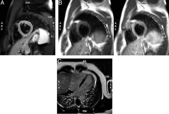
Figure 1. Cardiovascular magnetic resonance of acute myocarditis. Panel A: T2-weighted image of LV myocardial edema showing global bright signal intensity (ratio 2.2) of the left ventricle relative to the myocardium. Panel B: Early enhancement (T1-weighted spin echo) before (left) and after (right) Gd administration; enhancement ratio 5.4. Panel C: Arrows indicating late enhancement (T1-weighted gradient echo sequence with myocardial nulling) 10 minutes after Gd. Gd indicates gadolinium; and LV, left ventricular.
such as transplant rejection, acute myocarditis, and acute myocardial infarction74 (Figure 1A).
Another noncontrast tissue characterization technique relates to the T2* relaxation of the tissue. T2* times are significantly altered by the myocardial iron content; their quantification provides an excellent marker for iron overload (see Section 3.5.8, Hemochromatosis).
Contrast agents such as gadolinium (Gd) chelates shorten the T1 relaxation time within the surrounding tissue and increase the signal intensity of regions with high Gd concentration during T1-weighted imaging. In essence, Gd chelates facilitate water visualization in the intravascular (blood) or in the extravascular organ tissue space. This can be used to selectively identify areas with reduced or increased “uptake” of Gd (Figure 1B). Regional differences of Gd inflow characteristics after intravenous injection (“first-pass imaging”) can be used to assess myocardial perfusion. T1-weighted sequences with 3 to 5 slices per heartbeat have been used in the diagnostic workup for CAD with a very high negativepredictive value.52,75 Early after the first pass of Gd, a significant fraction of the injected Gd enters the interstitial space. Several minutes after intravenous administration of Gd, the larger volume of distribution available in necrotic or fibrotic myocardium results in a higher concentration of contrast agent than what is present in viable myocardium. This is typically referred to as “delayed (hyper)enhancement” or “late gadolinium enhancement” (LGE).76 The transmural extent of myocardial scars as defined by LGE predicts functional recovery after revascularization77 and is related to prognosis.78
Patterns other than the endocardial accumulation of LGE can occur. For example, LV epicardial and midwall enhancement are known to be associated with infectious causes of myocardial inflammation (Figure 1C). Also, inflammatory conditions involving the heart, such as with sarcoidosis, are associated with midwall accumulation of LGE. A special mechanism may be the cause for Gd accumulation in cardiac amyloidosis. Data indicate that a molecular binding of Gd to amyloid may lead to the extensive uptake of the agent in myocardial tissue, typically associated with a very rapid washout from blood.79
3.Important Applications
3.1.Heart Failure
CMR may be used for assessment of LV and RV size and morphology, systolic and diastolic function, and for characterizing myocardial tissue for the purpose of understanding the etiology of LV systolic or diastolic dysfunction. The writing committee recognizes the potential capabilities of spectroscopic techniques for acquiring metabolic information of the heart when evaluating individuals with heart failure.
When assessing patients with heart failure, CMR is useful in several aspects.80 Questions that may be answered by CMR include understanding of the presence and severity of morphological and functional abnormalities of the LV or RV myocardium, determining the underlying etiology (eg, isch-