


42 |
M. Díez Ferrer and A. Rosell |
|
|
Fig. 3.4 Two bronchoscopists and one trained nurse performing ultrathin bronchoscopy with virtual bronchoscopic navigation (LungPoint®) in the endoscopy suite. Note that fuoroscopy is also used
Optional equipment:
•\ Cone-beam CT to corroborate the position of the ultrathin bronchoscope and the sampling instrument relative to the lesion.
•\ Virtual bronchoscopy, virtual bronchoscopic navigation, or electromagnetic navigation to assist procedure planning and guiding of the ultrathin bronchoscope to the peripheral pulmonary lesion.
In Fig. 3.4, ultrathin bronchoscopy with virtual bronchoscopic navigation is performed under general anesthesia in the endoscopy suite.
choscope and to assist sampling, although CT or cone-beam CT can also be used to corroborate the position of the ultrathin bronchoscope and the sampling instrument relative to the lesion. Sampling is performed with instruments of limited size that can be passed through the working channel of the ultrathin bronchoscope.
To develop all the aforementioned aspects and to provide an integrated understanding of the technologies that can be coupled with ultrathin bronchoscopy, we have structured ultrathin bronchoscopy in the following steps: procedure planning, target approximation, position veri cation, and sampling.
Procedure Description |
Procedure Planning |
In general terms, the procedure starts by conducting an accurate planning of the bronchial route leading to the peripheral lesion, either through ne reading of a high-resolution chest CT or supported by a planning software. During the procedure of ultrathin bronchoscopy, target approximation can be accomplished after memorization of the planned bronchial route or assisted by navigation software. Fluoroscopy is usually used to track the position of the ultrathin bron-
First thing to consider when planning the procedure on a CT image is the presence of a bronchus or artery afferent or, within the peripheral lesion, the so called bronchus sign and artery sign. When present, the diagnostic yield of the procedure is signi cantly higher [10–12]. Examples of bronchus and artery signs are shown in Fig. 3.5.
If the slice thickness of the CT is suf cient to allow identi cation of small bronchi and if a bronchus leading to the lesions is present, then it
Данная книга находится в списке для перевода на русский язык сайта https://meduniver.com/
3 Ultrathin Bronchoscopy: Indications and Technique |
43 |
|
|
will be feasible to reconstruct a three-dimensional bronchial route to the peripheral nodule. With this information alone, highly trained bronchoscopists have the ability to reconstruct the bronchial route from the CT and mentally reproduce the route during the procedure. Fortunately, complementary technologies have been developed to assist procedure planning. The mainstay technology for procedure planning is virtual bronchoscopy. Dedicated software is used for multiplanar
Fig. 3.5 Chest CT showing both a bronchus and artery leading to a PPL (bronchus and artery signs, respectively)
reconstruction of CT images and segmentation of the airways. After manual identi cation of the nodule in the CT, the bronchial route to the nodule can be followed through the inner lumen of the segmented airways. This route can be memorized by the bronchoscopist to be reproduced in the bronchoscopy suite. Otherwise, a virtual bronchoscopic navigation platform can be used to perform virtual bronchoscopy in the endoscopy suite and match virtual and endoscopic images during the procedure to facilitate orientation. A screenshot of a virtual bronchoscopy is shown in Fig. 3.6.
Target Approximation
In order to guarantee patient stillness during the procedure, the authors of the present text prefer performing ultrathin bronchoscopy under general anesthesia and laryngeal mask or endotracheal intubation. This facilitates bronchoscope manipulation in the smaller subsegmental bronchi, allows application of short controlled apneas to gain greater operator control during sampling, and may also avoid accidental damaging of theber bronchoscope due to abrupt patient movements. It has to be pointed out that, in some cases, cutting some centimeters of the proximal end of the orotracheal tube may be necessary to warrant full insertion of the 600 mm working length of the ultrathin bronchoscope and avoid falling
Fig. 3.6 Procedure plani cation with a virtual bronchoscopy navigation system (LungPoint®). This system allows matching the virtual bronchoscopy seen in the Figure with the endoscopic images during the procedure
44 |
M. Díez Ferrer and A. Rosell |
|
|
short to the lung periphery, especially in relatively tall patients. However, general anesthesia is not mandatory and moderate sedation is preferred in many centers.
As previously mentioned, target approximation can be accomplished after memorization of the planned bronchial route or assisted by virtual bronchoscopic navigation software. This software allows matching virtual and endoscopic images during the procedure to guide the ultrathin bronchoscope through every bifurcation leading to the afferent bronchus. In either case, fuoroscopy is usually used to track the real-time position of the ultrathin bronchoscope.
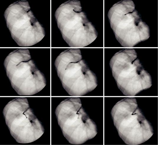
Fluoroscopy was the rst imaging technique used for guiding the ultrathin bronchoscope to the nodule [1]. Biplanar fuoroscopy is desirable but, when not accessible, the C-arm must be rotated adequately. Although it is not a guidance tool per se, it can be useful to con rm the position and direction of the bronchoscope throughout the procedure and relative to the peripheral lesion, as long as the lesion is fuoroscopically visible (see Fig. 3.7). Unfortunately, peripheral pulmonary nodules are not always visible with fuoroscopy. In one study, the authors reported that from a population of 1369 individuals at high risk for lung cancer, 15 small peripheral lung
a |
b |
c |
d |
e |
f |
g |
h |
i |
Fig. 3.7 Fluoroscopy Fluoroscopy can be used to track down the path leading to the peripheral pulmonary lesion. To assure that the right direction is followed, fuoroscopy
is performed in every bifurcation (a–i). Whenever misdirected, the bronchoscope is pulled backward to the previous bifurcation and another bronchus followed
Данная книга находится в списке для перевода на русский язык сайта https://meduniver.com/
3 Ultrathin Bronchoscopy: Indications and Technique |
45 |
|
|
cancers were detected with low-dose CT. Of these, 73% had negative chest radiography [13]. Because CT allows for nodule detection independent of size, localization, and characteristics of the PPL, it has also been used for verifying the position of the instrument when exploring the peripheral airways [14].
Virtual bronchoscopic navigation can also be used to assist in lesion approximation. When using virtual bronchoscopic navigation systems, a trained bronchoscopist is needed to perform the virtual bronchoscopy through the previously selected path. The assistant bronchoscopist will guide the operator through the airways and will indicate the right direction in each encountered bifurcation. To date, there is only one large randomized trial comparing ultrathin bronchoscopy with and without use of virtual bronchoscopic navigation. This study by Asano et al. showed no signi cant differences in diagnostic yield on both groups (67.1% vs. 59.9%; p = 0.173) in 350 patients with peripheral nodules ≤3 cm. However, subgroup analysis of these data showed that the navigation system could be helpful for achieving nodules located in the peripheral third of the lung, those invisible in the postero-ante- rior radiographs and when located in the upper right lobe. Fluoroscopy was used in both groups to ensure location of the ultrathin bronchoscope and sampling of the desired location [15]. In a later study, we compared ultrathin bronchoscopy with and without virtual bronchoscopic navigation and evaluated the infuence of segmentation on diagnostic yield [16]. We compared 55 cases of virtual bronchoscopic navigationguided ultrathin bronchoscopy to 110 unguided controls. Although the diagnostic yield did not differ between both arms (47% and 40%, respectively; p = 0.354), an 85% diagnostic yield was observed when segmentation was optimal and the peripheral nodule was endobronchial, compared to a 30% diagnostic yield when the segmentation was suboptimal and a 20% diagnostic yield when segmentation was optimal but the lesion extrabronchial. In fact, the position of the lesion relative to the bronchus, that is, if the lesion is endoor extrabronchial, will determine the diagnostic yield of the procedure. This point is fur-
ther commented in the next section on position veri cation.
During target approximation with an ultrathin bronchoscope, several considerations should be made. First of all is the limited suction capability of the ultrathin bronchoscope due to the small working channel. Therefore, if abundant or thick secretions are present, it might be recommended that bronchial hygiene with a conventional bronchoscope is performed either prior to starting or during the procedure. Also, angulation of the tip of the ultrathin bronchoscope can be challenging in the upper lobes. Sometimes it might be simply impossible to overcome some anatomical angulations with the ultrathin bronchoscope. Leaving the biopsy forceps inside the working channel may provide greater stiffness to the bronchoscope in some cases. Finally, another disadvantage of ultrathin compared to conventional bronchoscopes is the quality of the endoscopic vision, not only in regard to technical manufacturing details but also to bronchial anatomy in the lung periphery. Particularly, a greater collapsibility is found in the periphery due to progressive loss of stiffness in the intrapulmonary airways. To overcome bronchial collapsibility and improve endoscopic vision in the peripheral airways, it is recommended that secretions are not aspirated and saline is continuously instilled instead. In our institution, a 50 mL syringe with room-temperature saline is connected to the working channel of the ultrathin bronchoscope. The assistant instills saline as requested, thus facilitating bypassing of secretions and bronchial lumen widening. A view of ultrathin bronchoscopy in peripheral airways under saline infusion is shown in Fig. 3.8.
Position Verifcation
Once the peripheral pulmonary lesion is approximated and no endobronchial abnormality is visualized, then two issues might have occurred: either approximation to the lesion has not been accurate enough, or the lesion is extrabronchial. At this point, fuoroscopy, CT, or cone-beam CT can be used to verify the position of the ultrathin bronchoscope relative to the lesion. Also, a radial

46 |
M. Díez Ferrer and A. Rosell |
|
|
a
b
Fig. 3.8 Ultrathin bronchoscopy in peripheral airways: (a) 50 mL aliquot with saline connected to the working channel. (b) Views before and after saline infusion
Данная книга находится в списке для перевода на русский язык сайта https://meduniver.com/