


Part ten. Summary of upper limb innervation
Brachial plexus
The roots of the plexus (the anterior rami of C5–T1 nerves) are between the scalene muscles, the trunks in the posterior triangle, the divisions behind the clavicle, and the cords arranged round the second part of the axillary artery. About 10% of plexuses are prefixed (from C4–C8) and 10% postfixed (C6–T2).
The preganglionic sympathetic fibres for the upper limb originate mainly from the second to fifth thoracic spinal cord segments, and ascend along the sympathetic trunk. Grey rami communicantes carrying postganglionic fibres from the middle and inferior cervical and the first thoracic sympathetic ganglia join the roots of the brachial plexus. They hitch-hike through the plexus and its branches, remaining in the nerves until very near their area of supply. Thus, the brachial artery receives sympathetic fibres from the median nerve in the arm and arterioles in a finger receive filaments from digital nerves. In the skin, in addition to arterioles, sweat glands and the arrectores pilorum muscles receive sympathetic innervation.
Branches of the roots
C5 Dorsal scapular
C5, 6 Nerve to subclavius
C5–7 Long thoracic.
T he dorsal scapular nerve (C5) runs down deep to levator scapulae and the two rhomboids, supplying all three muscles. Lying on serratus posterior superior, it forms a neurovascular bundle with the descending scapular vessels alongside the vertebral border of the scapula (Fig. 2.5).
The nerve to subclavius (C5, 6) passes down over the trunks of the plexus and in front of the subclavian vein. It frequently contains accessory phrenic fibres which join the phrenic nerve in the superior mediastinum.
The long thoracic nerve (C5, 6, 7) forms on the first digitation of the serratus anterior muscle and runs vertically downwards just behind the midaxillary line, deep to the fascia over the muscle.
Branch of the upper trunk
The suprascapular nerve (C5, 6), runs backwards beneath the fascial floor of the posterior triangle, then passes beneath the transverse scapular ligament and round the lateral border of the scapular spine. The nerve supplies supraspinatus, infraspinatus, and the shoulder and acromioclavicular joints.
Branches of the lateral cord
C5–7 Lateral pectoral
C5–7 Musculocutaneous
C5–7 Lateral root of median.
The lateral pectoral nerve (C5, 6, 7) passes through the clavipectoral fascia and supplies the upper
fibres of pectoralis major. A communicating branch to the medial pectoral nerve crosses in front of the first part of the axillary artery and contributes to the supply of pectoralis minor (Fig. 2.48).
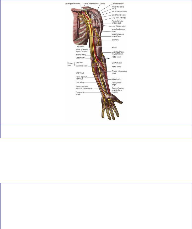
Figure 2.48 Nerves on the anterior aspect of the left upper limb.
The musculocutaneous nerve (C5–7) is muscular to the flexors in the arm and cutaneous in the forearm. Emerging from the medial cord high in the axilla, it supplies coracobrachialis, then pierces that muscle to slope down between biceps and brachialis, supplying both muscles. Emerging at the lateral border of the biceps tendon, it pierces the deep fascia at the flexure crease of the elbow. Now called the lateral cutaneous nerve of the forearm, it supplies skin from elbow to wrist by an anterior and a posterior branch along the radial border of the forearm (Fig. 2.49).
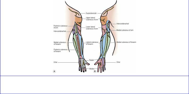
Figure 2.49 Cutaneous nerves of the right upper limb, A from behind, B from the front. Compare with the dermatomes on Figure 1.9, page 14.
The lateral root of the median nerve (C5–7) is joined by the medial root at the lateral side of the axillary artery to form the main nerve (see below).
Branches of the medial cord
C8, T1 Medial pectoral
C8, T1 Medial root of median
C8, T1 Medial cutaneous of arm
C8, T1 Medial cutaneous of forearm
C7, 8, T1 Ulnar.
The medial pectoral nerve (C8, T1) supplies pectoralis minor and then pierces it to supply the lower (sternocostal) fibres of pectoralis major.
The medial root of the median nerve (C8, T1) crosses the front of the axillary artery to join its companion and form the median nerve (C5–T1) at the lateral side of the artery.
The median nerve (C5–8, T1; Fig. 2.48) supplies most of the flexor muscles of the forearm, but only the three thenar muscles and two lumbricals in the hand. It is cutaneous to the flexor surfaces and nail beds of the three and a half radial digits and a corresponding area of palm. Formed lateral to the axillary artery, the nerve leaves the axilla and crosses in front of the brachial artery at the middle of the arm. At the elbow it lies medial to the artery beneath the bicipital aponeurosis. It passes between the two heads of pronator teres and deep to the fibrous arch of flexor digitorum superficialis. Adherent to the deep surface of the muscle, it emerges on the radial side of its tendons, lying deep to the palmaris longus tendon before passing through the carpal tunnel into the hand.
Branches. In the arm the nerve gives sympathetic filaments to the brachial artery, a twig to the elbow joint and may supply pronator teres above the elbow. In the cubital fossa, it supplies pronator teres,
palmaris longus, flexor carpi radialis and flexor digitorum superficialis. In the forearm it gives off the anterior interosseous nerve, which descends on the interosseous membrane to the wrist. The anterior interosseous is the nerve of the deep flexor compartment; it supplies the radial half (usually) of flexor digitorum profundus, all of flexor pollicis longus and pronator quadratus and is sensory to the wrist and carpal joints. The palmar cutaneous branch of the median nerve pierces the deep fascia just above the flexor retinaculum and supplies more than half of the thumb side of the palm.
In the hand the median nerve gives a muscular (recurrent) branch , which recurves around the distal border of the flexor retinaculum to supply the three thenar muscles (abductor and flexor pollicis brevis, and opponens pollicis), and palmar digital branches; these supply both sides of the thumb, index and middle fingers, the radial side of the ring finger and characteristically the two radial lumbricals. The palmar digital branches supply the flexor skin of the radial three and a half digits, and the nail beds and the dorsal skin over the distal and middle phalanges of these digits.
T he medial cutaneous nerve of the arm (C8, T1) is the smallest branch of the plexus. It is sometimes replaced entirely by the intercostobrachial nerve. It runs down with the axillary vein to pierce the deep fascia and supply skin on the medial aspect of the arm.
The medial cutaneous nerve of the forearm (C8, T1) is a much bigger nerve than the last. It runs down between axillary artery and vein and pierces the deep fascia half way to the elbow, often in common with the basilic vein. It supplies the lower part of the front of the arm above the elbow and then divides into anterior and posterior branches to supply the skin along the ulnar border of the forearm down to the wrist. In the forearm it is symmetrical with the lateral cutaneous nerve (musculocutaneous) and the two meet without overlap along the anterior axial line. Their territories are separated posteriorly by the posterior cutaneous branch of the radial nerve.
The ulnar nerve (C7, 8, T1) is the direct continuation of the medial cord (C8, T1), with additional C7 fibres picked up in the axilla, usually from the lateral cord. The nerve supplies some flexor muscles on the ulnar side of the forearm, most of the intrinsic muscles of the hand and the skin of the ulnar one and a half digits.
Running down between the axillary artery and vein, behind the medial cutaneous nerve of the forearm, the ulnar nerve pierces the medial intermuscular septum and descends in the groove on the back of the base of the medial epicondyle. It passes between the two heads of flexor carpi ulnaris and enters the flexor compartment of the forearm. It descends on flexor digitorum profundus, under cover of flexor carpi ulnaris. Here it is joined on its lateral side by the ulnar artery. The two emerge from beneath the tendon of flexor carpi ulnaris just above the wrist and cross the flexor retinaculum lateral to the pisiform bone.
Branches. Articular twigs are given to the elbow joint as the nerve lies on its medial collateral ligament. In the forearm the nerve supplies flexor carpi ulnaris and the ulnar half (usually) of flexor digitorum profundus. It has a palmar cutaneous branch which pierces the deep fascia above the flexor retinaculum to supply skin over the hypothenar muscles. A dorsal cutaneous branch winds around the lower end of the ulna deep to the tendon of flexor carpi ulnaris and is distributed to the dorsal skin of one and a half fingers (except that over the distal phalanx of the little finger, and the middle and distal phalanges of the ring finger) and a corresponding area of the back of the hand. Not uncommonly it supplies two and a half instead of one and a half fingers.
The ulnar nerve divides on the flexor retinaculum alongside the pisiform bone. The superficial branch runs distally beneath palmaris brevis (which it supplies) and is distributed by two digital branches to the ulnar one and a half fingers, including their nail beds and the skin on the dorsum not supplied by the dorsal branch.
The deep branch passes deeply between abductor and flexor digiti minimi then through opponens. It supplies all three hypothenar muscles. It grooves the distal border of the hook of the hamate and crosses the palm in the concavity of the deep palmar arch, supplying the two ulnar lumbricals and all the interossei, both palmar and dorsal. It ends by supplying adductor pollicis.
Branches of the posterior cord
C5, 6 Upper subscapular
C6–8 Thoracodorsal
C5, 6 Lower subscapular
C5, 6 Axillary
C5–8, T1 Radial.
The upper and lower subscapular nerves (C5, 6) supply the respective parts of subscapularis, with the lower nerve also innervating teres major.
The thoracodorsal nerve (nerve to latissimus dorsi) (C6–8) inclines forwards and enters the deep surface of latissimus dorsi just behind the anterior border. Its terminal part lies anterior to the thoracodorsal artery and it is vulnerable in operations on the axillary lymph nodes.
The axillary nerve (C5, 6) passes backwards through the quadrangular space (Fig. 2.9), lying above the posterior circumflex humeral vessels and the glistening tendon of latissimus dorsi (as it overlaps teres major) just below the capsule of the shoulder joint, which it supplies. In the quadrangular space it divides. The posterior branch supplies teres minor and winds around the posterior border of deltoid (Fig. 2.17), supplying it, and continuing as the upper lateral cutaneous nerve of the arm to supply skin over the lower half of deltoid and the upper part of the back of the arm. The anterior branch curves round the surgical neck of the humerus, deep to deltoid (Fig. 2.5) which it supplies as well as a small area of overlying skin.
The radial nerve (C5–8, T1) is the nerve of the extensor compartments of the arm and forearm, supplying skin over them and on the dorsum of the hand. A direct continuation of the posterior cord, the radial nerve passes beyond the posterior wall of the axilla, below the easily identifiable tendon of latissimus dorsi, running dorsally downwards between the long and medial heads of triceps. It spirals across the back of the humerus, between the lateral and medial heads of triceps, lying on the radial groove of the bone, deep to the lateral head (Fig. 2.17). It pierces the lateral intermuscular septum one-third of the way down from the deltoid tuberosity to the lateral epicondyle. In the flexor compartment of the lower arm it descends in the intermuscular slit between brachialis and brachioradialis. After giving off the posterior interosseous branch, the rather slender remnant, purely cutaneous now, retains the name of radial nerve. It runs down the flexor compartment of the forearm, winds around the lower end of the radius deep to the tendon of brachioradialis and crosses abductor pollicis longus, extensor pollicis brevis and extensor pollicis longus (as one of the contents of the
anatomical snuffbox) to reach the back of the hand. Here it supplies the skin of the radial two and a half or three and a half digits (falling short of the nail beds and distal and middle phalanges) and a corresponding area of the dorsum of the hand.
Branches. The posterior cutaneous nerve of the arm arises in the axilla and pierces the deep fascia to supply a strip of skin along the extensor surface of the arm down to the elbow. The triceps is supplied by four radial nerve branches. They arise as nerves to the long, medial, lateral and medial heads, the first two being given off in the axilla and the last two behind the humerus. The first branch to the medial head (the ulnar collateral nerve) runs down with the ulnar nerve to enter the lower part of the medial head. The second branch to the medial head continues deep to triceps to supply anconeus.
The lower lateral cutaneous nerve of the arm pierces the lateral head of triceps to supply skin over the lateral surface of the arm down to the elbow. In common with it arises the posterior cutaneous nerve of the forearm which runs straight down behind the elbow to supply a strip of skin over the extensor surface of the forearm as far as the wrist.
While lying in the flexor compartment of the forearm between brachialis and brachioradialis, the main trunk gives a small branch to the lateral part of brachialis and supplies brachioradialis and extensor carpi radialis longus. At the level of the lateral epicondyle it gives off the posterior interosseous branch, and then continues on as the terminal cutaneous branch already described.
The posterior interosseous nerve supplies extensor carpi radialis brevis and supinator in the cubital fossa, and then spirals down around the upper end of the radius between the two layers of supinator to enter the extensor compartment of the forearm. It crosses abductor pollicis longus, dips down to the interosseous membrane and runs to the back of the wrist. In the extensor compartment it supplies seven more muscles; three extensors from the common extensor origin (extensor digitorum, extensor digiti minimi, and extensor carpi ulnaris), the three thumb muscles (abductor pollicis longus, extensor pollicis brevis and extensor pollicis longus) and extensor indicis. It is sensory to the wrist and carpal joints.