

Guidelines-peripheral-AD-FT
.pdfESC Guidelines |
2881 |
|
|
bypasses revealed lower risk for primary occlusion with Dacron grafts (HR 0.71 vs. polytetrafluoroethylene, P ¼ 0.003), but longterm results are awaited. The pooled weighted data for 1-, 3-, and 5-year primary patency rates for femorodistal (tibial or pedal) bypasses are, respectively, reported at 85, 80, and 70% for venous bypass and 70, 35, and 25% with a prosthetic graft.6 In one trial with above-knee grafting, the 4-year primary and secondary patency rates were significantly better with the use of the saphenous vein (73% and 90%, respectively) compared with polytetrafluoroethylene (47% and 47%, both P ,0.05) and Dacron (54% and 60%, both P ,0.01). Two trials comparing in situ and reversed saphenous vein grafts to the aboveand belowknee popliteal artery revealed no differences in primary and secondary patency as well as survival with an intact limb. Three trials comparing polytetrafluoroethylene with human umbilical vein showed significantly higher secondary patency rates with the latter.300 Comparison of polytetrafluoroethylene grafts with and without a vein cuff found no difference in above-knee grafts. However, primary patency for below-knee bypass was higher
with a polytetrafluoroethylene prosthesis with vein cuff bypass at 2 years.296,301
Only one randomized trial has compared angioplasty with infrainguinal bypass. In the Bypass versus Angioplasty in Severe Ischaemia of the Leg (BASIL) trial, 452 patients with severe limb ischaemia due to infrainguinal disease were randomized to angioplasty or infrainguinal bypass. The primary endpoint was amputation-free survival. Secondary endpoints included all-cause mortality, morbidity, reintervention, quality of life, and hospital costs.302 The 30-day mortality was similar in both groups (5% for surgery and 3% for angioplasty). However, surgery was associated with a higher morbidity (57% vs. 41%), mainly due to myocardial infarction and wound infection. Moreover, surgery was more expensive during the first year, due to the longer hospital stay. The 6-month amputation-free survival was similar in both strategies. Angioplasty patients presented higher failure rates (20% vs. 3% at 1 year), resulting in higher reintervention rates (27% vs. 17%). These results suggest that surgical revascularization is superior to angioplasty in patients with good quality veins for bypass. Recently additional data with a longer follow-up period (.3 years) have been published211,303: overall, there was no significant difference in amputation-free or overall survival between the two strategies. However, for patients who survived for at least 2 years after randomization, the surgery-first revascularization strategy was associated with a significant increase in subsequent overall survival and a trend towards improved amputation-free survival.
One small, randomized trial comparing stenting with femoral-to-above-knee prosthetic bypass found no difference in primary and secondary patency rates at 12 months.290 Further trials are required comparing infrainguinal stenting with surgery.
Another infrainguinal surgical reconstruction is the profundoplasty, the correction of a stenosis at the origin of the deep femoral artery. It may be considered as an inflow procedure, instead of a distal bypass, in the presence of an excellent proximal inflow, .50% stenosis of the proximal third of the profunda femoris artery, and the presence of excellent collateral flow to the tibial vessels.
Secondary amputation should be performed when revascularization has failed and reintervention is no longer possible or when the
limb continues to deteriorate because of infection or necrosis despite a patent graft. The goals of secondary amputation are: ischaemic pain relief, complete removal of diseased, necrotic, or infected tissue, and construction of a stump suitable for ambulation with prosthesis.
Recommendation for surgical revascularization in patients with LEAD
|
|
|
|
|
|
|
|
|
|
|
|
|
Recommendations |
Classa |
Levelb |
Ref c |
|
|
When surgery is considered |
|
|
|
|
|
to revascularize infrailiac |
|
|
|
|
|
lesions, the autologous |
I |
A |
296, 304 |
|
|
saphenous vein is the bypass |
|
|
|
|
|
graft of choice. |
|
|
|
|
|
|
|
|
|
|
|
|
|
|
|
|
aClass of recommendation. bLevel of evidence. cReferences.
LEAD ¼ lower extremity artery disease.
4.5.3.3.3 Surveillance
Clinical surveillance including clinical assessment and ankle pressure follow-up should be performed following any revascularization procedure. Although there is no consensual protocol of surveillance, regular monitoring of revascularized limbs can permit a prompt prophylactic intervention (e.g. repair of an arterial bypass at high risk of occlusion according to DUS criteria) and improve long-term patency.305 However, in a multicentre randomized trial including 594 patients with vein grafts, a systematic duplex surveillance programme was not found to be beneficial in terms of graft patency and limb survival rates, and was less cost-effective than clinical surveillance.306 DUS could be useful to select high-risk prosthetic grafts, which may require long-term anticoagulation to reduce the risk of graft thrombosis,307 but these data are based on observational series and require confirmation in trials.
4.5.3.3.4 Antiplatelet and anticoagulant therapy after revascularization
Beyond potential benefits of antiplatelet agents to reduce fatal or non-fatal CVD events in patients with LEAD, these drugs are also specifically proposed after revascularization to improve patency rates. In a meta-analysis of 16 studies, the effect of antiplatelet therapy administered post-operatively was evaluated in patients receiving infrainguinal bypasses.308 Antiplatelet treatment with aspirin or a combination of aspirin and dipyridamole had an overall positive effect on primary patency 12 months after the procedure (OR 0.59, 95% CI 0.45–0.79). Subgroup analysis indicated that patients receiving a prosthetic graft were more likely to benefit from administration of platelet inhibitors than patients treated with venous grafts.308 The multicentre, prospective Dutch Bypass Oral Anticoagulants or Aspirin (BOA) trial309 randomized 2690 lower extremity bypass patients into two groups: anticoagulation (with the international normalized ratio targeted within the 3.0–4.5 interval) vs. antiplatelet therapy (aspirin 80 mg/day). Overall patency rates did not differ, but the results of a subgroup analysis suggested that oral anticoagulation improved vein graft patency compared with aspirin. Conversely, aspirin

2882 |
ESC Guidelines |
|
|
improved prosthetic graft patency vs. anticoagulation. Notably, the risk of major bleeding was two-fold higher in the anticoagulation group. In another trial,310 665 patients undergoing femoropopliteal bypass were randomized to aspirin (325 mg/day) plus warfarin (goal international normalized ratio 1.4–2.8) vs. aspirin (325 mg/ day) alone. This trial failed to demonstrate any improvement in terms of graft patency with dual therapy. However, the results were in favour of combination therapy for patients with prosthetic bypasses. The haemorrhagic risk doubled when warfarin was added to aspirin. In another randomized study,311 warfarin (international normalized ratio 2.0–3.0) plus aspirin (325 mg/day) was compared with aspirin (325 mg/day) alone in 56 patients with highrisk vein grafts (defined as poor arterial run-off, suboptimal vein conduit, and repeat interventions). At 3 years, patency and limb salvage rates were significantly higher in those receiving warfarin and aspirin, with in turn higher bleeding rates with this combination. Recently, the Clopidogrel and Acetylsalicylic Acid in Bypass Surgery for Peripheral ARterial disease (CASPAR) randomized double-blind trial assessed the efficacy of aspirin plus clopidogrel vs. aspirin alone to increase primary patency, limb salvage, and survival in patients receiving a below-knee bypass graft.312 Among the 851 patients enrolled, almost 70% had a venous graft and 30% a prosthetic graft. After a mean follow-up of 1 year, no overall difference was found regarding the combined primary outcome between the two groups. Subgroup analysis was in favour of a beneficial effect of clopidogrel in association with aspirin in prosthetic grafts. The number needed to treat using the dual antiplatelet therapy to save one limb after below-knee surgery was dramatically low, estimated at 10.2 patients.
The role of anticoagulation after infrainguinal balloon PTA and stenting has been assessed in three prospective randomized trials.313 None of these trials showed any significant improvement in arterial patency with the use of anticoagulation therapy, while bleeding complications increased.313 Yet, anticoagulation therapy cannot be recommended routinely after lower extremity PTA or stenting.
4.5.3.4 Stem cell and gene therapy for revascularization
The development of novel therapies to stimulate neovascularization, known as therapeutic angiogenesis, is based on the use of angiogenic factors or stem cells to promote revascularization and remodelling of collaterals with the aim of ameliorating symptoms and preventing amputation.
While several trials reported relief of ischaemic symptoms, functional improvement, and prevention of amputation,314 – 317 others failed to confirm this early promise of efficacy.318 – 320
For autologous cell transplantation in humans, bone marrow and peripheral blood are rich sources of stem and progenitor cells. Bone marrow is currently the most frequent source of cells used for clinical repair trials, because it is easy to obtain and no complex purification steps are required. Another advantage is that it contains a variety of stem and progenitor cells with suggested superiority over one selected type of progenitor cell. With the many different cell types that can be used for stem cell therapy, it is not yet clear which ones are the most promising.321 In a recent meta-analysis of 37 trials, autologous cell therapy was effective in improving surrogate indexes of ischaemia, subjective
Recommendations for antiplatelet and anticoagulant therapy after revascularization
|
|
|
|
|
|
|
|
|
|
|
|
|
Recommendations |
Classa |
Levelb |
Ref c |
|
|
Antiplatelet therapy with |
|
|
|
|
|
aspirin is recommended in all |
|
|
|
|
|
patients with angioplasty for |
I |
C |
|
|
|
LEAD to reduce the risk of |
|
|
|
|
|
systemic vascular events. |
|
|
|
|
|
|
|
|
|
|
|
Dual antiplatelet therapy with |
|
|
|
|
|
aspirin and a thienopyridine |
|
|
|
|
|
for at least one month is |
I |
C |
|
|
|
recommended after |
|
|
||
|
|
|
|
|
|
|
infrainguinal bare-metal-stent |
|
|
|
|
|
implantation. |
|
|
|
|
|
|
|
|
|
|
|
Antiplatelet treatment with |
|
|
|
|
|
aspirin or a combination |
|
|
|
|
|
of aspirin and dipyridamole |
I |
A |
308 |
|
|
is recommended after |
|
|
|
|
|
infrainguinal bypass surgery. |
|
|
|
|
|
|
|
|
|
|
|
Antithrombotic treatment |
|
|
|
|
|
with vitamin K antagonists |
|
|
|
|
|
may be considered after |
IIb |
B |
309 |
|
|
autogenous vein infrainguinal |
|
|
|
|
|
bypass. |
|
|
|
|
|
|
|
|
|
|
|
Dual antiplatelet therapy |
|
|
|
|
|
combining aspirin and |
|
|
|
|
|
clopidogrel may be considered |
IIb |
B |
312 |
|
|
in the case of below-knee |
|
|
|
|
|
bypass with a prosthetic graft. |
|
|
|
|
|
|
|
|
|
|
|
|
|
|
|
|
aClass of recommendation. bLevel of evidence. cReferences.
LEAD ¼ lower extremity artery disease.
symptoms, and hard endpoints (ulcer healing and amputation). Patients with thromboangiitis obliterans showed larger benefits than patients with atherosclerotic LEAD. The TAMARIS study is the largest randomized placebo-controlled trial of gene therapy in CLI, including .520 patients from 30 countries with CLI and skin lesions, unsuitable for standard revascularization. This study found no statistical difference between the two groups regarding the primary efficacy endpoint of death or first major amputation on the treated leg, whichever came first (37.0% vs. 33.2%, P ¼ 0.48).322 At present angiogenic gene and stem cell therapy are still being investigated and it is too early to give firm recommendations.
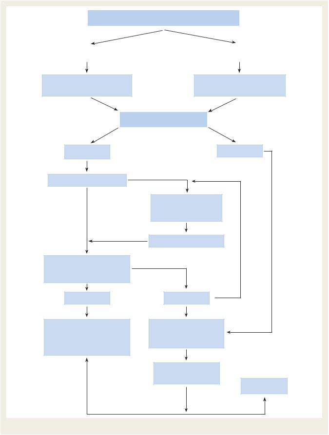
4.5.4 Management of intermittent claudication
The management of intermittent claudication consists of optimal risk factor control in order to improve the vital prognosis (see Section 3.4) and the symptoms. Therapeutic options to relieve symptoms are non-invasive (mostly exercise therapy and drug therapy) or invasive (revascularization). An algorithm for the management of intermittent claudication is proposed in Figure 3. With the increasing use of endovascular therapy to improve walking distance, there is an apparent need to compare it with ‘supervised exercise training’. In a study of 51 patients with intermittent

ESC Guidelines |
2883 |
|
|
Management of intermittent claudication
Conservative therapy
(Risk factors control, exercise training, pharmacotherapy 3–6 months)
Favourable results |
|
No favourable results |
|
|
|
Image lesions
Endovascular therapy feasible?
yes
no
Endovascular therapy
Bypass surgery
Follow up: - Symptoms
- CV risk control
Figure 3 Algorithm for the management of intermittent claudication. CV ¼ cardiovascular.
claudication, there was no significant difference in walking distance or quality of life 2 years after treatment.323 More recently, a randomized controlled study initiated in 151 patients with intermittent claudication confirmed no difference in quality of life 12 months after intervention. However, this study showed a higher cost for the endovascular intervention group.279 The adjuvant benefit of endovascular therapy to ‘supervised exercise training’ associated with best medical therapy has been assessed in patients with mild to moderate intermittent claudication.324 Although no difference in quality of life was reported in this study, at 24 months the improvement in walking distance in the angioplasty group was 38% greater than that in the control group in the case of femoropopliteal lesions, and 78% in the case of aortoiliac lesions. The ongoing Claudication: Exercise Versus Endoluminal Revascularization (CLEVER) trial will provide important insights into the indications of these therapeutic options in the management of patients with intermittent claudication.325
4.5.4.1 Medical treatment
In patients with intermittent claudication, the primary goal is to reduce the risk of CVD morbidity and mortality. This risk is present in all patients with LEAD, including those with mild, atypical, and even no symptoms.2,326 Therefore, the management and control of risk factors is necessary in every patient with LEAD,
to reach the goals of secondary prevention. Among them, smoking cessation also provides the most noticeable improvement in walking distance when combined with regular exercise training, especially when lesions are located below the femoral arteries.
Symptoms can be improved by exercise training (preferably supervised) and drug therapy. Walking tests on the treadmill should be performed regularly to assess the evolution objectively. Patients should also be advised to keep a logbook to follow their home training and the evolution of their walking distance and symptoms. The logbook can help the patient adhere to medical advice. In the case of typical claudication, drug therapy to improve walking distance can be initiated.
In many patients with mild to moderate symptoms, these first steps will lead to a significant improvement in claudication and in quality of life. In this case, training (and eventually drug therapy) should be continued and the patients should be evaluated at regular intervals. ABI should be controlled periodically, although substantial functional improvement may not necessarily follow significant ABI change. The risk factor profile should be checked regularly and treatment adapted accordingly.
4.5.4.2 Interventional therapy
In severe cases with disabling claudication, medical therapy including ‘supervised exercise training’ is often insufficient to improve

2884 |
ESC Guidelines |
|
|
symptoms, and imaging of the lesions should be performed to define the exact location and characteristics of the lesions. This will help to decide whether interventional treatment is indicated and/or possible.
Evidence for any long-term benefit of revascularization over supervised exercise and best medical therapy is inconclusive, especially in patients with mild to moderate claudication.324 However, the expansion of endovascular therapy has prompted many physicians to consider more liberal indications for percutaneous intervention. The indications for endovascular revascularization also depend on the level of daily disability related to claudication, when clinical and imaging features suggest a reasonable likelihood of symptomatic improvement and there is insufficient response to exercise or pharmacological therapy. Owing to the limited probability of improvement in symptoms with exercise therapy in the case of aortoiliac lesions, revascularization should be considered without initial conservative treatment. Surgery is limited to extensive lesions without the possibility for endovascular treatment. The management of patients with intermittent claudication is summarized in Figure 3.
Recommendations for patients with intermittent claudication
|
|
|
|
|
|
|
|
|
|
|
|
|
|
|
|
|
|
|
|
|
Recommendations |
Classa |
Levelb |
Ref c |
|
|
|
|
|
Supervised exercise therapy is |
I |
A |
255 |
|
|
|
|
|
indicated. |
|
|
|||
|
|
|
|
|
|
|
|
|
|
|
|
|
|
|
|
|
|
|
|
|
Non-supervised exercise |
|
|
|
|
|
|
|
|
therapy is indicated when |
I |
C |
- |
|
|
|
|
|
supervised exercise therapy is |
|
|
|||
|
|
|
|
|
|
|
|
|
|
|
|
not feasible or available. |
|
|
|
|
|
|
|
|
|
|
|
|
|
|
|
|
|
In patients with intermittent |
|
|
|
|
|
|
|
|
claudication with symptoms |
IIb |
A |
260-265, |
|
|
|
|
|
affecting daily life activity, drug |
269 |
|
|
||
|
|
|
|
|
|
|
||
|
|
|
therapy may be considered. |
|
|
|
|
|
|
|
|
|
|
|
|
|
|
|
|
|
In the case of intermittent |
|
|
|
|
|
|
|
|
claudication with poor |
|
|
|
|
|
|
|
|
improvement after |
IIa |
C |
- |
|
|
|
|
|
conservative therapy, |
|
|
|||
|
|
|
|
|
|
|
|
|
|
|
|
revascularization should be |
|
|
|
|
|
|
|
|
considered. |
|
|
|
|
|
|
|
|
|
|
|
|
|
|
|
|
|
In patients with disabling |
|
|
|
|
|
|
|
|
intermittent claudication |
|
|
|
|
|
|
|
|
that impacts their activities |
|
|
|
|
|
|
|
|
of daily living, with culprit |
|
|
|
|
|
|
|
|
lesions located at the aorta/ |
|
|
|
|
|
|
|
|
iliac arteries, revascularization |
IIa |
C |
- |
|
|
|
|
|
(endovascular or surgical) |
|
|
|
|
|
|
|
|
should be considered as first- |
|
|
|
|
|
|
|
|
choice therapeutic option, |
|
|
|
|
|
|
|
|
along with the risk factor |
|
|
|
|
|
|
|
|
management. |
|
|
|
|
|
|
|
|
|
|
|
|
|
|
|
|
|
Stem cell/gene therapy is not |
III |
C |
- |
|
|
|
|
|
indicated. |
|
|
|||
|
|
|
|
|
|
|
|
|
|
|
|
|
|
|
|
|
|
|
|
|
|
|
|
|
|
|
|
|
|
|
|
|
|
|
|
|
a |
Class of recommendation. |
|
|
|
|
|
|
|
|
|
|
|
|
|||
|
b |
Level of evidence. |
|
|
|
|
|
|
|
c |
References. |
|
|
|
|
|
|
|
|
|
|
|
|
|
|
|
4.5.5 Critical limb ischaemia (CLI)
4.5.5.1 Definition and clinical presentation
CLI is the most severe clinical manifestation of LEAD, defined as the presence of ischaemic rest pain, and ischaemic lesions or gangrene objectively attributable to arterial occlusive disease. It implies a chronic condition, to be distinguished from acute limb ischaemia (ALI) (see Section 4.5.6). An ankle pressure ,50 mmHg is usually recommended as a diagnostic criterion because it includes most patients for whom rest pain or ischaemic lesions do not improve spontaneously without intervention. Since healing needs additional perfusion above that required for supporting intact skin, the ankle and toe pressure levels needed for healing are higher than the pressures found in ischaemic rest pain. For patients with ischaemic lesions or gangrene, CLI is suggested by an ankle pressure of ,70 mmHg. Toe pressure ,30 mmHg replaces the ankle pressure criteria in case of medial calcinosis.6 The investigation of the microcirculation (i.e. transcutaneous oxygen pressure) is also helpful in some cases, not only for diagnostic and prognostic purpose, but also sometimes to determine the level of amputation (Table 7).
Primary amputation rates range from 5% to 20%, mainly in patients unsuitable for revascularization, who are neurologically impaired or non-ambulatory.6,327 CLI is also a marker for generalized, severe atherosclerosis, with a three-fold risk excess of future myocardial infarction, stroke, and vascular death compared with patients with intermittent claudication.6
4.5.5.2 Therapeutic options
Comprehensive management requires multidisciplinary care to control atherosclerotic risk factors, provide revascularization as far as possible, optimize wound care, adapt shoe wear, treat infection, and initiate rehabilitation therapy (Figure 4).
The cornerstone of the management is arterial reconstruction and limb salvage.328 Revascularization should be attempted without delay in all patients presenting CLI, whenever technically possible. The screening or assessment of coronary or cerebrovascular diseases should not delay the management of patients with CLI if clinically stable. Medical baseline therapy including at least platelet inhibitors and statins should be initiated.329,330
All patients with CLI should be referred to a vascular specialist early in the course of their disease, to plan revascularization. The most significant change in the treatment of CLI is the increasing tendency to shift from bypass surgery to less invasive endovascular procedures as an accepted first-choice revascularization strategy including tibial arteries, with bypass surgery reserved as a back-up option if necessary.6 The main advantages of endovascular revascularization are the low complication rates, ranging from 0.5% to 4.0%, high technical success rates (even in long occlusions) approaching 90%, and an acceptable short-term clinical outcome. The BASIL trial demonstrated that rates of amputation-free survival are similar for surgery and balloon angioplasty for at least 2 years after the procedure.302,331 The endovascular approach, including liberal use of stents above the knee level, is justified as long as low rates of complications are encountered and the surgical landing zone for the distal anastomosis of a potential secondary bypass remains unaffected by the interventional procedure. In patients with extensive foot gangrene or sepsis, open procedures possibly deliver more immediate pulsatile flow to the limb; however, the higher morbidity of surgery and the risk of graft

ESC Guidelines |
|
|
2885 |
||||
|
|
|
|
|
|
|
|
|
|
|
|
|
|
|
|
|
Table 7 Presentation of a patient with CLI |
|
|
|
|
||
|
|
|
|
|
|
|
|
|
|
|
|
|
|
|
|
|
|
Assessment |
Feature |
Presentation to define CLI |
Remarks |
|
|
|
|
|
|
|
|
|
|
|
|
History |
Duration of symptoms and clinical signs |
>2 weeks |
Needs morphine analgesics to be controlled |
|
|
|
|
|
of CLI |
|
|
|
|
|
|
|
|
|
|
|
|
|
|
Symptoms |
Rest pain |
Toe, forefoot |
Especially with elevation of limb (i.e. during night |
|
|
|
|
|
|
|
sleep). Calf pain/cramps do not constitute clinical |
|
|
|
|
|
|
|
presentation of CLI |
|
|
|
|
|
|
|
|
|
|
|
|
|
Ischaemic lesions |
Periungual, toes, heel, over-bone |
|
|
|
|
|
|
|
prominences |
|
|
|
|
|
|
|
|
|
|
|
|
|
|
Infection |
|
Secondary complication: inflammation and |
|
|
|
|
|
|
|
infection |
|
|
|
|
|
|
|
|
|
|
|
|
|
Probe-to-bone test |
|
Positive test identifies osteomyelitis with high |
|
|
|
|
|
|
|
specificity and sensitivity |
|
|
|
|
|
|
|
|
|
|
|
|
Haemodynamics |
Absolute ankle pressure |
<50 mmHg |
Plus rest pain |
|
|
|
|
|
|
or <70 mmHg |
Plus ischaemic lesion(s) |
|
|
|
|
|
|
|
|
|
|
|
|
|
Absolute great toe pressure |
<30 mmHg |
To be measured in the presence of medial |
|
|
|
|
|
|
|
calcinosis (incompressible or falsely elevated ankle |
|
|
|
|
|
|
|
pressure, ABI >1.40) |
|
|
|
|
|
|
|
|
|
|
|
|
|
Transcutaneous partial oxygen pressure |
<30 mmHg |
Estimation of wound healing, considerable |
|
|
|
|
|
|
|
variability |
|
|
|
|
|
|
|
|
|
|
|
|
|
|
|
|
|
|
ABI ¼ ankle–brachial index; CLI ¼ critical limb ischaemia.
infection must be kept in mind.332 Very distal venous bypass grafts to
the pedal arteries are feasible and are characterized by an excellent patency rate of 88% at 4 years.333,334
There are large discrepancies between the reported results of arterial reconstruction,335 mostly because of the inappropriate inclusion of patients with non-critical limbs in studies on CLI. Of note, there is a lower risk group consisting of patients with rest pain, and a higher risk group consisting of patients with true limb ischaemia with major tissue loss. At 1 year, 73% of patients in the low-risk group lost their leg or died if treated conservatively. For those patients fitting the high-risk criteria, 95% of those treated conservatively required amputation within a year. In comparison, for those high-risk patients who received reconstruction, only 25% required major amputation.336 The primary efficacy endpoint of therapy is vascular reconstruction patency and limb salvage, whereas the patient-related main successful outcome includes maintenance of ambulation and independence. Despite acceptable patency and limb salvage rates, reinterventions within 3 months and readmission to the hospital within 6 months occur in over a half of patients. Independent predictors of failure include impaired ambulatory status at presentation (HR 6.44), presence of infrainguinal disease (HR 3.93), ESRD (HR 2.48), and presence of gangrene (OR 2.40).337
In patients with CLI unsuitable for revascularization, the only drugs with some positive results within randomized studies are prostanoids.338,339 However, due to some divergent results in other studies, there is no conclusive evidence on effectiveness.340 The safety and efficacy of various forms of therapeutic angiogenesis (gene or stem cell therapy) are promising, but robust evidence from RCTs is needed. The benefits of spinal cord stimulation are still debated, but a Cochrane review published in 2005 suggests some efficacy.341
The management of patients with CLI is summarized in Figure 4.
Recommendations for the management of critical limb ischaemia
|
|
|
|
|
|
|
|
|
|
|
|
|
Recommendations |
Classa |
Levelb |
Ref c |
|
|
For limb salvage, |
|
|
302, 331, |
|
|
revascularization is indicated |
I |
A |
|
|
|
336 |
|
|||
|
whenever technically feasible. |
|
|
|
|
|
|
|
|
|
|
|
|
|
|
|
|
|
When technically feasible, |
|
|
|
|
|
endovascular therapy may be |
IIb |
B |
302, 331 |
|
|
considered as the first-line |
|
|||
|
|
|
|
|
|
|
option. |
|
|
|
|
|
|
|
|
|
|
|
If revascularization is |
|
|
|
|
|
impossible, prostanoids may be |
IIb |
B |
338, 339 |
|
|
considered. |
|
|
|
|
|
|
|
|
|
|
|
|
|
|
|
|
aClass of recommendation. bLevel of evidence. cReferences.
4.5.6 Acute limb ischaemia (ALI)
ALI is related to a sudden decrease in arterial perfusion in the limb. Thrombotic or embolic causes can be involved. Artery disease progression, cardiac embolization, aortic dissection or embolization, graft thrombosis, thrombosis of a popliteal aneurysm, entrapment or cyst, trauma, phlegmasia cerulea, ergotism, hypercoagulable states, and iatrogenic complications related to cardiac catheterization, endovascular procedures, intra-aortic balloon pump, extra-corporeal cardiac assistance, as well as
2886 |
ESC Guidelines |
|
|
Management of critical limb ischaemia
Rest pains |
|
Ischaemic lesion, gangrene |
|
|
|
Pain control (morphine)
Pain control (morphine), wound care, treatment of infection (antibiotics)
Urgent revascularization
Feasible
Endovascular revascularization
Clinical and non-invasive assessment of haemodynamic result (Table 8 )
Favourable
Control CVD risk factors, debridement, shoe adaptation (removal of weight-bearing stress to lesion), surveillance
Technical failure, endovascular revascularization unsuitable
Surgical revascularization
Unfavourable
Control CVD risk factors, pain control (morphine), wound care
Prostaglandins, consider spinal cord stimulation
Unfeasible
procedure do-re surgical) or (endovascular
Amputation, rehabilitation
Figure 4 Management of critical limb ischaemia. CVD ¼ cardiovascular disease.

ESC Guidelines |
2887 |
|
|
vessel closure devices are the potential causes of ALI. The viability of the limb is mostly threatened in this context. Quick and proper management is needed for limb salvage.
Once the clinical diagnosis is established, treatment with unfractionated heparin should be given.6,342 Analgesic treatment is often necessary. The level of emergency and the choice of therapeutic strategy depend on the clinical presentation, mainly the presence of neurological deficiencies, and the thrombotic vs. embolic cause. The clinical categories are presented in Table 8.
An irreversible or unsalvageable extremity may require amputation before deterioration of the patient’s clinical condition, although attempts are usually made to save the limb, or at least to limit the level of amputation. A viable limb mandates urgent imaging as well as the assessment of major co-morbidities. In the case of severely deteriorated renal function, detailed DUS imaging may replace angiography. In some cases, a clear cardiac embolization in potentially normal arteries can be treated by surgical embolectomy without previous angiographic imaging. Otherwise, given the emergency level of care, angiography can be performed with no previous vascular ultrasound to avoid therapeutic delays.
Different revascularization modalities can be applied (Figure 5). The options for quick revascularization consist of percutaneous catheterdirected thrombolytic therapy, percutaneous mechanical thrombus extraction or thromboaspiration (with or without thrombolytic therapy), and surgical thrombectomy, bypass, and/or arterial repair. The therapeutic strategy will depend on the type of occlusion (thrombus or embolus) and its location, duration of ischaemia, co-morbidities, type of conduit (artery or graft), and therapy-related risks and outcomes. Owing to reduced morbidity and mortality compared with open surgery, endovascular therapy is the initial treatment of choice, especially in patients with severe co-morbidities, if the degree of severity allows time for revascularization, and pending local availability of an emergency interventional team. Treatment results are best with an ALI duration ,14 days.6 Intra-arterial thrombolysis is the classic endovascular technique for thrombus removal. Various techniques and different thrombolytic agents are currently
Table 8 Clinical categories of acute limb ischaemia 
|
|
|
|
|
|
|
|
|
|
|
|
|
|
|
|
|
|
|
|
Grade |
Category |
Sensory |
Motor |
Prognosis |
|
|
|
|
loss |
deficit |
|
|
|||
|
|
|
|
|
|
|
||
|
|
|
|
|
|
|
|
|
|
|
I |
Viable |
None |
None |
No immediate |
|
|
|
|
|
|
|
|
threat |
|
|
|
|
|
|
|
|
|
|
|
|
|
IIA |
Marginally |
None or |
None |
Salvageable if |
|
|
|
|
|
threatened |
minimal |
|
promptly treated |
|
|
|
|
|
|
(toes) |
|
|
|
|
|
|
|
|
|
|
|
|
|
|
|
IIB |
Immediately |
More than |
Mild/ |
Salvageable |
|
|
|
|
|
threatened |
toes |
moderate |
if promptly |
|
|
|
|
|
|
|
|
revascularized |
|
|
|
|
|
|
|
|
|
|
|
|
|
III |
Irreversible |
Profound, |
Profound, |
Major tissue loss |
|
|
|
|
|
|
anaesthetic |
paralysis |
Amputation. |
|
|
|
|
|
|
|
(rigor) |
Permanent nerve |
|
|
|
|
|
|
|
|
damage inevitable |
|
|
|
|
|
|
|
|
|
|
|
|
|
|
|
|
|
|
|
|
|
|
|
|
|
|
|
|
|
|
Adapted from Rutherford et al., with permission.328 |
|
|
|
||||
|
|
|
|
|
|
|
|
|
used.6 Intrathrombotic delivery of the thrombolytic agent is more effective than non-selective catheter-directed infusion. Different devices aiming at mechanical removal of the clot have been developed and are commonly used alone or in combination with thrombolysis, with the main advantage of decreasing delay to reperfusion. The modern concept of the combination of intra-arterial thrombolysis and catheter-based clot removal is associated with 6-month amputation rates ,10%.6 Systemic thrombolysis has no role in the treatment of patients with ALI.
Based on the results of old randomized trials,343 – 345 there is no clear superiority of thrombolysis vs. surgery on 30-day mortality or limb salvage. Thrombolysis offers better results when applied within the first 14 days after the onset of symptoms. Thrombectomy devices have been proposed to treat ALI, but the benefits are not well documented. After thrombus removal, the pre-existing arterial lesion should be treated by endovascular methods or open surgery. Based on clinical presentation and availability of an emergency centre, surgical revascularization should be preferred when limb ischaemia is highly threatening and catheter-based treatment attempts may delay revascularization. Lower extremity fourcompartment fasciotomies are sometimes performed to prevent a post-reperfusion compartment syndrome, especially in the setting of class IIb and III ischaemia with surgical revascularization. In cases of viable limb, open or endovascular revascularization may not be possible, especially in the case of absent distal arteries, even after primary in situ thrombolysis; the only option then is to stabilize the ischaemic status with medical therapy (anticoagulation, prostanoids).
Recommendations for acute limb ischaemia
|
|
|
Recommendations |
Classa |
Levelb |
Ref c |
|
|
|
|
|
|
Urgent revascularization |
|
|
|
|
|
|
|
|
|
is indicated for ALI with |
I |
A |
6, 342 |
|
|
|
|
|
|
threatened viability (stage II). |
|
|
|
|
|
|
|
|
|
|
|
|
|
|
|
|
|
|
|
In the case of urgent |
|
|
|
|
|
|
|
|
|
endovascular therapy, |
|
|
|
|
|
|
|
|
|
catheter-based thrombolysis in |
|
|
|
|
|
|
|
|
|
combination with mechanical |
I |
B |
6, 304 |
|
|
|
|
|
|
clot removal is indicated |
|
|
|
|
|
|
|
|
|
to decrease the time to |
|
|
|
|
|
|
|
|
|
reperfusion. |
|
|
|
|
|
|
|
|
|
|
|
|
|
|
|
|
|
|
|
Surgery is indicated in ALI |
|
|
|
|
|
|
|
|
|
patients with motor or severe |
I |
B |
304 |
|
|
|
|
|
|
sensory deficit (stage IIB). |
|
|
|
|
|
|
|
|
|
|
|
|
|
|
|
|
|
|
|
In all patients with ALI, heparin |
|
|
|
|
|
|
|
|
|
treatment should be instituted |
I |
C |
- |
|
|
|
|
|
|
as soon as possible. |
|
|
|
|
|
|
|
|
|
|
|
|
|
|
|
|
|
|
|
Endovascular therapy should |
|
|
|
|
|
|
|
|
|
be considered for ALI patients |
|
|
|
|
|
|
|
|
|
with symptom onset |
IIa |
A |
6, 304 |
|
|
|
|
|
|
<14 days without motor |
|
|
|
|
|
|
|
|
|
deficit (stage IIA). |
|
|
|
|
|
|
|
|
|
|
|
|
|
|
|
|
|
|
|
|
|
|
|
|
|
|
|
|
|
|
|
|
|
|
|
|
|
a |
Class of recommendation. |
|
|
|
|
|
|
|
|
b |
Level of evidence. |
|
|
|
|
|
|
|
|
c |
|
|
|
|
|
|
||
|
References. |
|
|
|
|
|
|
||
|
ALI ¼ acute limb ischaemia. |
|
|
|
|
|
|
||
|
|
|
|
|
|
|
|
|
|

2888 ESC Guidelines
Acute limb ischaemia
Viable |
|
|
|
Limb Threatening |
|
|
|
|
Irreversible |
|||||
|
|
|
|
|
|
|
|
|
|
|
|
|
|
|
|
|
|
|
|
|
|
|
|
|
|
|
|
|
|
|
|
|
|
|
|
|
|
|
|
|
|
|
|
|
|
|
|
|
|
|
|
|
|
|
|
|
|
|
|
Heparin |
|
|
|
|
|
Heparin |
|
|
|
|
Amputation* |
|||
|
|
|
|
|
|
|
|
|
|
|
|
|
|
|
|
|
|
|
|
|
|
|
|
|
|
|
|
|
|
|
|
|
|
|
|
|
|
|
|
|
|
|
|
|
|
|
|
|
|
|
|
|
|
|
|
|
|
|
|
Work-up |
|
|
|
|
Emergent |
|
|
|
|
|
|
|||
Risk evaluation |
|
|
|
Imaging technique |
|
|
|
|
|
|
||||
|
|
|
|
|
|
|
|
|
|
|
|
|
|
|
|
|
|
|
|
|
|
|
|
|
|
|
|
|
|
|
|
|
|
|
|
|
|
|
|
|
|
|
|
|
Semi-urgent |
|
|
|
Decision making |
|
|
|
|
|
|
||||
Imaging technique |
|
|
|
|
|
|
|
|
|
|||||
|
|
|
|
|
|
|
|
|
|
|
|
|
||
|
|
|
|
|
|
|
|
|
|
|
|
|
|
|
|
|
|
|
|
|
|
|
|
|
|
|
|
|
|
|
|
|
|
|
|
|
|
|
|
|
|
|
|
|
|
|
|
|
|
|
|
|
|
|
|
|
|
|
|
|
|
|
|
|
|
|
|
Catheter directed |
|
|
|
|||
|
|
|
|
|
|
|
Thrombolysis–thrombectomy |
|
|
|
||||
|
|
|
|
|
|
|
|
|
|
|
|
|
|
|
|
|
|
|
|
|
|
|
|
|
|
|
|
|
|
|
|
|
|
|
|
|
|
|
|
|
|
|
|
|
|
Feasible—proceed |
|
Unfeasible |
|
|
|
|
|
|
|
|
|
|
|
|
|
|
|
|
|
|
|
|
|
|
|
|
Underlying lesion |
|
|
|
|
|
|
|
|
|
|
||
|
|
|
|
|
|
|
|
|
|
|
|
|
|
|
|
|
|
|
|
|
|
|
|
|
|
|
|
|
|
|
|
|
|
|
|
|
|
|
|
|
|
|
|
|
|
|
|
|
|
|
|
|
|
|
|
|
|
|
|
|
|
|
|
|
|
|
|
|
|
|
|
|
|
|
|
|
|
|
|
No |
|
|
|
|
|
Yes |
|
|
|
|
|
|
|||
|
|
|
|
|
|
|
|
|
|
|
|
|
|
|
|
|
|
|
|
|
|
|
|
|
|
|
|
|
|
|
|
|
|
|
|
|
|
|
|
|
|
|
|
|
|
|
|
|
|
|
|
|
|
|
|
|
|
|
|
|
|
|
|
|
|
|
|
|
|
|
|
|
|
|
|
|
|
|
|
|
|
|
Endovascular |
|
|
|
|
|
|
|
Open |
||||
|
|
|
revascularization |
|
|
|
|
|
|
|
revascularization |
||||
|
|
|
|
|
|
|
|
|
|
||||||
|
|
|
|
|
|
|
|
|
|
|
|
|
|
|
|
|
|
|
|
|
|
|
|
|
|
|
|
|
|
|
|
|
|
|
|
|
|
|
|
|
|
|
|
|
|
|
|
Feasible—proceed |
|
Unfeasible |
|
|
|
Feasible—proceed |
|
Unfeasible |
|
|
|
|
|
||||
|
|
|
|
|
|
|
|
|
Medical
Treatment
* Sometimes, differentiation between a salvageable and unsalvageable extremity is almost impossible. If the doubt is raised, any surgical or endovascular revascularization action is justified even in advanced profound ischaemia.
Figure 5 Decision-making algorithm in acute limb ischaemia.

ESC Guidelines |
2889 |
|
|
4.6 Multisite artery disease
4.6.1 Definition
Multisite artery disease is defined as the simultaneous presence of clinically relevant atherosclerotic lesions in at least two major vascular territories. Although patients with multisite artery disease are encountered regularly in clinical practice, no randomized trials have been designed to compare different treatment strategies, and the available data originate only from subgroup analyses or consecutive patient series.
The recent ESC/European Association for Cardio-Thoracic Surgery guidelines on myocardial revascularization offer for the first time specific recommendations for the management of patients suffering from CAD associated with carotid artery disease, renal artery disease, or LEAD.346
When dealing with a patient with multisite artery disease, one must focus attention not only on lesion sites and inherent technical difficulties related to specific treatment options, but also on the overall clinical status of the patient, taking into account the presence of cardiovascular risk factors and co-morbidities. Consequently, the treatment strategy should be chosen individually, based more on clinical rather than technical issues. A multidisciplinary team approach is required.
The present guidelines address the impact of multisite artery disease on prognosis, as well as the screening for and management of multisite artery disease, taking into account the combinations most relevant for clinical practice.
4.6.2 Impact of multisite artery disease on prognosis
In patients with atherosclerotic disease in one vascular site, the presence of co-existing disease in a different vascular bed is associated with a higher risk of recurrent symptoms and complications in the first site. In fact, among 828 patients enrolled in the Framingham Study who had a myocardial infarction, those with a history of stroke or symptomatic LEAD had a two-fold increase in the risk of recurrent myocardial infarction.347 The REACH Registry enrolled
68 236 |
patients |
with |
either established |
atherosclerotic arterial |
|
|
|
|
|
|
or |
disease |
(CAD, |
|
LEAD, |
cerebrovascular |
disease; n ¼ 55 814) 348 |
three or more |
risk factors for atherothrombosis (n ¼ 12 422). |
||||
The incidence of cardiovascular death, myocardial infarction, stroke, or hospitalization for atherothrombotic events at 1 year increased with the number of symptomatic sites, ranging from 5.3% for patients with risk factors only to 12.6, 21.1, and 26.3% for patients with one, two, and three symptomatic sites, respectively (P ,0.001 for trend).1 At 3 years, the rates of myocardial infarction/stroke/vascular death/rehospitalization were 25.5% for patients with symptomatic vascular disease in one vascular site vs. 40.5% for patients symptomatic in multiple vascular sites (P ,0.001).348 In a survey on 7783 outpatients who had experienced an atherothrombotic event, the rate of a first recurrent event at 1 year was almost doubled for patients with multisite disease vs. single disease location.349
4.6.3 Screening for and management of multisite artery disease
4.6.3.1 Peripheral artery disease in patients presenting with coronary artery disease
Screening for and management of carotid artery disease, renal artery disease, and LEAD in patients presenting with CAD are addressed below.
4.6.3.1.1 Carotid artery disease in patients presenting with coronary artery disease
4.6.3.1.1.1 Carotid artery stenosis in patients not scheduled for coronary artery bypass grafting
In patients with CAD, the prevalence of severe carotid stenosis increases concurrently with the severity of CAD and is a known predictor of worse cardiovascular prognosis. Furthermore, a complex morphology of carotid plaque, such as echolucent plaque, is associated with heterogeneous coronary plaques and clinically unstable CAD. In a general review of cohorts with consecutive CAD patients enrolled without exclusion criteria,350 an average prevalence of .50, .60, .70, and .80% carotid stenosis was reported in 14.5, 8.7, 5.0, and 4.5% of patients, respectively. Thus, although the association between carotid artery stenosis and CAD is evident, the prevalence of significant carotid stenosis over the entire cohort is relatively low. Therefore, systematic carotid duplex screening is of limited value.
4.6.3.1.1.2 Carotid artery stenosis in patients scheduled for coronary artery bypass grafting
The question of prophylactic carotid revascularization in patients needing coronary artery bypass grafting (CABG) who also have a severe carotid artery stenosis arises from the higher risk of stroke in this population (Table 9).
Table 9 Risk of stroke related to CABG
|
Patient category |
Stroke risk (%) |
|
|
|
|
|
|
No carotid stenosis |
1.4–3.8 |
|
|
|
|
|
|
Unilateral >50% carotid stenosis |
3.0 |
|
|
|
|
|
|
Bilateral >50% carotid stenosis |
5.0 |
|
|
|
|
|
|
Carotid occlusion |
7.0 |
|
|
|
|
|
|
Previous stroke or TIA |
8.5 |
|
|
|
|
|
|
|
|
|
CABG ¼ coronary artery bypass grafting; TIA ¼ transient ischaemic attack. Modified from Blacker et al.351
4.6.3.1.1.2.1 Screening for carotid stenosis in patients undergoing coronary artery bypass grafting
The prevalence of carotid stenosis in patients undergoing CABG varies in the literature, because of patient specificities, selection bias, DUS diagnostic criteria, and the extent of stenosis considered. Several studies attempted to identify clinical risk factors for the presence of significant carotid artery stenosis among patients undergoing planned CABG.352 The most frequent risk factors are increasing age, history of cerebrovascular disease, or the co-existence of LEAD. Other risk factors mostly reported are female sex, multivessel CAD, and smoking. These risk factors are taken into consideration in the ESC/EACTS guidelines on myocardial revascularization.346 The criteria for screening carotid artery disease in patients undergoing CABG differ slightly from their expert-based recommendation, based on data from a study which assessed the efficacy of a clinical score to propose carotid DUS scanning in patients undergoing CABG.352 The authors

2890 |
ESC Guidelines |
|
|
identified four independent risk factors for carotid stenosis in candidates for CABG: age .70 years, neck bruit, history of cerebrovascular disease, and presence of clinical or subclinical LEAD. In a prospective assessment, they found that performing DUS scanning only in patients with at least one of these risk factors detected 100% of those with a carotid stenosis .70%, and decreased the number of useless scans by 40%. This approach does, however, need validation in a multicentre study.
Recommendations for screening for carotid artery stenosis in patients undergoing CABG
|
Recommendations |
Classa |
Levelb |
Ref c |
|
|
In patients undergoing |
|
|
|
|
|
CABG, DUS scanning is |
|
|
|
|
|
recommended in patients with |
|
|
|
|
|
a history of cerebrovascular |
I |
B |
352 |
|
|
disease, carotid bruit, |
|
|
|
|
|
age ≥70 years, multivessel |
|
|
|
|
|
CAD, or LEAD. |
|
|
|
|
|
|
|
|
|
|
|
Screening for carotid stenosis |
|
|
|
|
|
is not indicated in patients |
|
|
|
|
|
with unstable CAD requiring |
III |
B |
352 |
|
|
emergent CABG with no |
|
|
|
|
|
recent stroke/TIA. |
|
|
|
|
|
|
|
|
|
|
|
|
|
|
|
|
aClass of recommendation. bLevel of evidence. cReferences.
CABG ¼ coronary artery bypass grafting; CAD ¼ coronary artery disease; DUS ¼ duplex ultrasonography; LEAD ¼ lower extremity artery disease; TIA ¼ transient ischaemic attack.
4.6.3.1.1.2.2 Management of carotid artery disease in patients undergoing coronary artery bypass grafting
It is unclear whether the benefits expected from CEA in the case of asymptomatic carotid artery stenosis are similar in those with concomitant CAD, and no specific randomized trial has been conducted in CAD patients with asymptomatic carotid stenosis. The Asymptomatic Carotid Atherosclerosis Study (ACAS) trial53 found no interaction between perioperative outcomes after CEA and a history of myocardial infarction. A subgroup analysis of the ACST54 observed long-term benefits with carotid surgery similar to those for the overall sample in the subset of 830 patients with CAD. However, the occurrence of stroke after CABG is multifactorial. In patients with carotid stenosis who undergo CABG without intervention on the carotid arteries, only 40% of postoperative strokes are ipsilateral to the carotid lesion. Besides, only a quarter of the strokes in patients with combined carotid and coronary surgery are exclusively ipsilateral to the stenotic carotid artery.353 In fact, the most common single cause of stroke after CABG is embolization with atherothrombotic debris from the aortic arch, while atrial fibrillation, low cardiac output, and hypercoagulation states resulting from tissue injury also contribute to the risk of stroke. Thus, the presence of carotid stenosis appears more as a marker of high risk of stroke after CABG rather
than the causal factor. Only those patients who have symptomatic carotid artery disease and those with asymptomatic bilateral carotid artery stenosis or unilateral carotid occlusion are definitely at higher risk of stroke during cardiac surgery, compared with patients without carotid artery stenosis.351,354
Owing to the multitude of causes of stroke during CABG, prophylactic carotid revascularization before coronary surgery offers a partial solution for stroke risk reduction, at the expense of the risk related to the carotid revascularization itself, including the risk of myocardial infarction if carotid surgery is considered before coronary surgery in patients who often have severe CAD. Irrespective of whether the patient will undergo prophylactic carotid revascularization, the risk of stroke in these patients is overall higher than in the absence of CAD. The 30-day rate of stroke/death after combined (either synchronous or staged) CABG + CEA353,355 – 363 or CABG + CAS363 – 368 is .9% in most reports (ranging from 4.0% to 19.2%). On the other hand, a recent study reported a 5-year rate of death/stroke or myocardial infarction as low as 8% after isolated CABG in low-risk patients with asymptomatic carotid stenosis .70%.369 Thus, in the absence of clear proof that CEA or CAS is beneficial in patients undergoing CABG, all patients should be assessed on an individual basis, by a multidisciplinary team including a neurologist. Based on trials in patients with symptomatic carotid disease, it is reasonable to propose carotid revascularization (see Section 4.1.1.3.2) in patients scheduled for non-emergency CABG with recent (,6 months) TIA/stroke and symptomatic carotid stenosis, although those trials do not address the specific issue of patients undergoing coronary bypass.
Management of asymptomatic carotid stenosis should be delayed in cases of acute coronary events, because of increased rates of unstable carotid plaques concomitant to unstable CAD, with high perioperative risk of stroke in the case of carotid intervention.350 Selected patients with high-grade, asymptomatic carotid stenosis, particularly in the case of bilateral stenosis, may benefit from prophylactic carotid revascularization. The preoperative evaluation of such patients should include a detailed neurological examination, history aimed at unreported TIA symptoms, and a brain CT or MRI study to assess the presence of ‘silent’ ipsilateral infarcts.
Choice of carotid revascularization method in patients scheduled for coronary artery bypass grafting
Timaran et al. compared the in-hospital outcome of patients who underwent CAS before CABG with those who were treated by combined CEA and CABG between 2000 and 2004.363 During this 5-year period, 27 084 concurrent carotid revascularizations and CABGs were done. Of these, 96.7% underwent CEA–CABG, whereas only 3.3% (887 patients) had CAS– CABG. Patients undergoing CAS–CABG had significantly lower rates of post-operative stroke (2.4% vs. 3.9%; P ,0.001) and tended to have lower rates of combined stroke and death (6.9% vs. 8.6%; P ¼ 0.1) compared with patients undergoing CEA– CABG, although in-hospital death rates were similar (5.2% vs. 5.4%, respectively). After risk stratification, CEA–CABG patients had a 65% increased risk of post-operative stroke compared with patients undergoing CAS–CABG (OR 1.65, 95% CI 1.1– 2.6; P ¼ 0.02). However, no differences in the risk of combined